Jin Ye
SegRap2025: A Benchmark of Gross Tumor Volume and Lymph Node Clinical Target Volume Segmentation for Radiotherapy Planning of Nasopharyngeal Carcinoma
Jan 28, 2026Abstract:Accurate delineation of Gross Tumor Volume (GTV), Lymph Node Clinical Target Volume (LN CTV), and Organ-at-Risk (OAR) from Computed Tomography (CT) scans is essential for precise radiotherapy planning in Nasopharyngeal Carcinoma (NPC). Building upon SegRap2023, which focused on OAR and GTV segmentation using single-center paired non-contrast CT (ncCT) and contrast-enhanced CT (ceCT) scans, the SegRap2025 challenge aims to enhance the generalizability and robustness of segmentation models across imaging centers and modalities. SegRap2025 comprises two tasks: Task01 addresses GTV segmentation using paired CT from the SegRap2023 dataset, with an additional external testing set to evaluate cross-center generalization, and Task02 focuses on LN CTV segmentation using multi-center training data and an unseen external testing set, where each case contains paired CT scans or a single modality, emphasizing both cross-center and cross-modality robustness. This paper presents the challenge setup and provides a comprehensive analysis of the solutions submitted by ten participating teams. For GTV segmentation task, the top-performing models achieved average Dice Similarity Coefficient (DSC) of 74.61% and 56.79% on the internal and external testing cohorts, respectively. For LN CTV segmentation task, the highest average DSC values reached 60.24%, 60.50%, and 57.23% on paired CT, ceCT-only, and ncCT-only subsets, respectively. SegRap2025 establishes a large-scale multi-center, multi-modality benchmark for evaluating the generalization and robustness in radiotherapy target segmentation, providing valuable insights toward clinically applicable automated radiotherapy planning systems. The benchmark is available at: https://hilab-git.github.io/SegRap2025_Challenge.
Optimized scheduling of electricity-heat cooperative system considering wind energy consumption and peak shaving and valley filling
Nov 19, 2025


Abstract:With the global energy transition and rapid development of renewable energy, the scheduling optimization challenge for combined power-heat systems under new energy integration and multiple uncertainties has become increasingly prominent. Addressing this challenge, this study proposes an intelligent scheduling method based on the improved Dual-Delay Deep Deterministic Policy Gradient (PVTD3) algorithm. System optimization is achieved by introducing a penalty term for grid power purchase variations. Simulation results demonstrate that under three typical scenarios (10%, 20%, and 30% renewable penetration), the PVTD3 algorithm reduces the system's comprehensive cost by 6.93%, 12.68%, and 13.59% respectively compared to the traditional TD3 algorithm. Concurrently, it reduces the average fluctuation amplitude of grid power purchases by 12.8%. Regarding energy storage management, the PVTD3 algorithm reduces the end-time state values of low-temperature thermal storage tanks by 7.67-17.67 units while maintaining high-temperature tanks within the 3.59-4.25 safety operating range. Multi-scenario comparative validation demonstrates that the proposed algorithm not only excels in economic efficiency and grid stability but also exhibits superior sustainable scheduling capabilities in energy storage device management.
StockBench: Can LLM Agents Trade Stocks Profitably In Real-world Markets?
Oct 02, 2025Abstract:Large language models (LLMs) have recently demonstrated strong capabilities as autonomous agents, showing promise in reasoning, tool use, and sequential decision-making. While prior benchmarks have evaluated LLM agents in domains such as software engineering and scientific discovery, the finance domain remains underexplored, despite its direct relevance to economic value and high-stakes decision-making. Existing financial benchmarks primarily test static knowledge through question answering, but they fall short of capturing the dynamic and iterative nature of trading. To address this gap, we introduce StockBench, a contamination-free benchmark designed to evaluate LLM agents in realistic, multi-month stock trading environments. Agents receive daily market signals -- including prices, fundamentals, and news -- and must make sequential buy, sell, or hold decisions. Performance is assessed using financial metrics such as cumulative return, maximum drawdown, and the Sortino ratio. Our evaluation of state-of-the-art proprietary (e.g., GPT-5, Claude-4) and open-weight (e.g., Qwen3, Kimi-K2, GLM-4.5) models shows that while most LLM agents struggle to outperform the simple buy-and-hold baseline, several models demonstrate the potential to deliver higher returns and manage risk more effectively. These findings highlight both the challenges and opportunities in developing LLM-powered financial agents, showing that excelling at static financial knowledge tasks does not necessarily translate into successful trading strategies. We release StockBench as an open-source resource to support reproducibility and advance future research in this domain.
MedQ-Bench: Evaluating and Exploring Medical Image Quality Assessment Abilities in MLLMs
Oct 02, 2025


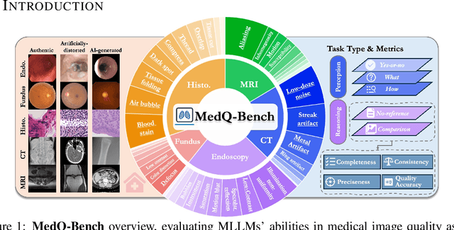
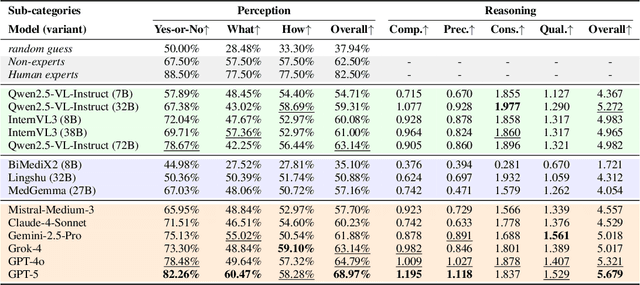
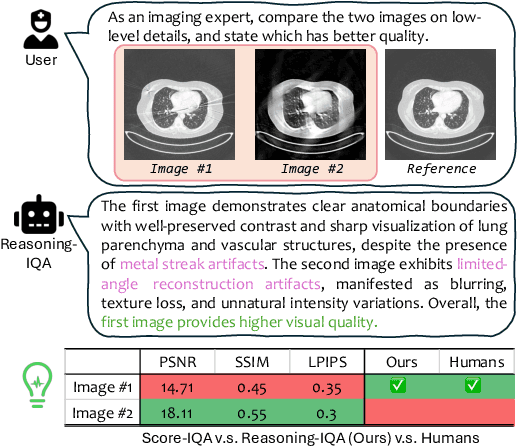
Abstract:Medical Image Quality Assessment (IQA) serves as the first-mile safety gate for clinical AI, yet existing approaches remain constrained by scalar, score-based metrics and fail to reflect the descriptive, human-like reasoning process central to expert evaluation. To address this gap, we introduce MedQ-Bench, a comprehensive benchmark that establishes a perception-reasoning paradigm for language-based evaluation of medical image quality with Multi-modal Large Language Models (MLLMs). MedQ-Bench defines two complementary tasks: (1) MedQ-Perception, which probes low-level perceptual capability via human-curated questions on fundamental visual attributes; and (2) MedQ-Reasoning, encompassing both no-reference and comparison reasoning tasks, aligning model evaluation with human-like reasoning on image quality. The benchmark spans five imaging modalities and over forty quality attributes, totaling 2,600 perceptual queries and 708 reasoning assessments, covering diverse image sources including authentic clinical acquisitions, images with simulated degradations via physics-based reconstructions, and AI-generated images. To evaluate reasoning ability, we propose a multi-dimensional judging protocol that assesses model outputs along four complementary axes. We further conduct rigorous human-AI alignment validation by comparing LLM-based judgement with radiologists. Our evaluation of 14 state-of-the-art MLLMs demonstrates that models exhibit preliminary but unstable perceptual and reasoning skills, with insufficient accuracy for reliable clinical use. These findings highlight the need for targeted optimization of MLLMs in medical IQA. We hope that MedQ-Bench will catalyze further exploration and unlock the untapped potential of MLLMs for medical image quality evaluation.
A Survey of Scientific Large Language Models: From Data Foundations to Agent Frontiers
Aug 28, 2025


Abstract:Scientific Large Language Models (Sci-LLMs) are transforming how knowledge is represented, integrated, and applied in scientific research, yet their progress is shaped by the complex nature of scientific data. This survey presents a comprehensive, data-centric synthesis that reframes the development of Sci-LLMs as a co-evolution between models and their underlying data substrate. We formulate a unified taxonomy of scientific data and a hierarchical model of scientific knowledge, emphasizing the multimodal, cross-scale, and domain-specific challenges that differentiate scientific corpora from general natural language processing datasets. We systematically review recent Sci-LLMs, from general-purpose foundations to specialized models across diverse scientific disciplines, alongside an extensive analysis of over 270 pre-/post-training datasets, showing why Sci-LLMs pose distinct demands -- heterogeneous, multi-scale, uncertainty-laden corpora that require representations preserving domain invariance and enabling cross-modal reasoning. On evaluation, we examine over 190 benchmark datasets and trace a shift from static exams toward process- and discovery-oriented assessments with advanced evaluation protocols. These data-centric analyses highlight persistent issues in scientific data development and discuss emerging solutions involving semi-automated annotation pipelines and expert validation. Finally, we outline a paradigm shift toward closed-loop systems where autonomous agents based on Sci-LLMs actively experiment, validate, and contribute to a living, evolving knowledge base. Collectively, this work provides a roadmap for building trustworthy, continually evolving artificial intelligence (AI) systems that function as a true partner in accelerating scientific discovery.
S2-UniSeg: Fast Universal Agglomerative Pooling for Scalable Segment Anything without Supervision
Aug 09, 2025Abstract:Recent self-supervised image segmentation models have achieved promising performance on semantic segmentation and class-agnostic instance segmentation. However, their pretraining schedule is multi-stage, requiring a time-consuming pseudo-masks generation process between each training epoch. This time-consuming offline process not only makes it difficult to scale with training dataset size, but also leads to sub-optimal solutions due to its discontinuous optimization routine. To solve these, we first present a novel pseudo-mask algorithm, Fast Universal Agglomerative Pooling (UniAP). Each layer of UniAP can identify groups of similar nodes in parallel, allowing to generate both semantic-level and instance-level and multi-granular pseudo-masks within ens of milliseconds for one image. Based on the fast UniAP, we propose the Scalable Self-Supervised Universal Segmentation (S2-UniSeg), which employs a student and a momentum teacher for continuous pretraining. A novel segmentation-oriented pretext task, Query-wise Self-Distillation (QuerySD), is proposed to pretrain S2-UniSeg to learn the local-to-global correspondences. Under the same setting, S2-UniSeg outperforms the SOTA UnSAM model, achieving notable improvements of AP+6.9 on COCO, AR+11.1 on UVO, PixelAcc+4.5 on COCOStuff-27, RQ+8.0 on Cityscapes. After scaling up to a larger 2M-image subset of SA-1B, S2-UniSeg further achieves performance gains on all four benchmarks. Our code and pretrained models are available at https://github.com/bio-mlhui/S2-UniSeg
MedITok: A Unified Tokenizer for Medical Image Synthesis and Interpretation
May 25, 2025Abstract:Advanced autoregressive models have reshaped multimodal AI. However, their transformative potential in medical imaging remains largely untapped due to the absence of a unified visual tokenizer -- one capable of capturing fine-grained visual structures for faithful image reconstruction and realistic image synthesis, as well as rich semantics for accurate diagnosis and image interpretation. To this end, we present MedITok, the first unified tokenizer tailored for medical images, encoding both low-level structural details and high-level clinical semantics within a unified latent space. To balance these competing objectives, we introduce a novel two-stage training framework: a visual representation alignment stage that cold-starts the tokenizer reconstruction learning with a visual semantic constraint, followed by a textual semantic representation alignment stage that infuses detailed clinical semantics into the latent space. Trained on the meticulously collected large-scale dataset with over 30 million medical images and 2 million image-caption pairs, MedITok achieves state-of-the-art performance on more than 30 datasets across 9 imaging modalities and 4 different tasks. By providing a unified token space for autoregressive modeling, MedITok supports a wide range of tasks in clinical diagnostics and generative healthcare applications. Model and code will be made publicly available at: https://github.com/Masaaki-75/meditok.
NeSyGeo: A Neuro-Symbolic Framework for Multimodal Geometric Reasoning Data Generation
May 21, 2025Abstract:Obtaining large-scale, high-quality data with reasoning paths is crucial for improving the geometric reasoning capabilities of multi-modal large language models (MLLMs). However, existing data generation methods, whether based on predefined templates or constrained symbolic provers, inevitably face diversity and numerical generalization limitations. To address these limitations, we propose NeSyGeo, a novel neuro-symbolic framework for generating geometric reasoning data. First, we propose a domain-specific language grounded in the entity-relation-constraint paradigm to comprehensively represent all components of plane geometry, along with generative actions defined within this symbolic space. We then design a symbolic-visual-text pipeline that synthesizes symbolic sequences, maps them to corresponding visual and textual representations, and generates diverse question-answer (Q&A) pairs using large language models (LLMs). To the best of our knowledge, we are the first to propose a neuro-symbolic approach in generating multimodal reasoning data. Based on this framework, we construct NeSyGeo-CoT and NeSyGeo-Caption datasets, containing 100k samples, and release a new benchmark NeSyGeo-Test for evaluating geometric reasoning abilities in MLLMs. Experiments demonstrate that the proposal significantly and consistently improves the performance of multiple MLLMs under both reinforcement and supervised fine-tuning. With only 4k samples and two epochs of reinforcement fine-tuning, base models achieve improvements of up to +15.8% on MathVision, +8.4% on MathVerse, and +7.3% on GeoQA. Notably, a 4B model can be improved to outperform an 8B model from the same series on geometric reasoning tasks.
GMAI-VL-R1: Harnessing Reinforcement Learning for Multimodal Medical Reasoning
Apr 02, 2025Abstract:Recent advances in general medical AI have made significant strides, but existing models often lack the reasoning capabilities needed for complex medical decision-making. This paper presents GMAI-VL-R1, a multimodal medical reasoning model enhanced by reinforcement learning (RL) to improve its reasoning abilities. Through iterative training, GMAI-VL-R1 optimizes decision-making, significantly boosting diagnostic accuracy and clinical support. We also develop a reasoning data synthesis method, generating step-by-step reasoning data via rejection sampling, which further enhances the model's generalization. Experimental results show that after RL training, GMAI-VL-R1 excels in tasks such as medical image diagnosis and visual question answering. While the model demonstrates basic memorization with supervised fine-tuning, RL is crucial for true generalization. Our work establishes new evaluation benchmarks and paves the way for future advancements in medical reasoning models. Code, data, and model will be released at \href{https://github.com/uni-medical/GMAI-VL-R1}{this link}.
Towards Interpretable Counterfactual Generation via Multimodal Autoregression
Mar 29, 2025Abstract:Counterfactual medical image generation enables clinicians to explore clinical hypotheses, such as predicting disease progression, facilitating their decision-making. While existing methods can generate visually plausible images from disease progression prompts, they produce silent predictions that lack interpretation to verify how the generation reflects the hypothesized progression -- a critical gap for medical applications that require traceable reasoning. In this paper, we propose Interpretable Counterfactual Generation (ICG), a novel task requiring the joint generation of counterfactual images that reflect the clinical hypothesis and interpretation texts that outline the visual changes induced by the hypothesis. To enable ICG, we present ICG-CXR, the first dataset pairing longitudinal medical images with hypothetical progression prompts and textual interpretations. We further introduce ProgEmu, an autoregressive model that unifies the generation of counterfactual images and textual interpretations. We demonstrate the superiority of ProgEmu in generating progression-aligned counterfactuals and interpretations, showing significant potential in enhancing clinical decision support and medical education. Project page: https://progemu.github.io.
 Add to Chrome
 Add to Firefox
 Add to Edge