 Add to Chrome
 Add to Firefox
 Add to Edge
RadThinking: A Dataset for Longitudinal Clinical Reasoning in Radiology
May 11, 2026Cancer screening is a reasoning task. A radiologist observes findings, compares them to prior scans, integrates clinical context, and reaches a diagnostic conclusion confirmed by pathology. We present RadThinking, a Visual Question Answering (VQA) dataset that makes this reasoning explicit and trainable. RadThinking releases VQA pairs at three difficulty tiers. Foundation VQAs are atomic perception questions. Single-step reasoning VQAs apply one clinical rule. Compositional VQAs require multi-step chain-of-thought to reach a guideline category such as LI-RADS-5. For every compositional VQA, we release the chain of foundation VQAs that solves it. The chain follows the rules of the governing clinical reporting standard. The dataset spans 20,362 CT scans from 9,131 patients across 43 cancer groups, plus 2,077 verified healthy controls with >1-year follow-up. To our knowledge, RadThinking is the first cancer-screening VQA corpus that stratifies questions by reasoning depth and grounds compositions in clinical reporting standards. The foundation tier supplies atomic perception supervision. The compositional tier supplies chain-of-thought data and verifiable rewards for reinforcement-learning recipes such as DeepSeek-R1 and OpenAI o1. RadThinking enables systematic training and evaluation of whether AI systems can reason about cancer, not merely detect it.
DeepTumorVQA: A Hierarchical 3D CT Benchmark for Stage-Wise Evaluation of Medical VLMs and Tool-Augmented Agents
May 10, 2026Medical vision-language models (VLMs) and AI agents have made significant progress in learning to analyze and reason about clinical images. However, existing medical visual question answering (VQA) benchmarks collapse model capabilities into a single accuracy score, obscuring where and why models fail. We propose DeepTumorVQA, a hierarchical benchmark that follows the multi-stage evidence chain in tumor diagnosis and decomposes 3D CT reasoning into four stages: recognition, measurement, visual reasoning, and medical reasoning. Higher-level questions remain independently scorable, while their ground-truth evidence chains are defined over lower-level primitives. The benchmark contains 476K questions across 42 clinical subtypes on 9,262 3D CT volumes. In addition to a direct reasoning mode for VLMs, DeepTumorVQA provides tool-interaction environments for agent evaluation, where a model can call external tools, including segmentation models, measurement programs, and medical knowledge modules, before answering the question. Evaluating over 30 model configurations, we find that reliable quantitative measurement is the primary bottleneck, making later-stage visual and medical reasoning harder for VLMs, while tool augmentation substantially mitigates this issue. When tools are available, leveraging medical knowledge and tools to reason about medical images becomes a new challenge. We further show that ground-truth step-by-step tool-use traces from DeepTumorVQA can supervise agents and reduce tool-use and reasoning failures. This stage-wise progression from recognition to measurement to visual and medical reasoning provides a concrete roadmap for future medical VLM and AI agent studies. All data and code are released at https://github.com/Schuture/DeepTumorVQA.
Early and Prediagnostic Detection of Pancreatic Cancer from Computed Tomography
Jan 29, 2026Pancreatic ductal adenocarcinoma (PDAC), one of the deadliest solid malignancies, is often detected at a late and inoperable stage. Retrospective reviews of prediagnostic CT scans, when conducted by expert radiologists aware that the patient later developed PDAC, frequently reveal lesions that were previously overlooked. To help detecting these lesions earlier, we developed an automated system named ePAI (early Pancreatic cancer detection with Artificial Intelligence). It was trained on data from 1,598 patients from a single medical center. In the internal test involving 1,009 patients, ePAI achieved an area under the receiver operating characteristic curve (AUC) of 0.939-0.999, a sensitivity of 95.3%, and a specificity of 98.7% for detecting small PDAC less than 2 cm in diameter, precisely localizing PDAC as small as 2 mm. In an external test involving 7,158 patients across 6 centers, ePAI achieved an AUC of 0.918-0.945, a sensitivity of 91.5%, and a specificity of 88.0%, precisely localizing PDAC as small as 5 mm. Importantly, ePAI detected PDACs on prediagnostic CT scans obtained 3 to 36 months before clinical diagnosis that had originally been overlooked by radiologists. It successfully detected and localized PDACs in 75 of 159 patients, with a median lead time of 347 days before clinical diagnosis. Our multi-reader study showed that ePAI significantly outperformed 30 board-certified radiologists by 50.3% (P < 0.05) in sensitivity while maintaining a comparable specificity of 95.4% in detecting PDACs early and prediagnostic. These findings suggest its potential of ePAI as an assistive tool to improve early detection of pancreatic cancer.
Auditing Significance, Metric Choice, and Demographic Fairness in Medical AI Challenges
Dec 22, 2025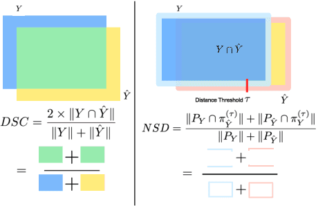
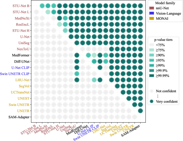
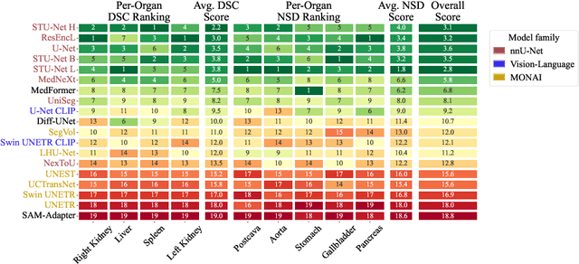
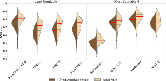
Open challenges have become the de facto standard for comparative ranking of medical AI methods. Despite their importance, medical AI leaderboards exhibit three persistent limitations: (1) score gaps are rarely tested for statistical significance, so rank stability is unknown; (2) single averaged metrics are applied to every organ, hiding clinically important boundary errors; (3) performance across intersecting demographics is seldom reported, masking fairness and equity gaps. We introduce RankInsight, an open-source toolkit that seeks to address these limitations. RankInsight (1) computes pair-wise significance maps that show the nnU-Net family outperforms Vision-Language and MONAI submissions with high statistical certainty; (2) recomputes leaderboards with organ-appropriate metrics, reversing the order of the top four models when Dice is replaced by NSD for tubular structures; and (3) audits intersectional fairness, revealing that more than half of the MONAI-based entries have the largest gender-race discrepancy on our proprietary Johns Hopkins Hospital dataset. The RankInsight toolkit is publicly released and can be directly applied to past, ongoing, and future challenges. It enables organizers and participants to publish rankings that are statistically sound, clinically meaningful, and demographically fair.
See More, Change Less: Anatomy-Aware Diffusion for Contrast Enhancement
Dec 08, 2025Image enhancement improves visual quality and helps reveal details that are hard to see in the original image. In medical imaging, it can support clinical decision-making, but current models often over-edit. This can distort organs, create false findings, and miss small tumors because these models do not understand anatomy or contrast dynamics. We propose SMILE, an anatomy-aware diffusion model that learns how organs are shaped and how they take up contrast. It enhances only clinically relevant regions while leaving all other areas unchanged. SMILE introduces three key ideas: (1) structure-aware supervision that follows true organ boundaries and contrast patterns; (2) registration-free learning that works directly with unaligned multi-phase CT scans; (3) unified inference that provides fast and consistent enhancement across all contrast phases. Across six external datasets, SMILE outperforms existing methods in image quality (14.2% higher SSIM, 20.6% higher PSNR, 50% better FID) and in clinical usefulness by producing anatomically accurate and diagnostically meaningful images. SMILE also improves cancer detection from non-contrast CT, raising the F1 score by up to 10 percent.
PanTS: The Pancreatic Tumor Segmentation Dataset
Jul 02, 2025PanTS is a large-scale, multi-institutional dataset curated to advance research in pancreatic CT analysis. It contains 36,390 CT scans from 145 medical centers, with expert-validated, voxel-wise annotations of over 993,000 anatomical structures, covering pancreatic tumors, pancreas head, body, and tail, and 24 surrounding anatomical structures such as vascular/skeletal structures and abdominal/thoracic organs. Each scan includes metadata such as patient age, sex, diagnosis, contrast phase, in-plane spacing, slice thickness, etc. AI models trained on PanTS achieve significantly better performance in pancreatic tumor detection, localization, and segmentation compared to those trained on existing public datasets. Our analysis indicates that these gains are directly attributable to the 16x larger-scale tumor annotations and indirectly supported by the 24 additional surrounding anatomical structures. As the largest and most comprehensive resource of its kind, PanTS offers a new benchmark for developing and evaluating AI models in pancreatic CT analysis.
Are Vision Language Models Ready for Clinical Diagnosis? A 3D Medical Benchmark for Tumor-centric Visual Question Answering
May 25, 2025Vision-Language Models (VLMs) have shown promise in various 2D visual tasks, yet their readiness for 3D clinical diagnosis remains unclear due to stringent demands for recognition precision, reasoning ability, and domain knowledge. To systematically evaluate these dimensions, we present DeepTumorVQA, a diagnostic visual question answering (VQA) benchmark targeting abdominal tumors in CT scans. It comprises 9,262 CT volumes (3.7M slices) from 17 public datasets, with 395K expert-level questions spanning four categories: Recognition, Measurement, Visual Reasoning, and Medical Reasoning. DeepTumorVQA introduces unique challenges, including small tumor detection and clinical reasoning across 3D anatomy. Benchmarking four advanced VLMs (RadFM, M3D, Merlin, CT-CHAT), we find current models perform adequately on measurement tasks but struggle with lesion recognition and reasoning, and are still not meeting clinical needs. Two key insights emerge: (1) large-scale multimodal pretraining plays a crucial role in DeepTumorVQA testing performance, making RadFM stand out among all VLMs. (2) Our dataset exposes critical differences in VLM components, where proper image preprocessing and design of vision modules significantly affect 3D perception. To facilitate medical multimodal research, we have released DeepTumorVQA as a rigorous benchmark: https://github.com/Schuture/DeepTumorVQA.
Text2CT: Towards 3D CT Volume Generation from Free-text Descriptions Using Diffusion Model
May 07, 2025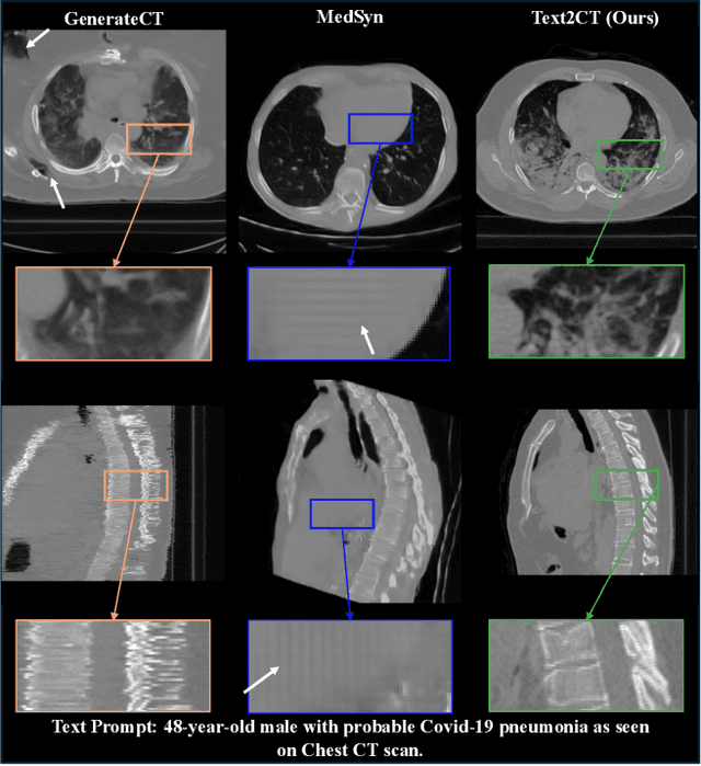
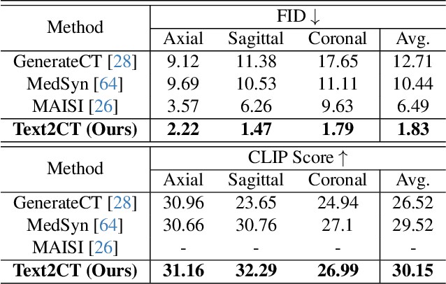
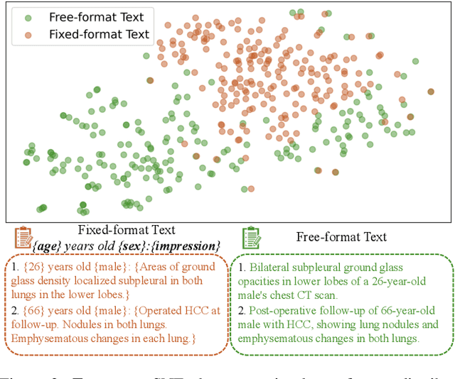
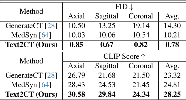
Generating 3D CT volumes from descriptive free-text inputs presents a transformative opportunity in diagnostics and research. In this paper, we introduce Text2CT, a novel approach for synthesizing 3D CT volumes from textual descriptions using the diffusion model. Unlike previous methods that rely on fixed-format text input, Text2CT employs a novel prompt formulation that enables generation from diverse, free-text descriptions. The proposed framework encodes medical text into latent representations and decodes them into high-resolution 3D CT scans, effectively bridging the gap between semantic text inputs and detailed volumetric representations in a unified 3D framework. Our method demonstrates superior performance in preserving anatomical fidelity and capturing intricate structures as described in the input text. Extensive evaluations show that our approach achieves state-of-the-art results, offering promising potential applications in diagnostics, and data augmentation.
RadGPT: Constructing 3D Image-Text Tumor Datasets
Jan 08, 2025With over 85 million CT scans performed annually in the United States, creating tumor-related reports is a challenging and time-consuming task for radiologists. To address this need, we present RadGPT, an Anatomy-Aware Vision-Language AI Agent for generating detailed reports from CT scans. RadGPT first segments tumors, including benign cysts and malignant tumors, and their surrounding anatomical structures, then transforms this information into both structured reports and narrative reports. These reports provide tumor size, shape, location, attenuation, volume, and interactions with surrounding blood vessels and organs. Extensive evaluation on unseen hospitals shows that RadGPT can produce accurate reports, with high sensitivity/specificity for small tumor (<2 cm) detection: 80/73% for liver tumors, 92/78% for kidney tumors, and 77/77% for pancreatic tumors. For large tumors, sensitivity ranges from 89% to 97%. The results significantly surpass the state-of-the-art in abdominal CT report generation. RadGPT generated reports for 17 public datasets. Through radiologist review and refinement, we have ensured the reports' accuracy, and created the first publicly available image-text 3D medical dataset, comprising over 1.8 million text tokens and 2.7 million images from 9,262 CT scans, including 2,947 tumor scans/reports of 8,562 tumor instances. Our reports can: (1) localize tumors in eight liver sub-segments and three pancreatic sub-segments annotated per-voxel; (2) determine pancreatic tumor stage (T1-T4) in 260 reports; and (3) present individual analyses of multiple tumors--rare in human-made reports. Importantly, 948 of the reports are for early-stage tumors.
ScaleMAI: Accelerating the Development of Trusted Datasets and AI Models
Jan 06, 2025


Building trusted datasets is critical for transparent and responsible Medical AI (MAI) research, but creating even small, high-quality datasets can take years of effort from multidisciplinary teams. This process often delays AI benefits, as human-centric data creation and AI-centric model development are treated as separate, sequential steps. To overcome this, we propose ScaleMAI, an agent of AI-integrated data curation and annotation, allowing data quality and AI performance to improve in a self-reinforcing cycle and reducing development time from years to months. We adopt pancreatic tumor detection as an example. First, ScaleMAI progressively creates a dataset of 25,362 CT scans, including per-voxel annotations for benign/malignant tumors and 24 anatomical structures. Second, through progressive human-in-the-loop iterations, ScaleMAI provides Flagship AI Model that can approach the proficiency of expert annotators (30-year experience) in detecting pancreatic tumors. Flagship Model significantly outperforms models developed from smaller, fixed-quality datasets, with substantial gains in tumor detection (+14%), segmentation (+5%), and classification (72%) on three prestigious benchmarks. In summary, ScaleMAI transforms the speed, scale, and reliability of medical dataset creation, paving the way for a variety of impactful, data-driven applications.