 Add to Chrome
 Add to Firefox
 Add to Edge
A Pathology Foundation Model for Gastric Cancer with Real-World Validation
Jun 03, 2026Gastric cancer remains a major cause of cancer mortality, yet its histological and molecular heterogeneity complicates diagnosis and risk stratification. General-purpose pathology foundation models (PFMs) often plateau on fine-grained endpoints central to gastric cancer care, and few have undergone rigorous prospective validation or clinical reader studies. We present GRACE, a Gastric-specific foundation model for Real-world Assessment and Clinical dEcision support. GRACE was developed from multicenter gastric pathology datasets totaling 48,364 primarily HE-stained whole-slide images from 37,493 patients. When evaluated on 28 clinically relevant tasks, GRACE consistently outperformed representative pancancer PFMs, achieving a macro-AUC of 0.9188, with strong performance for precancerous lesion diagnosis (macro-AUC 0.9322), tumor histopathological assessment (macro-AUC 0.9119), molecular profiling (macro-AUC 0.8682), and prognostic prediction. Beyond benchmarking, GRACE's translational value was substantiated through a rigorous evidence chain. Under safety-gated criteria requiring 100% NPV for rule-out and 100% PPV for rule-in, GRACE streamlined review for up to 69.6% of malignancy-diagnosis cases and triaged 46.8% of MMR-IHC follow-up requests. This translational feasibility was further strengthened by a randomized crossover reader study of pathologist-AI collaboration. With GRACE assistance, diagnostic accuracy improved from 82.0% to 89.9%, yielding nearly twofold higher adjusted odds of a correct diagnosis (OR 1.987) alongside concurrent gains in sensitivity and specificity. AI assistance also reduced diagnostic time by 14.9%, elevated diagnostic confidence by 9.0%, and markedly improved inter-rater agreement. When calibrated to maintain non-inferior performance to senior pathologists, the AI-assisted workflow could triage 60.7% of atrophy and 82.7% of intestinal metaplasia cases.
Spatial Transcriptomics-Guided Alignment Enhances Molecular Profiling in Pathology Foundation Model
May 29, 2026Comprehensive molecular profiling is essential for modern precision oncology but remains hindered by prohibitive costs, specimen exhaustion, and protracted turnaround times. While pathology foundation models (PFMs) have demonstrated potential for inferring molecular phenotypes from routine hematoxylin and eosin (H&E) whole-slide images (WSIs), current architectures primarily rely on vision-centric self-supervised learning or vision-language alignment, lacking the spatially resolved molecular supervision required to connect subtle morphological features with underlying genomic alterations. Spatial transcriptomics (ST) emerges as a transformative technology that enables transcriptomic quantification within intact tissue sections, thereby preserving the precise spatial link between histology and molecular profiles. In this study, we present a Spatial Transcriptomics-guided Alignment framework for Molecular Profiling (STAMP), which endows PFMs with intrinsic molecular awareness. To support this paradigm, we curated HumanST-1k, a human ST dataset spanning diverse anatomical organs and sequencing platforms. This atlas yields 1.8 million pairs of H&E patches and corresponding transcriptomic profiles, providing a corpus that links histological structures with their molecular states. To mitigate the technical noise inherent to raw transcriptomics, STAMP applies a pathway-informed alignment strategy that aggregates transcriptomic data into biologically functional pathways, which are subsequently integrated into PFMs via parameter-efficient fine-tuning. This alignment enriches the representation space of PFMs and unlocks their capacity to resolve sub-visual molecular signatures. The clinical utility of these augmented representations was validated through a multi-tier evaluation framework.
A Clinically Validated Foundation Model for Comprehensive Lung Pathology Interpretation
May 25, 2026Pathological assessment guides lung cancer diagnosis, treatment selection, and prognostic evaluation, yet current CPath approaches rely on task-specific models for isolated objectives. Although pan-cancer foundation models offer versatility, they lack subspecialty-level depth and have not been evaluated across clinical workflows or prospectively validated in real-world settings. We introduce PulmoFoundation, a multi-center, prospectively validated, randomized controlled trial (RCT)-evaluated foundation model for comprehensive lung pathology assessment across pre-operative, intra-operative, and post-operative care. Built upon Virchow2 via subspecialty-specific pretraining using ~40,000 diagnostic H&E-stained whole-slide images (WSIs), PulmoFoundation was systematically evaluated on ~26,000 WSIs across 32 clinically relevant tasks. In addition to accurately predicting molecular markers and patient survival, our model achieves clinical-grade performance in core diagnostic tasks across biopsy, frozen section, and surgical resection slides. In a registered prospective study of 1,357 patients across 11 diagnostic tasks, our model achieved an average AUC of 92.3%. Using pre-specified triage thresholds, PulmoFoundation could reduce additional second-review burden for 68.8% of biopsies and 83.0% of frozen sections, and defer 44.5% of IHC stain orders, with PPVs of 1.0, 0.991, and 0.966. Beyond prospective validation, we conducted a crossover RCT with eight pathologists, in which AI assistance improved diagnostic accuracy across 4,928 case-reader pairs (91.7% w/ AI vs. 83.8% w/o AI). AI assistance also reduced median diagnostic time by 19.6%, increased diagnostic confidence by 8.7%, and improved inter-rater agreement from moderate (kappa = 0.56) to substantial (kappa = 0.76). Together, these evaluations support PulmoFoundation as a clinically validated decision-support system for lung pathology.
A Deployment-Friendly Foundational Framework for Efficient Computational Pathology
Feb 15, 2026Pathology foundation models (PFMs) have enabled robust generalization in computational pathology through large-scale datasets and expansive architectures, but their substantial computational cost, particularly for gigapixel whole slide images, limits clinical accessibility and scalability. Here, we present LitePath, a deployment-friendly foundational framework designed to mitigate model over-parameterization and patch level redundancy. LitePath integrates LiteFM, a compact model distilled from three large PFMs (Virchow2, H-Optimus-1 and UNI2) using 190 million patches, and the Adaptive Patch Selector (APS), a lightweight component for task-specific patch selection. The framework reduces model parameters by 28x and lowers FLOPs by 403.5x relative to Virchow2, enabling deployment on low-power edge hardware such as the NVIDIA Jetson Orin Nano Super. On this device, LitePath processes 208 slides per hour, 104.5x faster than Virchow2, and consumes 0.36 kWh per 3,000 slides, 171x lower than Virchow2 on an RTX3090 GPU. We validated accuracy using 37 cohorts across four organs and 26 tasks (26 internal, 9 external, and 2 prospective), comprising 15,672 slides from 9,808 patients disjoint from the pretraining data. LitePath ranks second among 19 evaluated models and outperforms larger models including H-Optimus-1, mSTAR, UNI2 and GPFM, while retaining 99.71% of the AUC of Virchow2 on average. To quantify the balance between accuracy and efficiency, we propose the Deployability Score (D-Score), defined as the weighted geometric mean of normalized AUC and normalized FLOP, where LitePath achieves the highest value, surpassing Virchow2 by 10.64%. These results demonstrate that LitePath enables rapid, cost-effective and energy-efficient pathology image analysis on accessible hardware while maintaining accuracy comparable to state-of-the-art PFMs and reducing the carbon footprint of AI deployment.
A Multimodal Foundation Model to Enhance Generalizability and Data Efficiency for Pan-cancer Prognosis Prediction
Sep 16, 2025Multimodal data provides heterogeneous information for a holistic understanding of the tumor microenvironment. However, existing AI models often struggle to harness the rich information within multimodal data and extract poorly generalizable representations. Here we present MICE (Multimodal data Integration via Collaborative Experts), a multimodal foundation model that effectively integrates pathology images, clinical reports, and genomics data for precise pan-cancer prognosis prediction. Instead of conventional multi-expert modules, MICE employs multiple functionally diverse experts to comprehensively capture both cross-cancer and cancer-specific insights. Leveraging data from 11,799 patients across 30 cancer types, we enhanced MICE's generalizability by coupling contrastive and supervised learning. MICE outperformed both unimodal and state-of-the-art multi-expert-based multimodal models, demonstrating substantial improvements in C-index ranging from 3.8% to 11.2% on internal cohorts and 5.8% to 8.8% on independent cohorts, respectively. Moreover, it exhibited remarkable data efficiency across diverse clinical scenarios. With its enhanced generalizability and data efficiency, MICE establishes an effective and scalable foundation for pan-cancer prognosis prediction, holding strong potential to personalize tailored therapies and improve treatment outcomes.
Genome-Anchored Foundation Model Embeddings Improve Molecular Prediction from Histology Images
Jun 24, 2025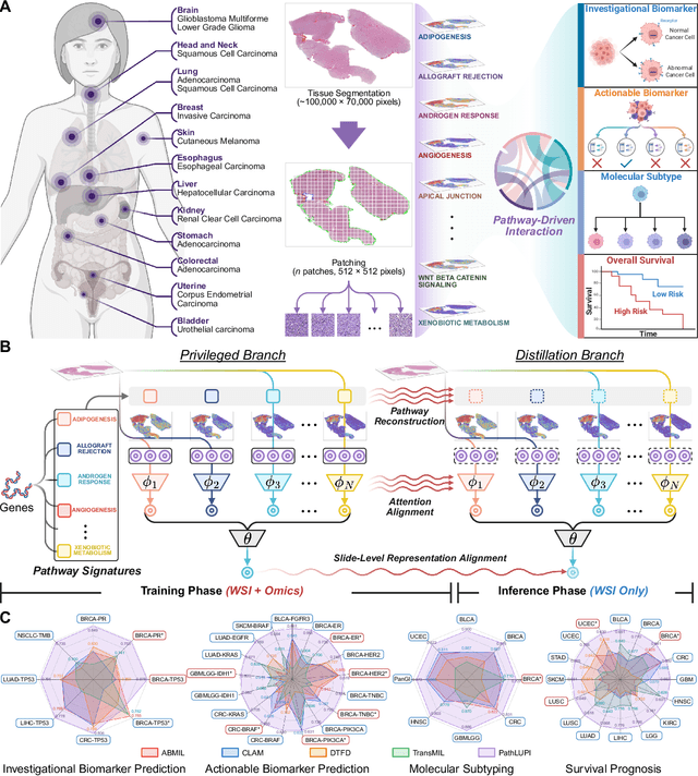
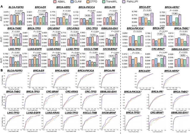
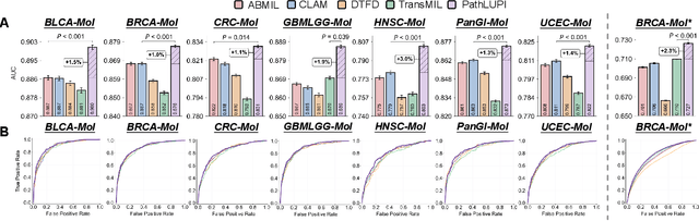
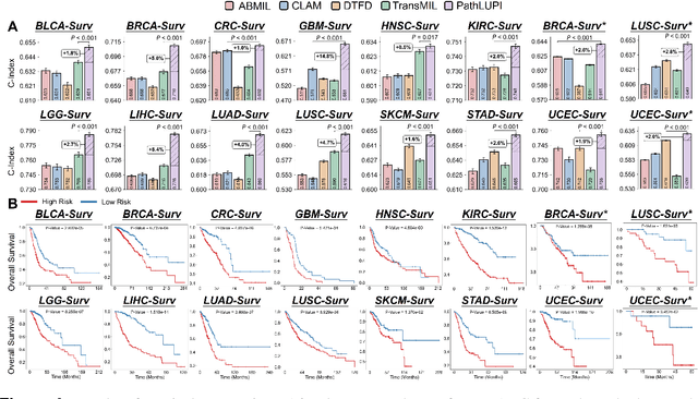
Precision oncology requires accurate molecular insights, yet obtaining these directly from genomics is costly and time-consuming for broad clinical use. Predicting complex molecular features and patient prognosis directly from routine whole-slide images (WSI) remains a major challenge for current deep learning methods. Here we introduce PathLUPI, which uses transcriptomic privileged information during training to extract genome-anchored histological embeddings, enabling effective molecular prediction using only WSIs at inference. Through extensive evaluation across 49 molecular oncology tasks using 11,257 cases among 20 cohorts, PathLUPI demonstrated superior performance compared to conventional methods trained solely on WSIs. Crucially, it achieves AUC $\geq$ 0.80 in 14 of the biomarker prediction and molecular subtyping tasks and C-index $\geq$ 0.70 in survival cohorts of 5 major cancer types. Moreover, PathLUPI embeddings reveal distinct cellular morphological signatures associated with specific genotypes and related biological pathways within WSIs. By effectively encoding molecular context to refine WSI representations, PathLUPI overcomes a key limitation of existing models and offers a novel strategy to bridge molecular insights with routine pathology workflows for wider clinical application.
PathBench: A comprehensive comparison benchmark for pathology foundation models towards precision oncology
May 26, 2025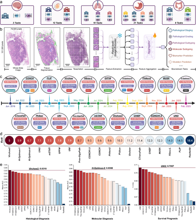


The emergence of pathology foundation models has revolutionized computational histopathology, enabling highly accurate, generalized whole-slide image analysis for improved cancer diagnosis, and prognosis assessment. While these models show remarkable potential across cancer diagnostics and prognostics, their clinical translation faces critical challenges including variability in optimal model across cancer types, potential data leakage in evaluation, and lack of standardized benchmarks. Without rigorous, unbiased evaluation, even the most advanced PFMs risk remaining confined to research settings, delaying their life-saving applications. Existing benchmarking efforts remain limited by narrow cancer-type focus, potential pretraining data overlaps, or incomplete task coverage. We present PathBench, the first comprehensive benchmark addressing these gaps through: multi-center in-hourse datasets spanning common cancers with rigorous leakage prevention, evaluation across the full clinical spectrum from diagnosis to prognosis, and an automated leaderboard system for continuous model assessment. Our framework incorporates large-scale data, enabling objective comparison of PFMs while reflecting real-world clinical complexity. All evaluation data comes from private medical providers, with strict exclusion of any pretraining usage to avoid data leakage risks. We have collected 15,888 WSIs from 8,549 patients across 10 hospitals, encompassing over 64 diagnosis and prognosis tasks. Currently, our evaluation of 19 PFMs shows that Virchow2 and H-Optimus-1 are the most effective models overall. This work provides researchers with a robust platform for model development and offers clinicians actionable insights into PFM performance across diverse clinical scenarios, ultimately accelerating the translation of these transformative technologies into routine pathology practice.
SEW: Self-calibration Enhanced Whole Slide Pathology Image Analysis
Dec 14, 2024


Pathology images are considered the "gold standard" for cancer diagnosis and treatment, with gigapixel images providing extensive tissue and cellular information. Existing methods fail to simultaneously extract global structural and local detail f
Efficient and Comprehensive Feature Extraction in Large Vision-Language Model for Clinical Pathology Analysis
Dec 12, 2024Pathological diagnosis is vital for determining disease characteristics, guiding treatment, and assessing prognosis, relying heavily on detailed, multi-scale analysis of high-resolution whole slide images (WSI). However, traditional pure vision models face challenges of redundant feature extraction, whereas existing large vision-language models (LVLMs) are limited by input resolution constraints, hindering their efficiency and accuracy. To overcome these issues, we propose two innovative strategies: the mixed task-guided feature enhancement, which directs feature extraction toward lesion-related details across scales, and the prompt-guided detail feature completion, which integrates coarse- and fine-grained features from WSI based on specific prompts without compromising inference speed. Leveraging a comprehensive dataset of 490,000 samples from diverse pathology tasks-including cancer detection, grading, vascular and neural invasion identification, and so on-we trained the pathology-specialized LVLM, OmniPath. Extensive experiments demonstrate that this model significantly outperforms existing methods in diagnostic accuracy and efficiency, offering an interactive, clinically aligned approach for auxiliary diagnosis in a wide range of pathology applications.
DiffusionRig: Learning Personalized Priors for Facial Appearance Editing
Apr 13, 2023
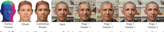

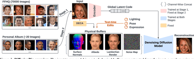
We address the problem of learning person-specific facial priors from a small number (e.g., 20) of portrait photos of the same person. This enables us to edit this specific person's facial appearance, such as expression and lighting, while preserving their identity and high-frequency facial details. Key to our approach, which we dub DiffusionRig, is a diffusion model conditioned on, or "rigged by," crude 3D face models estimated from single in-the-wild images by an off-the-shelf estimator. On a high level, DiffusionRig learns to map simplistic renderings of 3D face models to realistic photos of a given person. Specifically, DiffusionRig is trained in two stages: It first learns generic facial priors from a large-scale face dataset and then person-specific priors from a small portrait photo collection of the person of interest. By learning the CGI-to-photo mapping with such personalized priors, DiffusionRig can "rig" the lighting, facial expression, head pose, etc. of a portrait photo, conditioned only on coarse 3D models while preserving this person's identity and other high-frequency characteristics. Qualitative and quantitative experiments show that DiffusionRig outperforms existing approaches in both identity preservation and photorealism. Please see the project website: https://diffusionrig.github.io for the supplemental material, video, code, and data.