Xiulan Zhang
VisionUnite: A Vision-Language Foundation Model for Ophthalmology Enhanced with Clinical Knowledge
Aug 05, 2024


Abstract:The need for improved diagnostic methods in ophthalmology is acute, especially in the less developed regions with limited access to specialists and advanced equipment. Therefore, we introduce VisionUnite, a novel vision-language foundation model for ophthalmology enhanced with clinical knowledge. VisionUnite has been pretrained on an extensive dataset comprising 1.24 million image-text pairs, and further refined using our proposed MMFundus dataset, which includes 296,379 high-quality fundus image-text pairs and 889,137 simulated doctor-patient dialogue instances. Our experiments indicate that VisionUnite outperforms existing generative foundation models such as GPT-4V and Gemini Pro. It also demonstrates diagnostic capabilities comparable to junior ophthalmologists. VisionUnite performs well in various clinical scenarios including open-ended multi-disease diagnosis, clinical explanation, and patient interaction, making it a highly versatile tool for initial ophthalmic disease screening. VisionUnite can also serve as an educational aid for junior ophthalmologists, accelerating their acquisition of knowledge regarding both common and rare ophthalmic conditions. VisionUnite represents a significant advancement in ophthalmology, with broad implications for diagnostics, medical education, and understanding of disease mechanisms.
PALM: Open Fundus Photograph Dataset with Pathologic Myopia Recognition and Anatomical Structure Annotation
May 13, 2023Abstract:Pathologic myopia (PM) is a common blinding retinal degeneration suffered by highly myopic population. Early screening of this condition can reduce the damage caused by the associated fundus lesions and therefore prevent vision loss. Automated diagnostic tools based on artificial intelligence methods can benefit this process by aiding clinicians to identify disease signs or to screen mass populations using color fundus photographs as inputs. This paper provides insights about PALM, our open fundus imaging dataset for pathological myopia recognition and anatomical structure annotation. Our databases comprises 1200 images with associated labels for the pathologic myopia category and manual annotations of the optic disc, the position of the fovea and delineations of lesions such as patchy retinal atrophy (including peripapillary atrophy) and retinal detachment. In addition, this paper elaborates on other details such as the labeling process used to construct the database, the quality and characteristics of the samples and provides other relevant usage notes.
Multi-Scale Multi-Target Domain Adaptation for Angle Closure Classification
Aug 25, 2022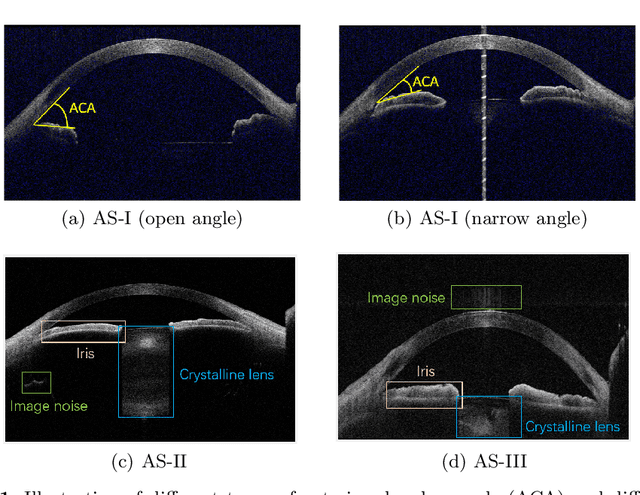

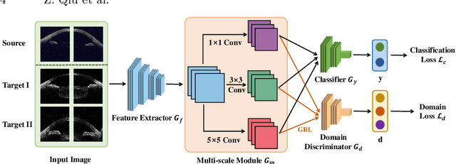
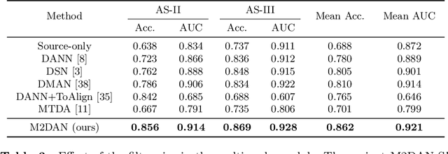
Abstract:Deep learning (DL) has made significant progress in angle closure classification with anterior segment optical coherence tomography (AS-OCT) images. These AS-OCT images are often acquired by different imaging devices/conditions, which results in a vast change of underlying data distributions (called "data domains"). Moreover, due to practical labeling difficulties, some domains (e.g., devices) may not have any data labels. As a result, deep models trained on one specific domain (e.g., a specific device) are difficult to adapt to and thus may perform poorly on other domains (e.g., other devices). To address this issue, we present a multi-target domain adaptation paradigm to transfer a model trained on one labeled source domain to multiple unlabeled target domains. Specifically, we propose a novel Multi-scale Multi-target Domain Adversarial Network (M2DAN) for angle closure classification. M2DAN conducts multi-domain adversarial learning for extracting domain-invariant features and develops a multi-scale module for capturing local and global information of AS-OCT images. Based on these domain-invariant features at different scales, the deep model trained on the source domain is able to classify angle closure on multiple target domains even without any annotations in these domains. Extensive experiments on a real-world AS-OCT dataset demonstrate the effectiveness of the proposed method.
Dataset and Evaluation algorithm design for GOALS Challenge
Jul 29, 2022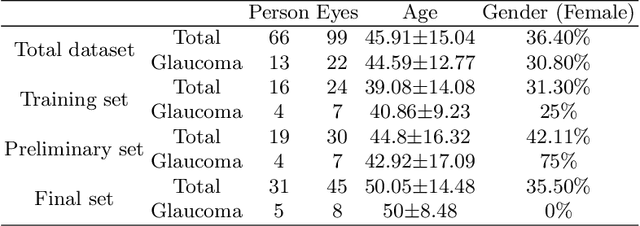
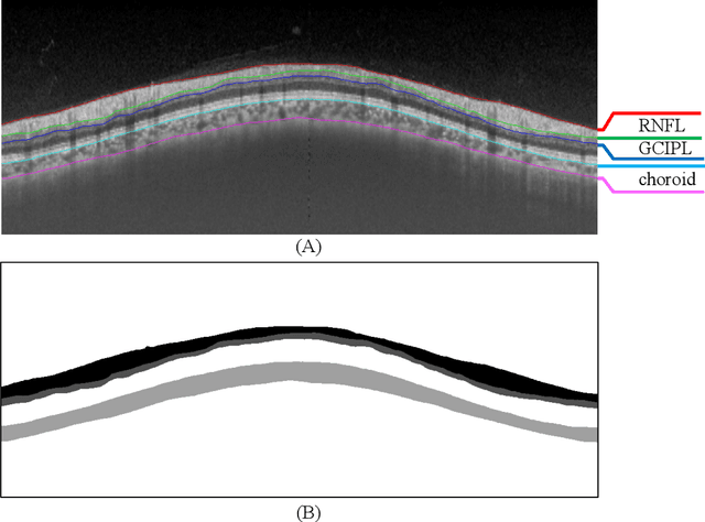
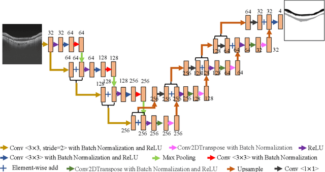
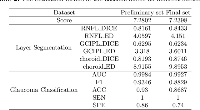
Abstract:Glaucoma causes irreversible vision loss due to damage to the optic nerve, and there is no cure for glaucoma.OCT imaging modality is an essential technique for assessing glaucomatous damage since it aids in quantifying fundus structures. To promote the research of AI technology in the field of OCT-assisted diagnosis of glaucoma, we held a Glaucoma OCT Analysis and Layer Segmentation (GOALS) Challenge in conjunction with the International Conference on Medical Image Computing and Computer Assisted Intervention (MICCAI) 2022 to provide data and corresponding annotations for researchers studying layer segmentation from OCT images and the classification of glaucoma. This paper describes the released 300 circumpapillary OCT images, the baselines of the two sub-tasks, and the evaluation methodology. The GOALS Challenge is accessible at https://aistudio.baidu.com/aistudio/competition/detail/230.
REFUGE2 Challenge: Treasure for Multi-Domain Learning in Glaucoma Assessment
Feb 24, 2022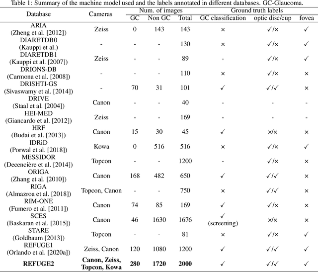
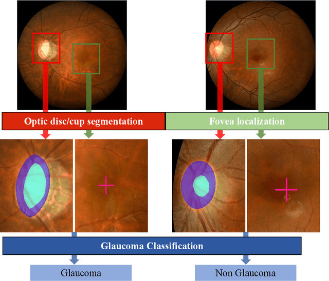
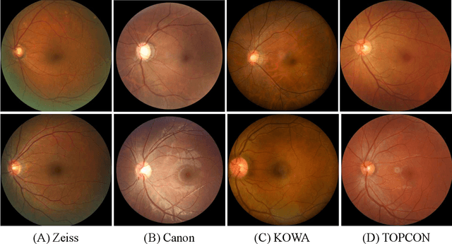

Abstract:Glaucoma is the second leading cause of blindness and is the leading cause of irreversible blindness disease in the world. Early screening for glaucoma in the population is significant. Color fundus photography is the most cost effective imaging modality to screen for ocular diseases. Deep learning network is often used in color fundus image analysis due to its powful feature extraction capability. However, the model training of deep learning method needs a large amount of data, and the distribution of data should be abundant for the robustness of model performance. To promote the research of deep learning in color fundus photography and help researchers further explore the clinical application signification of AI technology, we held a REFUGE2 challenge. This challenge released 2,000 color fundus images of four models, including Zeiss, Canon, Kowa and Topcon, which can validate the stabilization and generalization of algorithms on multi-domain. Moreover, three sub-tasks were designed in the challenge, including glaucoma classification, cup/optic disc segmentation, and macular fovea localization. These sub-tasks technically cover the three main problems of computer vision and clinicly cover the main researchs of glaucoma diagnosis. Over 1,300 international competitors joined the REFUGE2 challenge, 134 teams submitted more than 3,000 valid preliminary results, and 22 teams reached the final. This article summarizes the methods of some of the finalists and analyzes their results. In particular, we observed that the teams using domain adaptation strategies had high and robust performance on the dataset with multi-domain. This indicates that UDA and other multi-domain related researches will be the trend of deep learning field in the future, and our REFUGE2 datasets will play an important role in these researches.
ADAM Challenge: Detecting Age-related Macular Degeneration from Fundus Images
Feb 18, 2022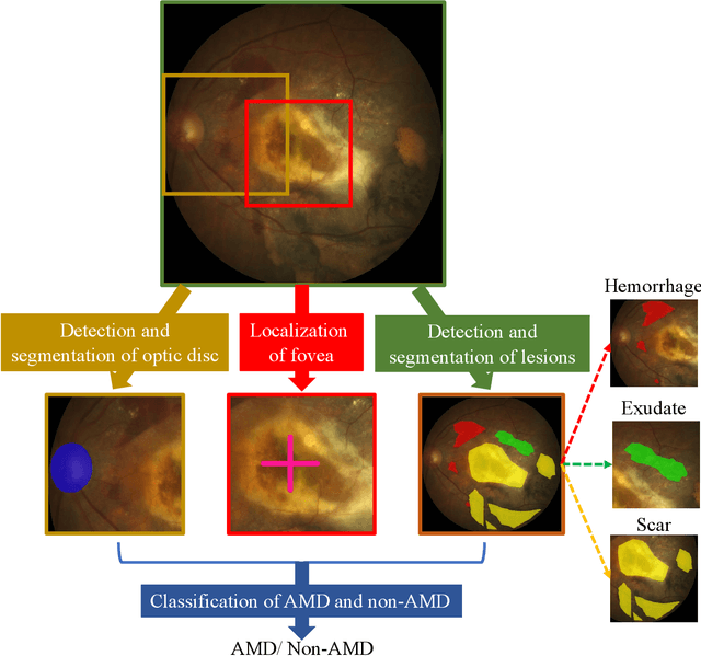

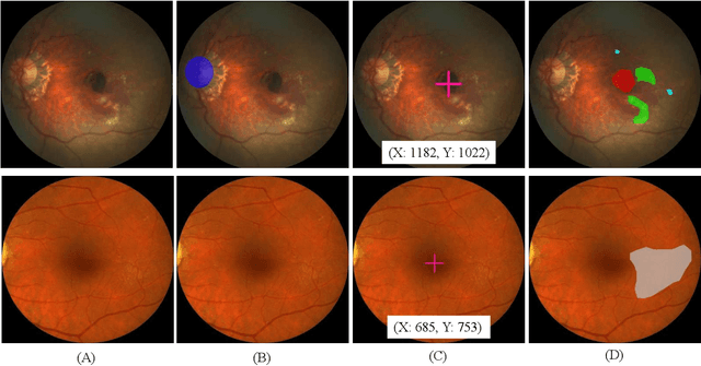
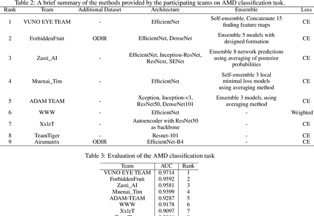
Abstract:Age-related macular degeneration (AMD) is the leading cause of visual impairment among elderly in the world. Early detection of AMD is of great importance as the vision loss caused by AMD is irreversible and permanent. Color fundus photography is the most cost-effective imaging modality to screen for retinal disorders. \textcolor{red}{Recently, some algorithms based on deep learning had been developed for fundus image analysis and automatic AMD detection. However, a comprehensive annotated dataset and a standard evaluation benchmark are still missing.} To deal with this issue, we set up the Automatic Detection challenge on Age-related Macular degeneration (ADAM) for the first time, held as a satellite event of the ISBI 2020 conference. The ADAM challenge consisted of four tasks which cover the main topics in detecting AMD from fundus images, including classification of AMD, detection and segmentation of optic disc, localization of fovea, and detection and segmentation of lesions. The ADAM challenge has released a comprehensive dataset of 1200 fundus images with the category labels of AMD, the pixel-wise segmentation masks of the full optic disc and lesions (drusen, exudate, hemorrhage, scar, and other), as well as the location coordinates of the macular fovea. A uniform evaluation framework has been built to make a fair comparison of different models. During the ADAM challenge, 610 results were submitted for online evaluation, and finally, 11 teams participated in the onsite challenge. This paper introduces the challenge, dataset, and evaluation methods, as well as summarizes the methods and analyzes the results of the participating teams of each task. In particular, we observed that ensembling strategy and clinical prior knowledge can better improve the performances of the deep learning models.
GAMMA Challenge:Glaucoma grAding from Multi-Modality imAges
Feb 16, 2022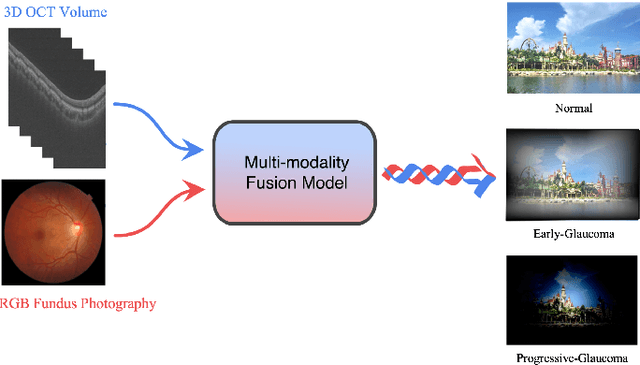
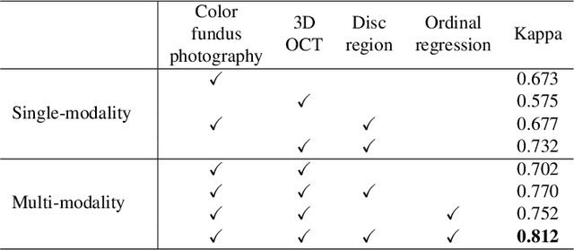
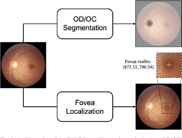
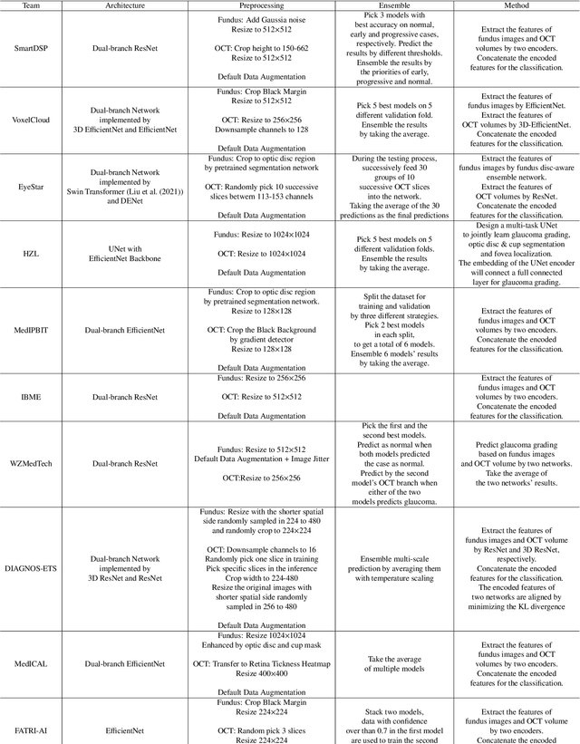
Abstract:Color fundus photography and Optical Coherence Tomography (OCT) are the two most cost-effective tools for glaucoma screening. Both two modalities of images have prominent biomarkers to indicate glaucoma suspected. Clinically, it is often recommended to take both of the screenings for a more accurate and reliable diagnosis. However, although numerous algorithms are proposed based on fundus images or OCT volumes in computer-aided diagnosis, there are still few methods leveraging both of the modalities for the glaucoma assessment. Inspired by the success of Retinal Fundus Glaucoma Challenge (REFUGE) we held previously, we set up the Glaucoma grAding from Multi-Modality imAges (GAMMA) Challenge to encourage the development of fundus \& OCT-based glaucoma grading. The primary task of the challenge is to grade glaucoma from both the 2D fundus images and 3D OCT scanning volumes. As part of GAMMA, we have publicly released a glaucoma annotated dataset with both 2D fundus color photography and 3D OCT volumes, which is the first multi-modality dataset for glaucoma grading. In addition, an evaluation framework is also established to evaluate the performance of the submitted methods. During the challenge, 1272 results were submitted, and finally, top-10 teams were selected to the final stage. We analysis their results and summarize their methods in the paper. Since all these teams submitted their source code in the challenge, a detailed ablation study is also conducted to verify the effectiveness of the particular modules proposed. We find many of the proposed techniques are practical for the clinical diagnosis of glaucoma. As the first in-depth study of fundus \& OCT multi-modality glaucoma grading, we believe the GAMMA Challenge will be an essential starting point for future research.
Open-Narrow-Synechiae Anterior Chamber Angle Classification in AS-OCT Sequences
Jun 09, 2020
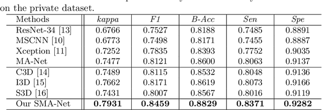
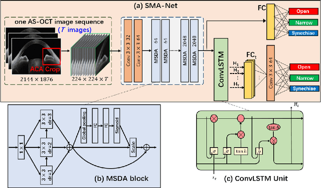

Abstract:Anterior chamber angle (ACA) classification is a key step in the diagnosis of angle-closure glaucoma in Anterior Segment Optical Coherence Tomography (AS-OCT). Existing automated analysis methods focus on a binary classification system (i.e., open angle or angle-closure) in a 2D AS-OCT slice. However, clinical diagnosis requires a more discriminating ACA three-class system (i.e., open, narrow, or synechiae angles) for the benefit of clinicians who seek better to understand the progression of the spectrum of angle-closure glaucoma types. To address this, we propose a novel sequence multi-scale aggregation deep network (SMA-Net) for open-narrow-synechiae ACA classification based on an AS-OCT sequence. In our method, a Multi-Scale Discriminative Aggregation (MSDA) block is utilized to learn the multi-scale representations at slice level, while a ConvLSTM is introduced to study the temporal dynamics of these representations at sequence level. Finally, a multi-level loss function is used to combine the slice-based and sequence-based losses. The proposed method is evaluated across two AS-OCT datasets. The experimental results show that the proposed method outperforms existing state-of-the-art methods in applicability, effectiveness, and accuracy. We believe this work to be the first attempt to classify ACAs into open, narrow, or synechia types grading using AS-OCT sequences.
Reconstruction and Quantification of 3D Iris Surface for Angle-Closure Glaucoma Detection in Anterior Segment OCT
Jun 09, 2020

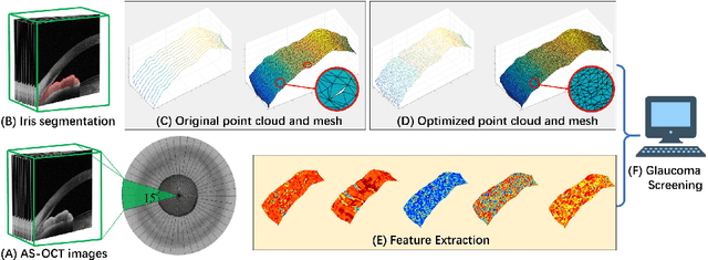
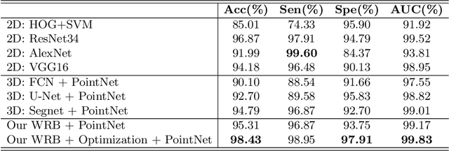
Abstract:Precise characterization and analysis of iris shape from Anterior Segment OCT (AS-OCT) are of great importance in facilitating diagnosis of angle-closure-related diseases. Existing methods focus solely on analyzing structural properties identified from the 2D slice, while accurate characterization of morphological changes of iris shape in 3D AS-OCT may be able to reveal in addition the risk of disease progression. In this paper, we propose a novel framework for reconstruction and quantification of 3D iris surface from AS-OCT imagery. We consider it to be the first work to detect angle-closure glaucoma by means of 3D representation. An iris segmentation network with wavelet refinement block (WRB) is first proposed to generate the initial shape of the iris from single AS-OCT slice. The 3D iris surface is then reconstructed using a guided optimization method with Poisson-disk sampling. Finally, a set of surface-based features are extracted, which are used in detecting of angle-closure glaucoma. Experimental results demonstrate that our method is highly effective in iris segmentation and surface reconstruction. Moreover, we show that 3D-based representation achieves better performance in angle-closure glaucoma detection than does 2D-based feature.
AGE Challenge: Angle Closure Glaucoma Evaluation in Anterior Segment Optical Coherence Tomography
May 05, 2020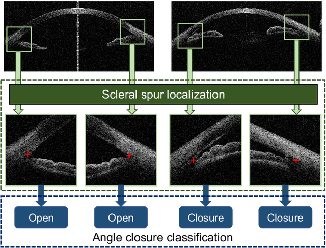

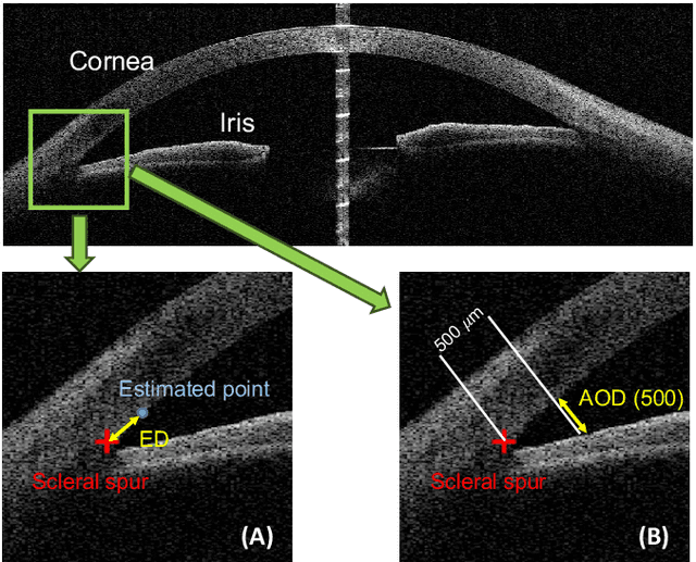

Abstract:Angle closure glaucoma (ACG) is a more aggressive disease than open-angle glaucoma, where the abnormal anatomical structures of the anterior chamber angle (ACA) may cause an elevated intraocular pressure and gradually leads to glaucomatous optic neuropathy and eventually to visual impairment and blindness. Anterior Segment Optical Coherence Tomography (AS-OCT) imaging provides a fast and contactless way to discriminate angle closure from open angle. Although many medical image analysis algorithms have been developed for glaucoma diagnosis, only a few studies have focused on AS-OCT imaging. In particular, there is no public AS-OCT dataset available for evaluating the existing methods in a uniform way, which limits the progress in the development of automated techniques for angle closure detection and assessment. To address this, we organized the Angle closure Glaucoma Evaluation challenge (AGE), held in conjunction with MICCAI 2019. The AGE challenge consisted of two tasks: scleral spur localization and angle closure classification. For this challenge, we released a large data of 4800 annotated AS-OCT images from 199 patients, and also proposed an evaluation framework to benchmark and compare different models. During the AGE challenge, over 200 teams registered online, and more than 1100 results were submitted for online evaluation. Finally, eight teams participated in the onsite challenge. In this paper, we summarize these eight onsite challenge methods and analyze their corresponding results in the two tasks. We further discuss limitations and future directions. In the AGE challenge, the top-performing approach had an average Euclidean Distance of 10 pixel in scleral spur localization, while in the task of angle closure classification, all the algorithms achieved the satisfactory performances, especially, 100% accuracy rate for top-two performances.
 Add to Chrome
 Add to Firefox
 Add to Edge