Huaying Hao
Sparse-based Domain Adaptation Network for OCTA Image Super-Resolution Reconstruction
Jul 25, 2022


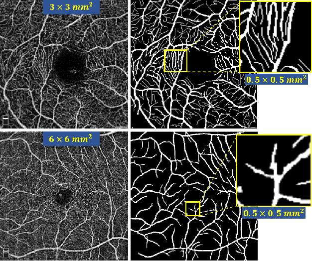
Abstract:Retinal Optical Coherence Tomography Angiography (OCTA) with high-resolution is important for the quantification and analysis of retinal vasculature. However, the resolution of OCTA images is inversely proportional to the field of view at the same sampling frequency, which is not conducive to clinicians for analyzing larger vascular areas. In this paper, we propose a novel Sparse-based domain Adaptation Super-Resolution network (SASR) for the reconstruction of realistic 6x6 mm2/low-resolution (LR) OCTA images to high-resolution (HR) representations. To be more specific, we first perform a simple degradation of the 3x3 mm2/high-resolution (HR) image to obtain the synthetic LR image. An efficient registration method is then employed to register the synthetic LR with its corresponding 3x3 mm2 image region within the 6x6 mm2 image to obtain the cropped realistic LR image. We then propose a multi-level super-resolution model for the fully-supervised reconstruction of the synthetic data, guiding the reconstruction of the realistic LR images through a generative-adversarial strategy that allows the synthetic and realistic LR images to be unified in the feature domain. Finally, a novel sparse edge-aware loss is designed to dynamically optimize the vessel edge structure. Extensive experiments on two OCTA sets have shown that our method performs better than state-of-the-art super-resolution reconstruction methods. In addition, we have investigated the performance of the reconstruction results on retina structure segmentations, which further validate the effectiveness of our approach.
ROSE: A Retinal OCT-Angiography Vessel Segmentation Dataset and New Model
Jul 10, 2020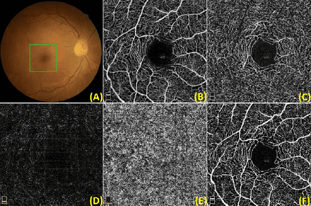
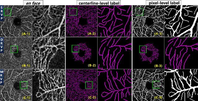
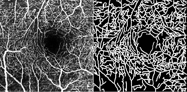
Abstract:Optical Coherence Tomography Angiography (OCT-A) is a non-invasive imaging technique, and has been increasingly used to image the retinal vasculature at capillary level resolution. However, automated segmentation of retinal vessels in OCT-A has been under-studied due to various challenges such as low capillary visibility and high vessel complexity, despite its significance in understanding many eye-related diseases. In addition, there is no publicly available OCT-A dataset with manually graded vessels for training and validation. To address these issues, for the first time in the field of retinal image analysis we construct a dedicated Retinal OCT-A SEgmentation dataset (ROSE), which consists of 229 OCT-A images with vessel annotations at either centerline-level or pixel level. This dataset has been released for public access to assist researchers in the community in undertaking research in related topics. Secondly, we propose a novel Split-based Coarse-to-Fine vessel segmentation network (SCF-Net), with the ability to detect thick and thin vessels separately. In the SCF-Net, a split-based coarse segmentation (SCS) module is first introduced to produce a preliminary confidence map of vessels, and a split-based refinement (SRN) module is then used to optimize the shape/contour of the retinal microvasculature. Thirdly, we perform a thorough evaluation of the state-of-the-art vessel segmentation models and our SCF-Net on the proposed ROSE dataset. The experimental results demonstrate that our SCF-Net yields better vessel segmentation performance in OCT-A than both traditional methods and other deep learning methods.
Open-Narrow-Synechiae Anterior Chamber Angle Classification in AS-OCT Sequences
Jun 09, 2020
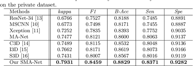
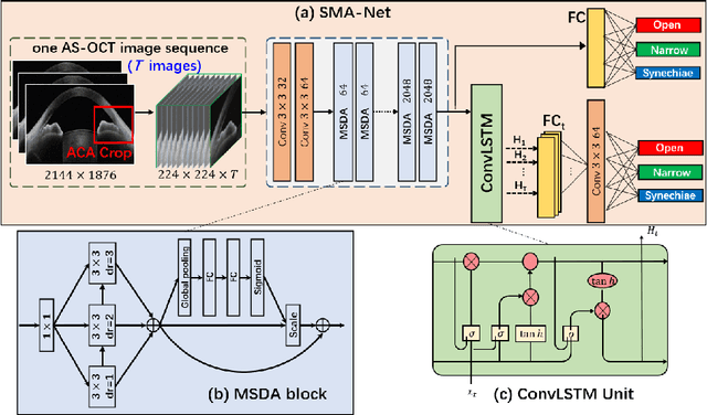

Abstract:Anterior chamber angle (ACA) classification is a key step in the diagnosis of angle-closure glaucoma in Anterior Segment Optical Coherence Tomography (AS-OCT). Existing automated analysis methods focus on a binary classification system (i.e., open angle or angle-closure) in a 2D AS-OCT slice. However, clinical diagnosis requires a more discriminating ACA three-class system (i.e., open, narrow, or synechiae angles) for the benefit of clinicians who seek better to understand the progression of the spectrum of angle-closure glaucoma types. To address this, we propose a novel sequence multi-scale aggregation deep network (SMA-Net) for open-narrow-synechiae ACA classification based on an AS-OCT sequence. In our method, a Multi-Scale Discriminative Aggregation (MSDA) block is utilized to learn the multi-scale representations at slice level, while a ConvLSTM is introduced to study the temporal dynamics of these representations at sequence level. Finally, a multi-level loss function is used to combine the slice-based and sequence-based losses. The proposed method is evaluated across two AS-OCT datasets. The experimental results show that the proposed method outperforms existing state-of-the-art methods in applicability, effectiveness, and accuracy. We believe this work to be the first attempt to classify ACAs into open, narrow, or synechia types grading using AS-OCT sequences.
AGE Challenge: Angle Closure Glaucoma Evaluation in Anterior Segment Optical Coherence Tomography
May 05, 2020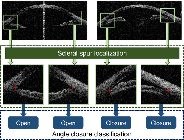

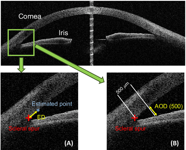

Abstract:Angle closure glaucoma (ACG) is a more aggressive disease than open-angle glaucoma, where the abnormal anatomical structures of the anterior chamber angle (ACA) may cause an elevated intraocular pressure and gradually leads to glaucomatous optic neuropathy and eventually to visual impairment and blindness. Anterior Segment Optical Coherence Tomography (AS-OCT) imaging provides a fast and contactless way to discriminate angle closure from open angle. Although many medical image analysis algorithms have been developed for glaucoma diagnosis, only a few studies have focused on AS-OCT imaging. In particular, there is no public AS-OCT dataset available for evaluating the existing methods in a uniform way, which limits the progress in the development of automated techniques for angle closure detection and assessment. To address this, we organized the Angle closure Glaucoma Evaluation challenge (AGE), held in conjunction with MICCAI 2019. The AGE challenge consisted of two tasks: scleral spur localization and angle closure classification. For this challenge, we released a large data of 4800 annotated AS-OCT images from 199 patients, and also proposed an evaluation framework to benchmark and compare different models. During the AGE challenge, over 200 teams registered online, and more than 1100 results were submitted for online evaluation. Finally, eight teams participated in the onsite challenge. In this paper, we summarize these eight onsite challenge methods and analyze their corresponding results in the two tasks. We further discuss limitations and future directions. In the AGE challenge, the top-performing approach had an average Euclidean Distance of 10 pixel in scleral spur localization, while in the task of angle closure classification, all the algorithms achieved the satisfactory performances, especially, 100% accuracy rate for top-two performances.
CE-Net: Context Encoder Network for 2D Medical Image Segmentation
Mar 07, 2019


Abstract:Medical image segmentation is an important step in medical image analysis. With the rapid development of convolutional neural network in image processing, deep learning has been used for medical image segmentation, such as optic disc segmentation, blood vessel detection, lung segmentation, cell segmentation, etc. Previously, U-net based approaches have been proposed. However, the consecutive pooling and strided convolutional operations lead to the loss of some spatial information. In this paper, we propose a context encoder network (referred to as CE-Net) to capture more high-level information and preserve spatial information for 2D medical image segmentation. CE-Net mainly contains three major components: a feature encoder module, a context extractor and a feature decoder module. We use pretrained ResNet block as the fixed feature extractor. The context extractor module is formed by a newly proposed dense atrous convolution (DAC) block and residual multi-kernel pooling (RMP) block. We applied the proposed CE-Net to different 2D medical image segmentation tasks. Comprehensive results show that the proposed method outperforms the original U-Net method and other state-of-the-art methods for optic disc segmentation, vessel detection, lung segmentation, cell contour segmentation and retinal optical coherence tomography layer segmentation.
 Add to Chrome
 Add to Firefox
 Add to Edge