 Add to Chrome
 Add to Firefox
 Add to Edge
EXACT: an explainable anomaly-aware vision foundation model for analysis of 3D chest CT
Apr 27, 2026Chest computed tomography (CT) is central to the detection and management of thoracic disease, yet the growing scale and complexity of volumetric imaging increasingly exceed what can be addressed by scan-level prediction alone. Clinically useful AI for CT must not only recognize disease across the whole volume, but also localize abnormalities and provide interpretable visual evidence. Existing vision-language foundation models typically compress scans and reports into global image-text representations, limiting their ability to preserve spatial evidence and support clinically meaningful interpretation. Here we developed EXACT, an explainable anomaly-aware foundation model for three-dimensional chest CT that learns spatially resolved representations from paired clinical scans and radiology reports. EXACT was pre-trained on 25,692 CT-reports pairs using anatomy-aware weak supervision, jointly learning organ segmentation and multi-instance anomaly localization without manual voxel-level annotations. The resulting organ-specific anomaly-aware maps assign each voxel a disease-specific anomaly score confined to its corresponding anatomy, jointly encoding lesion extent and organ-level context. In retrospective multinational and multi-center evaluations, EXACT showed broad and consistent improvements across clinically relevant CT tasks, spanning multi-disease diagnosis, zero-shot anomaly localization, downstream adaptation, and visually grounded report generation, outperforming existing three-dimensional medical foundation models. By transforming routine clinical CT scans and free-text reports into explainable voxel-level representations, EXACT establishes a scalable paradigm for trustworthy volumetric medical AI.
Adaptive Serverless Resource Management via Slot-Survival Prediction and Event-Driven Lifecycle Control
Apr 07, 2026Serverless computing eliminates infrastructure management overhead but introduces significant challenges regarding cold start latency and resource utilization. Traditional static resource allocation often leads to inefficiencies under variable workloads, resulting in performance degradation or excessive costs. This paper presents an adaptive engineering framework that optimizes serverless performance through event-driven architecture and probabilistic modeling. We propose a dual-strategy mechanism that dynamically adjusts idle durations and employs an intelligent request waiting strategy based on slot survival predictions. By leveraging sliding window aggregation and asynchronous processing, our system proactively manages resource lifecycles. Experimental results show that our approach reduces cold starts by up to 51.2% and improves cost-efficiency by nearly 2x compared to baseline methods in multi-cloud environments.
FetalAgents: A Multi-Agent System for Fetal Ultrasound Image and Video Analysis
Mar 10, 2026Fetal ultrasound (US) is the primary imaging modality for prenatal screening, yet its interpretation relies heavily on the expertise of the clinician. Despite advances in deep learning and foundation models, existing automated tools for fetal US analysis struggle to balance task-specific accuracy with the whole-process versatility required to support end-to-end clinical workflows. To address these limitations, we propose FetalAgents, the first multi-agent system for comprehensive fetal US analysis. Through a lightweight, agentic coordination framework, FetalAgents dynamically orchestrates specialized vision experts to maximize performance across diagnosis, measurement, and segmentation. Furthermore, FetalAgents advances beyond static image analysis by supporting end-to-end video stream summarization, where keyframes are automatically identified across multiple anatomical planes, analyzed by coordinated experts, and synthesized with patient metadata into a structured clinical report. Extensive multi-center external evaluations across eight clinical tasks demonstrate that FetalAgents consistently delivers the most robust and accurate performance when compared against specialized models and multimodal large language models (MLLMs), ultimately providing an auditable, workflow-aligned solution for fetal ultrasound analysis and reporting.
AnyAD: Unified Any-Modality Anomaly Detection in Incomplete Multi-Sequence MRI
Dec 24, 2025Reliable anomaly detection in brain MRI remains challenging due to the scarcity of annotated abnormal cases and the frequent absence of key imaging modalities in real clinical workflows. Existing single-class or multi-class anomaly detection (AD) models typically rely on fixed modality configurations, require repetitive training, or fail to generalize to unseen modality combinations, limiting their clinical scalability. In this work, we present a unified Any-Modality AD framework that performs robust anomaly detection and localization under arbitrary MRI modality availability. The framework integrates a dual-pathway DINOv2 encoder with a feature distribution alignment mechanism that statistically aligns incomplete-modality features with full-modality representations, enabling stable inference even with severe modality dropout. To further enhance semantic consistency, we introduce an Intrinsic Normal Prototypes (INPs) extractor and an INP-guided decoder that reconstruct only normal anatomical patterns while naturally amplifying abnormal deviations. Through randomized modality masking and indirect feature completion during training, the model learns to adapt to all modality configurations without re-training. Extensive experiments on BraTS2018, MU-Glioma-Post, and Pretreat-MetsToBrain-Masks demonstrate that our approach consistently surpasses state-of-the-art industrial and medical AD baselines across 7 modality combinations, achieving superior generalization. This study establishes a scalable paradigm for multimodal medical AD under real-world, imperfect modality conditions. Our source code is available at https://github.com/wuchangw/AnyAD.
No Modality Left Behind: Adapting to Missing Modalities via Knowledge Distillation for Brain Tumor Segmentation
Sep 18, 2025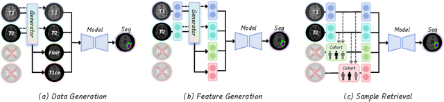
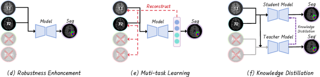
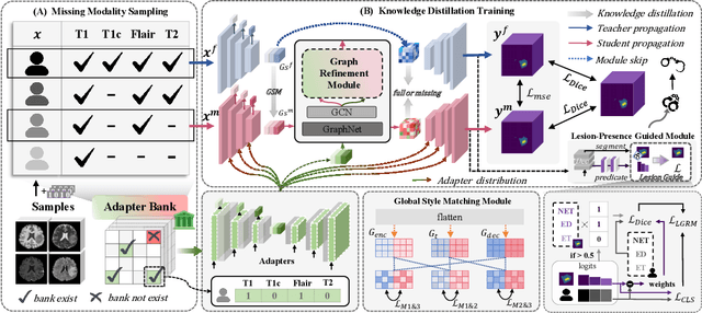
Accurate brain tumor segmentation is essential for preoperative evaluation and personalized treatment. Multi-modal MRI is widely used due to its ability to capture complementary tumor features across different sequences. However, in clinical practice, missing modalities are common, limiting the robustness and generalizability of existing deep learning methods that rely on complete inputs, especially under non-dominant modality combinations. To address this, we propose AdaMM, a multi-modal brain tumor segmentation framework tailored for missing-modality scenarios, centered on knowledge distillation and composed of three synergistic modules. The Graph-guided Adaptive Refinement Module explicitly models semantic associations between generalizable and modality-specific features, enhancing adaptability to modality absence. The Bi-Bottleneck Distillation Module transfers structural and textural knowledge from teacher to student models via global style matching and adversarial feature alignment. The Lesion-Presence-Guided Reliability Module predicts prior probabilities of lesion types through an auxiliary classification task, effectively suppressing false positives under incomplete inputs. Extensive experiments on the BraTS 2018 and 2024 datasets demonstrate that AdaMM consistently outperforms existing methods, exhibiting superior segmentation accuracy and robustness, particularly in single-modality and weak-modality configurations. In addition, we conduct a systematic evaluation of six categories of missing-modality strategies, confirming the superiority of knowledge distillation and offering practical guidance for method selection and future research. Our source code is available at https://github.com/Quanato607/AdaMM.
Diff5T: Benchmarking Human Brain Diffusion MRI with an Extensive 5.0 Tesla K-Space and Spatial Dataset
Dec 09, 2024

Diffusion magnetic resonance imaging (dMRI) provides critical insights into the microstructural and connectional organization of the human brain. However, the availability of high-field, open-access datasets that include raw k-space data for advanced research remains limited. To address this gap, we introduce Diff5T, a first comprehensive 5.0 Tesla diffusion MRI dataset focusing on the human brain. This dataset includes raw k-space data and reconstructed diffusion images, acquired using a variety of imaging protocols. Diff5T is designed to support the development and benchmarking of innovative methods in artifact correction, image reconstruction, image preprocessing, diffusion modelling and tractography. The dataset features a wide range of diffusion parameters, including multiple b-values and gradient directions, allowing extensive research applications in studying human brain microstructure and connectivity. With its emphasis on open accessibility and detailed benchmarks, Diff5T serves as a valuable resource for advancing human brain mapping research using diffusion MRI, fostering reproducibility, and enabling collaboration across the neuroscience and medical imaging communities.
Artificial Intelligence without Restriction Surpassing Human Intelligence with Probability One: Theoretical Insight into Secrets of the Brain with AI Twins of the Brain
Dec 04, 2024


Artificial Intelligence (AI) has apparently become one of the most important techniques discovered by humans in history while the human brain is widely recognized as one of the most complex systems in the universe. One fundamental critical question which would affect human sustainability remains open: Will artificial intelligence (AI) evolve to surpass human intelligence in the future? This paper shows that in theory new AI twins with fresh cellular level of AI techniques for neuroscience could approximate the brain and its functioning systems (e.g. perception and cognition functions) with any expected small error and AI without restrictions could surpass human intelligence with probability one in the end. This paper indirectly proves the validity of the conjecture made by Frank Rosenblatt 70 years ago about the potential capabilities of AI, especially in the realm of artificial neural networks. Intelligence is just one of fortuitous but sophisticated creations of the nature which has not been fully discovered. Like mathematics and physics, with no restrictions artificial intelligence would lead to a new subject with its self-contained systems and principles. We anticipate that this paper opens new doors for 1) AI twins and other AI techniques to be used in cellular level of efficient neuroscience dynamic analysis, functioning analysis of the brain and brain illness solutions; 2) new worldwide collaborative scheme for interdisciplinary teams concurrently working on and modelling different types of neurons and synapses and different level of functioning subsystems of the brain with AI techniques; 3) development of low energy of AI techniques with the aid of fundamental neuroscience properties; and 4) new controllable, explainable and safe AI techniques with reasoning capabilities of discovering principles in nature.
Enhance the Image: Super Resolution using Artificial Intelligence in MRI
Jun 19, 2024


This chapter provides an overview of deep learning techniques for improving the spatial resolution of MRI, ranging from convolutional neural networks, generative adversarial networks, to more advanced models including transformers, diffusion models, and implicit neural representations. Our exploration extends beyond the methodologies to scrutinize the impact of super-resolved images on clinical and neuroscientific assessments. We also cover various practical topics such as network architectures, image evaluation metrics, network loss functions, and training data specifics, including downsampling methods for simulating low-resolution images and dataset selection. Finally, we discuss existing challenges and potential future directions regarding the feasibility and reliability of deep learning-based MRI super-resolution, with the aim to facilitate its wider adoption to benefit various clinical and neuroscientific applications.
Artificial Intelligence for Neuro MRI Acquisition: A Review
Jun 10, 2024Magnetic resonance imaging (MRI) has significantly benefited from the resurgence of artificial intelligence (AI). By leveraging AI's capabilities in large-scale optimization and pattern recognition, innovative methods are transforming the MRI acquisition workflow, including planning, sequence design, and correction of acquisition artifacts. These emerging algorithms demonstrate substantial potential in enhancing the efficiency and throughput of acquisition steps. This review discusses several pivotal AI-based methods in neuro MRI acquisition, focusing on their technological advances, impact on clinical practice, and potential risks.
3D-EPI Blip-Up/Down Acquisition with CAIPI and Joint Hankel Structured Low-Rank Reconstruction for Rapid Distortion-Free High-Resolution T2* Mapping
Dec 01, 2022Purpose: This work aims to develop a novel distortion-free 3D-EPI acquisition and image reconstruction technique for fast and robust, high-resolution, whole-brain imaging as well as quantitative T2* mapping. Methods: 3D-Blip-Up and -Down Acquisition (3D-BUDA) sequence is designed for both single- and multi-echo 3D GRE-EPI imaging using multiple shots with blip-up and -down readouts to encode B0 field map information. Complementary k-space coverage is achieved using controlled aliasing in parallel imaging (CAIPI) sampling across the shots. For image reconstruction, an iterative hard-thresholding algorithm is employed to minimize the cost function that combines field map information informed parallel imaging with the structured low-rank constraint for multi-shot 3D-BUDA data. Extending 3D-BUDA to multi-echo imaging permits T2* mapping. For this, we propose constructing a joint Hankel matrix along both echo and shot dimensions to improve the reconstruction. Results: Experimental results on in vivo multi-echo data demonstrate that, by performing joint reconstruction along with both echo and shot dimensions, reconstruction accuracy is improved compared to standard 3D-BUDA reconstruction. CAIPI sampling is further shown to enhance the image quality. For T2* mapping, T2* values from 3D-Joint-CAIPI-BUDA and reference multi-echo GRE are within limits of agreement as quantified by Bland-Altman analysis. Conclusions: The proposed technique enables rapid 3D distortion-free high-resolution imaging and T2* mapping. Specifically, 3D-BUDA enables 1-mm isotropic whole-brain imaging in 22 s at 3 T and 9 s on a 7 T scanner. The combination of multi-echo 3D-BUDA with CAIPI acquisition and joint reconstruction enables distortion-free whole-brain T2* mapping in 47 s at 1.1x1.1x1.0 mm3 resolution.