Alireza Mehrtash
Deep Learning for Detection and Localization of B-Lines in Lung Ultrasound
Feb 15, 2023


Abstract:Lung ultrasound (LUS) is an important imaging modality used by emergency physicians to assess pulmonary congestion at the patient bedside. B-line artifacts in LUS videos are key findings associated with pulmonary congestion. Not only can the interpretation of LUS be challenging for novice operators, but visual quantification of B-lines remains subject to observer variability. In this work, we investigate the strengths and weaknesses of multiple deep learning approaches for automated B-line detection and localization in LUS videos. We curate and publish, BEDLUS, a new ultrasound dataset comprising 1,419 videos from 113 patients with a total of 15,755 expert-annotated B-lines. Based on this dataset, we present a benchmark of established deep learning methods applied to the task of B-line detection. To pave the way for interpretable quantification of B-lines, we propose a novel "single-point" approach to B-line localization using only the point of origin. Our results show that (a) the area under the receiver operating characteristic curve ranges from 0.864 to 0.955 for the benchmarked detection methods, (b) within this range, the best performance is achieved by models that leverage multiple successive frames as input, and (c) the proposed single-point approach for B-line localization reaches an F1-score of 0.65, performing on par with the inter-observer agreement. The dataset and developed methods can facilitate further biomedical research on automated interpretation of lung ultrasound with the potential to expand the clinical utility.
PEP: Parameter Ensembling by Perturbation
Oct 24, 2020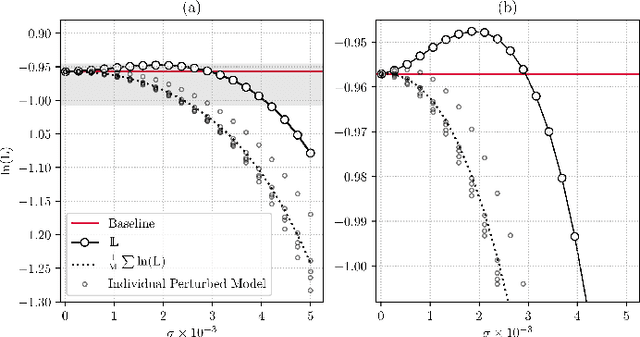
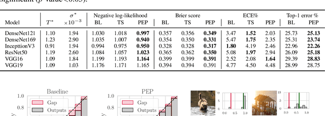
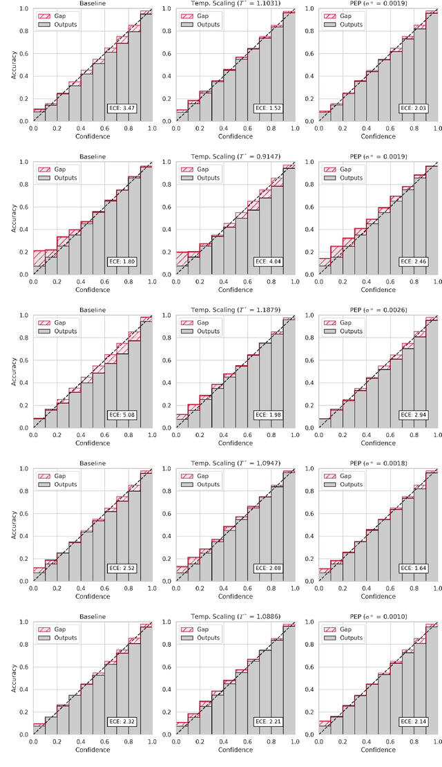
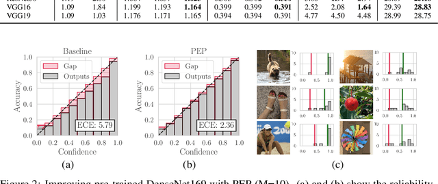
Abstract:Ensembling is now recognized as an effective approach for increasing the predictive performance and calibration of deep networks. We introduce a new approach, Parameter Ensembling by Perturbation (PEP), that constructs an ensemble of parameter values as random perturbations of the optimal parameter set from training by a Gaussian with a single variance parameter. The variance is chosen to maximize the log-likelihood of the ensemble average ($\mathbb{L}$) on the validation data set. Empirically, and perhaps surprisingly, $\mathbb{L}$ has a well-defined maximum as the variance grows from zero (which corresponds to the baseline model). Conveniently, calibration level of predictions also tends to grow favorably until the peak of $\mathbb{L}$ is reached. In most experiments, PEP provides a small improvement in performance, and, in some cases, a substantial improvement in empirical calibration. We show that this "PEP effect" (the gain in log-likelihood) is related to the mean curvature of the likelihood function and the empirical Fisher information. Experiments on ImageNet pre-trained networks including ResNet, DenseNet, and Inception showed improved calibration and likelihood. We further observed a mild improvement in classification accuracy on these networks. Experiments on classification benchmarks such as MNIST and CIFAR-10 showed improved calibration and likelihood, as well as the relationship between the PEP effect and overfitting; this demonstrates that PEP can be used to probe the level of overfitting that occurred during training. In general, no special training procedure or network architecture is needed, and in the case of pre-trained networks, no additional training is needed.
Confidence Calibration and Predictive Uncertainty Estimation for Deep Medical Image Segmentation
Nov 29, 2019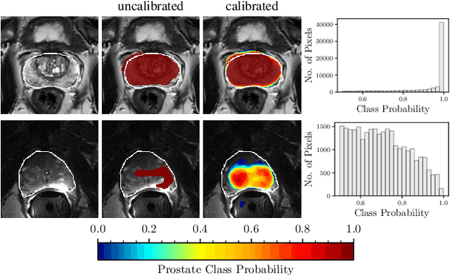
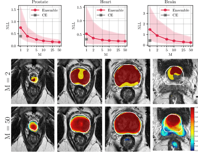
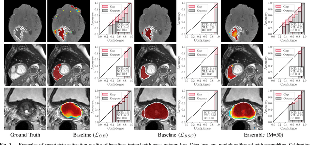
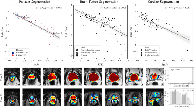
Abstract:Fully convolutional neural networks (FCNs), and in particular U-Nets, have achieved state-of-the-art results in semantic segmentation for numerous medical imaging applications. Moreover, batch normalization and Dice loss have been used successfully to stabilize and accelerate training. However, these networks are poorly calibrated i.e. they tend to produce overconfident predictions both in correct and erroneous classifications, making them unreliable and hard to interpret. In this paper, we study predictive uncertainty estimation in FCNs for medical image segmentation. We make the following contributions: 1) We systematically compare cross entropy loss with Dice loss in terms of segmentation quality and uncertainty estimation of FCNs; 2) We propose model ensembling for confidence calibration of the FCNs trained with batch normalization and Dice loss; 3) We assess the ability of calibrated FCNs to predict segmentation quality of structures and detect out-of-distribution test examples. We conduct extensive experiments across three medical image segmentation applications of the brain, the heart, and the prostate to evaluate our contributions. The results of this study offer considerable insight into the predictive uncertainty estimation and out-of-distribution detection in medical image segmentation and provide practical recipes for confidence calibration. Moreover, we consistently demonstrate that model ensembling improves confidence calibration.
Standardized Assessment of Automatic Segmentation of White Matter Hyperintensities and Results of the WMH Segmentation Challenge
Apr 01, 2019
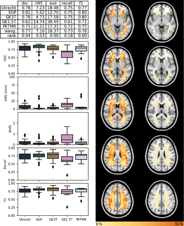
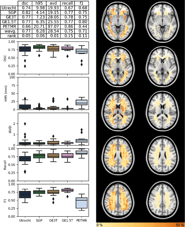
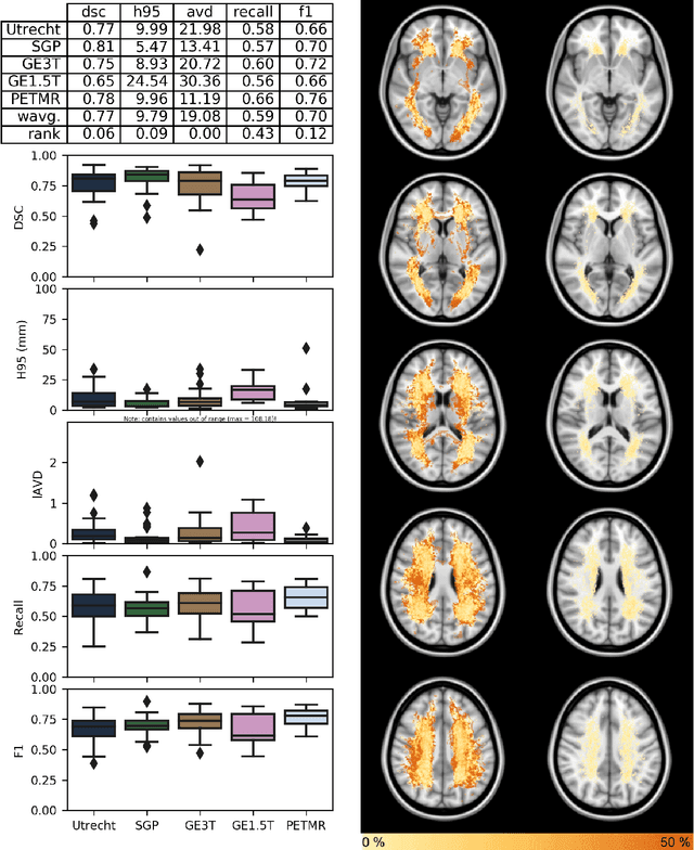
Abstract:Quantification of cerebral white matter hyperintensities (WMH) of presumed vascular origin is of key importance in many neurological research studies. Currently, measurements are often still obtained from manual segmentations on brain MR images, which is a laborious procedure. Automatic WMH segmentation methods exist, but a standardized comparison of the performance of such methods is lacking. We organized a scientific challenge, in which developers could evaluate their method on a standardized multi-center/-scanner image dataset, giving an objective comparison: the WMH Segmentation Challenge (https://wmh.isi.uu.nl/). Sixty T1+FLAIR images from three MR scanners were released with manual WMH segmentations for training. A test set of 110 images from five MR scanners was used for evaluation. Segmentation methods had to be containerized and submitted to the challenge organizers. Five evaluation metrics were used to rank the methods: (1) Dice similarity coefficient, (2) modified Hausdorff distance (95th percentile), (3) absolute log-transformed volume difference, (4) sensitivity for detecting individual lesions, and (5) F1-score for individual lesions. Additionally, methods were ranked on their inter-scanner robustness. Twenty participants submitted their method for evaluation. This paper provides a detailed analysis of the results. In brief, there is a cluster of four methods that rank significantly better than the other methods, with one clear winner. The inter-scanner robustness ranking shows that not all methods generalize to unseen scanners. The challenge remains open for future submissions and provides a public platform for method evaluation.
Deep Information Theoretic Registration
Dec 31, 2018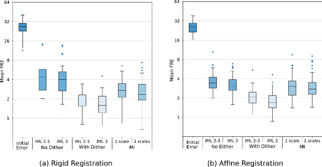
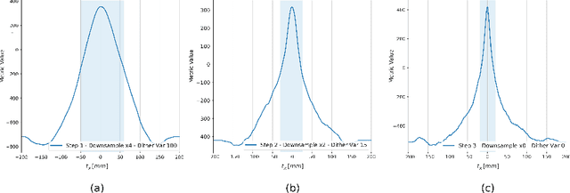
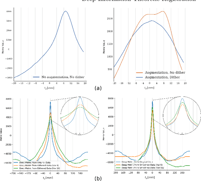
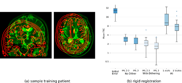
Abstract:This paper establishes an information theoretic framework for deep metric based image registration techniques. We show an exact equivalence between maximum profile likelihood and minimization of joint entropy, an important early information theoretic registration method. We further derive deep classifier-based metrics that can be used with iterated maximum likelihood to achieve Deep Information Theoretic Registration on patches rather than pixels. This alleviates a major shortcoming of previous information theoretic registration approaches, namely the implicit pixel-wise independence assumptions. Our proposed approach does not require well-registered training data; this brings previous fully supervised deep metric registration approaches to the realm of weak supervision. We evaluate our approach on several image registration tasks and show significantly better performance compared to mutual information, specifically when images have substantially different contrasts. This work enables general-purpose registration in applications where current methods are not successful.
Semi-Supervised Deep Metrics for Image Registration
Apr 04, 2018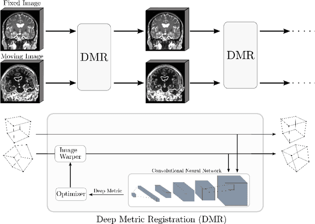

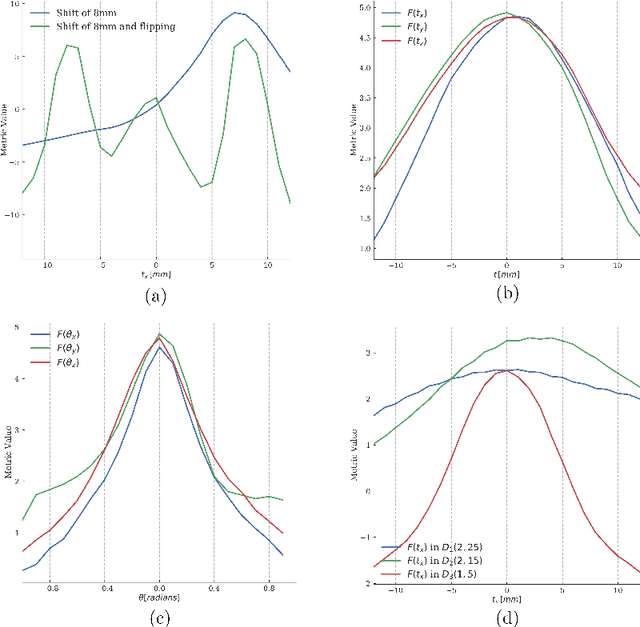
Abstract:Deep metrics have been shown effective as similarity measures in multi-modal image registration; however, the metrics are currently constructed from aligned image pairs in the training data. In this paper, we propose a strategy for learning such metrics from roughly aligned training data. Symmetrizing the data corrects bias in the metric that results from misalignment in the data (at the expense of increased variance), while random perturbations to the data, i.e. dithering, ensures that the metric has a single mode, and is amenable to registration by optimization. Evaluation is performed on the task of registration on separate unseen test image pairs. The results demonstrate the feasibility of learning a useful deep metric from substantially misaligned training data, in some cases the results are significantly better than from Mutual Information. Data augmentation via dithering is, therefore, an effective strategy for discharging the need for well-aligned training data; this brings deep metric registration from the realm of supervised to semi-supervised machine learning.
Transfer Learning for Domain Adaptation in MRI: Application in Brain Lesion Segmentation
Feb 25, 2017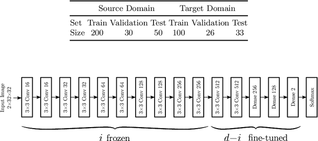
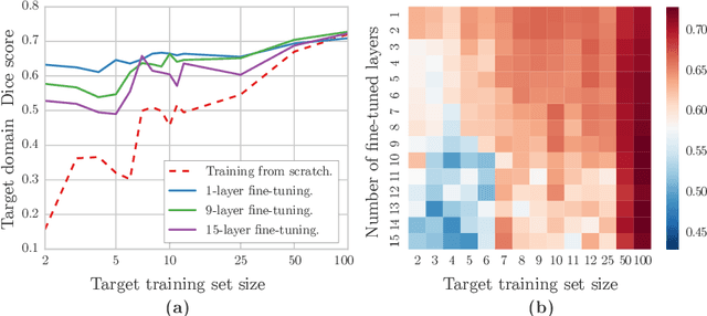
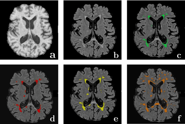
Abstract:Magnetic Resonance Imaging (MRI) is widely used in routine clinical diagnosis and treatment. However, variations in MRI acquisition protocols result in different appearances of normal and diseased tissue in the images. Convolutional neural networks (CNNs), which have shown to be successful in many medical image analysis tasks, are typically sensitive to the variations in imaging protocols. Therefore, in many cases, networks trained on data acquired with one MRI protocol, do not perform satisfactorily on data acquired with different protocols. This limits the use of models trained with large annotated legacy datasets on a new dataset with a different domain which is often a recurring situation in clinical settings. In this study, we aim to answer the following central questions regarding domain adaptation in medical image analysis: Given a fitted legacy model, 1) How much data from the new domain is required for a decent adaptation of the original network?; and, 2) What portion of the pre-trained model parameters should be retrained given a certain number of the new domain training samples? To address these questions, we conducted extensive experiments in white matter hyperintensity segmentation task. We trained a CNN on legacy MR images of brain and evaluated the performance of the domain-adapted network on the same task with images from a different domain. We then compared the performance of the model to the surrogate scenarios where either the same trained network is used or a new network is trained from scratch on the new dataset.The domain-adapted network tuned only by two training examples achieved a Dice score of 0.63 substantially outperforming a similar network trained on the same set of examples from scratch.
* 8 pages, 3 figures
 Add to Chrome
 Add to Firefox
 Add to Edge