Frank-Erik de Leeuw
Student Beats the Teacher: Deep Neural Networks for Lateral Ventricles Segmentation in Brain MR
Mar 03, 2018Abstract:Ventricular volume and its progression are known to be linked to several brain diseases such as dementia and schizophrenia. Therefore accurate measurement of ventricle volume is vital for longitudinal studies on these disorders, making automated ventricle segmentation algorithms desirable. In the past few years, deep neural networks have shown to outperform the classical models in many imaging domains. However, the success of deep networks is dependent on manually labeled data sets, which are expensive to acquire especially for higher dimensional data in the medical domain. In this work, we show that deep neural networks can be trained on much-cheaper-to-acquire pseudo-labels (e.g., generated by other automated less accurate methods) and still produce more accurate segmentations compared to the quality of the labels. To show this, we use noisy segmentation labels generated by a conventional region growing algorithm to train a deep network for lateral ventricle segmentation. Then on a large manually annotated test set, we show that the network significantly outperforms the conventional region growing algorithm which was used to produce the training labels for the network. Our experiments report a Dice Similarity Coefficient (DSC) of $0.874$ for the trained network compared to $0.754$ for the conventional region growing algorithm ($p < 0.001$).
* 7 pages, 4 figures, SPIE Medical Imaging 2018 Conference paper
Transfer Learning for Domain Adaptation in MRI: Application in Brain Lesion Segmentation
Feb 25, 2017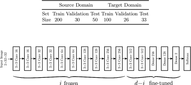
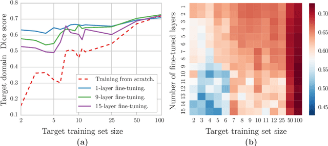
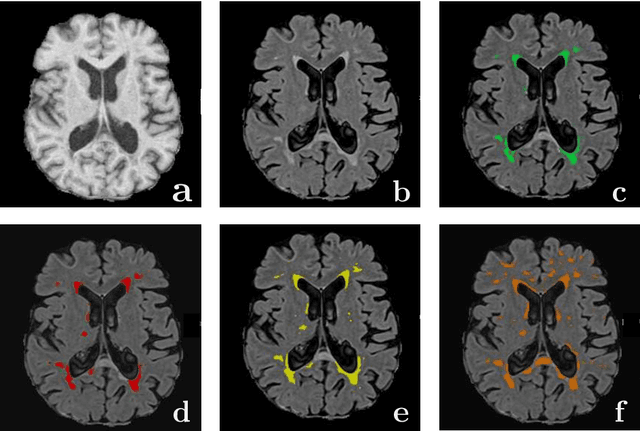
Abstract:Magnetic Resonance Imaging (MRI) is widely used in routine clinical diagnosis and treatment. However, variations in MRI acquisition protocols result in different appearances of normal and diseased tissue in the images. Convolutional neural networks (CNNs), which have shown to be successful in many medical image analysis tasks, are typically sensitive to the variations in imaging protocols. Therefore, in many cases, networks trained on data acquired with one MRI protocol, do not perform satisfactorily on data acquired with different protocols. This limits the use of models trained with large annotated legacy datasets on a new dataset with a different domain which is often a recurring situation in clinical settings. In this study, we aim to answer the following central questions regarding domain adaptation in medical image analysis: Given a fitted legacy model, 1) How much data from the new domain is required for a decent adaptation of the original network?; and, 2) What portion of the pre-trained model parameters should be retrained given a certain number of the new domain training samples? To address these questions, we conducted extensive experiments in white matter hyperintensity segmentation task. We trained a CNN on legacy MR images of brain and evaluated the performance of the domain-adapted network on the same task with images from a different domain. We then compared the performance of the model to the surrogate scenarios where either the same trained network is used or a new network is trained from scratch on the new dataset.The domain-adapted network tuned only by two training examples achieved a Dice score of 0.63 substantially outperforming a similar network trained on the same set of examples from scratch.
* 8 pages, 3 figures
Location Sensitive Deep Convolutional Neural Networks for Segmentation of White Matter Hyperintensities
Oct 29, 2016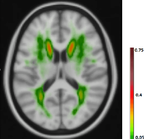

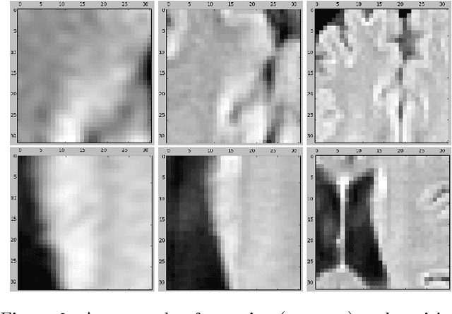

Abstract:The anatomical location of imaging features is of crucial importance for accurate diagnosis in many medical tasks. Convolutional neural networks (CNN) have had huge successes in computer vision, but they lack the natural ability to incorporate the anatomical location in their decision making process, hindering success in some medical image analysis tasks. In this paper, to integrate the anatomical location information into the network, we propose several deep CNN architectures that consider multi-scale patches or take explicit location features while training. We apply and compare the proposed architectures for segmentation of white matter hyperintensities in brain MR images on a large dataset. As a result, we observe that the CNNs that incorporate location information substantially outperform a conventional segmentation method with hand-crafted features as well as CNNs that do not integrate location information. On a test set of 46 scans, the best configuration of our networks obtained a Dice score of 0.791, compared to 0.797 for an independent human observer. Performance levels of the machine and the independent human observer were not statistically significantly different (p-value=0.17).
Deep Multi-scale Location-aware 3D Convolutional Neural Networks for Automated Detection of Lacunes of Presumed Vascular Origin
Oct 29, 2016


Abstract:Lacunes of presumed vascular origin (lacunes) are associated with an increased risk of stroke, gait impairment, and dementia and are a primary imaging feature of the small vessel disease. Quantification of lacunes may be of great importance to elucidate the mechanisms behind neuro-degenerative disorders and is recommended as part of study standards for small vessel disease research. However, due to the different appearance of lacunes in various brain regions and the existence of other similar-looking structures, such as perivascular spaces, manual annotation is a difficult, elaborative and subjective task, which can potentially be greatly improved by reliable and consistent computer-aided detection (CAD) routines. In this paper, we propose an automated two-stage method using deep convolutional neural networks (CNN). We show that this method has good performance and can considerably benefit readers. We first use a fully convolutional neural network to detect initial candidates. In the second step, we employ a 3D CNN as a false positive reduction tool. As the location information is important to the analysis of candidate structures, we further equip the network with contextual information using multi-scale analysis and integration of explicit location features. We trained, validated and tested our networks on a large dataset of 1075 cases obtained from two different studies. Subsequently, we conducted an observer study with four trained observers and compared our method with them using a free-response operating characteristic analysis. Shown on a test set of 111 cases, the resulting CAD system exhibits performance similar to the trained human observers and achieves a sensitivity of 0.974 with 0.13 false positives per slice. A feasibility study also showed that a trained human observer would considerably benefit once aided by the CAD system.
* 11 pages, 7 figures
 Add to Chrome
 Add to Firefox
 Add to Edge