Tobias Hepp
Adapted Foundation Models for Breast MRI Triaging in Contrast-Enhanced and Non-Contrast Enhanced Protocols
Nov 08, 2025Abstract:Background: Magnetic resonance imaging (MRI) has high sensitivity for breast cancer detection, but interpretation is time-consuming. Artificial intelligence may aid in pre-screening. Purpose: To evaluate the DINOv2-based Medical Slice Transformer (MST) for ruling out significant findings (Breast Imaging Reporting and Data System [BI-RADS] >=4) in contrast-enhanced and non-contrast-enhanced abbreviated breast MRI. Materials and Methods: This institutional review board approved retrospective study included 1,847 single-breast MRI examinations (377 BI-RADS >=4) from an in-house dataset and 924 from an external validation dataset (Duke). Four abbreviated protocols were tested: T1-weighted early subtraction (T1sub), diffusion-weighted imaging with b=1500 s/mm2 (DWI1500), DWI1500+T2-weighted (T2w), and T1sub+T2w. Performance was assessed at 90%, 95%, and 97.5% sensitivity using five-fold cross-validation and area under the receiver operating characteristic curve (AUC) analysis. AUC differences were compared with the DeLong test. False negatives were characterized, and attention maps of true positives were rated in the external dataset. Results: A total of 1,448 female patients (mean age, 49 +/- 12 years) were included. T1sub+T2w achieved an AUC of 0.77 +/- 0.04; DWI1500+T2w, 0.74 +/- 0.04 (p=0.15). At 97.5% sensitivity, T1sub+T2w had the highest specificity (19% +/- 7%), followed by DWI1500+T2w (17% +/- 11%). Missed lesions had a mean diameter <10 mm at 95% and 97.5% thresholds for both T1sub and DWI1500, predominantly non-mass enhancements. External validation yielded an AUC of 0.77, with 88% of attention maps rated good or moderate. Conclusion: At 97.5% sensitivity, the MST framework correctly triaged cases without BI-RADS >=4, achieving 19% specificity for contrast-enhanced and 17% for non-contrast-enhanced MRI. Further research is warranted before clinical implementation.
Conditional De-Identification of 3D Magnetic Resonance Images
Oct 18, 2021


Abstract:Privacy protection of medical image data is challenging. Even if metadata is removed, brain scans are vulnerable to attacks that match renderings of the face to facial image databases. Solutions have been developed to de-identify diagnostic scans by obfuscating or removing parts of the face. However, these solutions either fail to reliably hide the patient's identity or are so aggressive that they impair further analyses. We propose a new class of de-identification techniques that, instead of removing facial features, remodels them. Our solution relies on a conditional multi-scale GAN architecture. It takes a patient's MRI scan as input and generates a 3D volume conditioned on the patient's brain, which is preserved exactly, but where the face has been de-identified through remodeling. We demonstrate that our approach preserves privacy far better than existing techniques, without compromising downstream medical analyses. Analyses were run on the OASIS-3 and ADNI corpora.
Uncertainty-Guided Progressive GANs for Medical Image Translation
Jul 02, 2021


Abstract:Image-to-image translation plays a vital role in tackling various medical imaging tasks such as attenuation correction, motion correction, undersampled reconstruction, and denoising. Generative adversarial networks have been shown to achieve the state-of-the-art in generating high fidelity images for these tasks. However, the state-of-the-art GAN-based frameworks do not estimate the uncertainty in the predictions made by the network that is essential for making informed medical decisions and subsequent revision by medical experts and has recently been shown to improve the performance and interpretability of the model. In this work, we propose an uncertainty-guided progressive learning scheme for image-to-image translation. By incorporating aleatoric uncertainty as attention maps for GANs trained in a progressive manner, we generate images of increasing fidelity progressively. We demonstrate the efficacy of our model on three challenging medical image translation tasks, including PET to CT translation, undersampled MRI reconstruction, and MRI motion artefact correction. Our model generalizes well in three different tasks and improves performance over state of the art under full-supervision and weak-supervision with limited data. Code is released here: https://github.com/ExplainableML/UncerGuidedI2I
Uncertainty-Based Biological Age Estimation of Brain MRI Scans
Mar 15, 2021

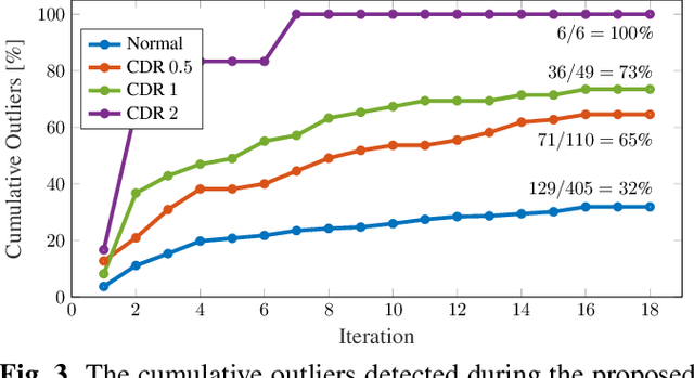
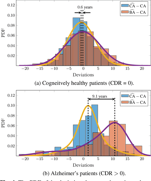
Abstract:Age is an essential factor in modern diagnostic procedures. However, assessment of the true biological age (BA) remains a daunting task due to the lack of reference ground-truth labels. Current BA estimation approaches are either restricted to skeletal images or rely on non-imaging modalities that yield a whole-body BA assessment. However, various organ systems may exhibit different aging characteristics due to lifestyle and genetic factors. In this initial study, we propose a new framework for organ-specific BA estimation utilizing 3D magnetic resonance image (MRI) scans. As a first step, this framework predicts the chronological age (CA) together with the corresponding patient-dependent aleatoric uncertainty. An iterative training algorithm is then utilized to segregate atypical aging patients from the given population based on the predicted uncertainty scores. In this manner, we hypothesize that training a new model on the remaining population should approximate the true BA behavior. We apply the proposed methodology on a brain MRI dataset containing healthy individuals as well as Alzheimer's patients. We demonstrate the correlation between the predicted BAs and the expected cognitive deterioration in Alzheimer's patients.
Overcoming Barriers to Data Sharing with Medical Image Generation: A Comprehensive Evaluation
Nov 29, 2020
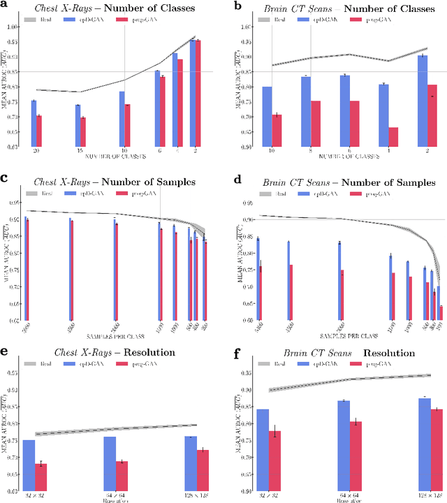
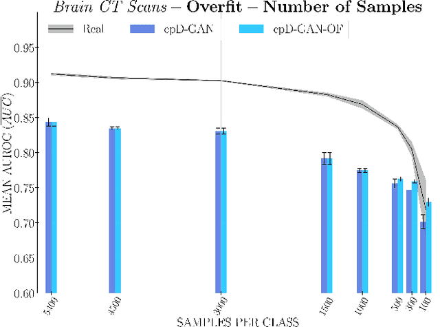
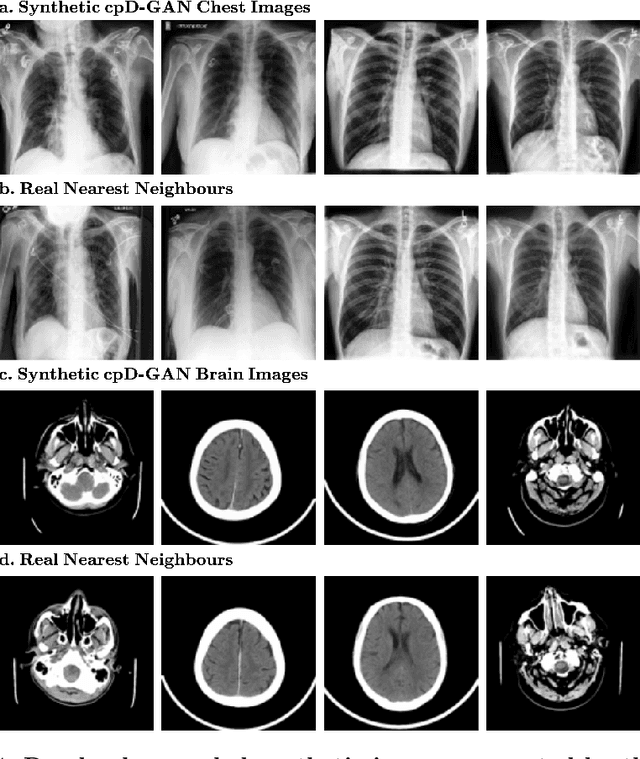
Abstract:Privacy concerns around sharing personally identifiable information are a major practical barrier to data sharing in medical research. However, in many cases, researchers have no interest in a particular individual's information but rather aim to derive insights at the level of cohorts. Here, we utilize Generative Adversarial Networks (GANs) to create derived medical imaging datasets consisting entirely of synthetic patient data. The synthetic images ideally have, in aggregate, similar statistical properties to those of a source dataset but do not contain sensitive personal information. We assess the quality of synthetic data generated by two GAN models for chest radiographs with 14 different radiology findings and brain computed tomography (CT) scans with six types of intracranial hemorrhages. We measure the synthetic image quality by the performance difference of predictive models trained on either the synthetic or the real dataset. We find that synthetic data performance disproportionately benefits from a reduced number of unique label combinations and determine at what number of samples per class overfitting effects start to dominate GAN training. Our open-source benchmark findings also indicate that synthetic data generation can benefit from higher levels of spatial resolution. We additionally conducted a reader study in which trained radiologists do not perform better than random on discriminating between synthetic and real medical images for both data modalities to a statistically significant extent. Our study offers valuable guidelines and outlines practical conditions under which insights derived from synthetic medical images are similar to those that would have been derived from real imaging data. Our results indicate that synthetic data sharing may be an attractive and privacy-preserving alternative to sharing real patient-level data in the right settings.
Age-Net: An MRI-Based Iterative Framework for Biological Age Estimation
Sep 22, 2020

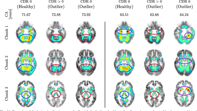
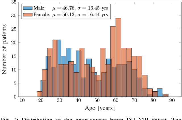
Abstract:The concept of biological age (BA) - although important in clinical practice - is hard to grasp mainly due to lack of a clearly defined reference standard. For specific applications, especially in pediatrics, medical image data are used for BA estimation in a routine clinical context. Beyond this young age group, BA estimation is restricted to whole-body assessment using non-imaging indicators such as blood biomarkers, genetic and cellular data. However, various organ systems may exhibit different aging characteristics due to lifestyle and genetic factors. Thus, a whole-body assessment of the BA does not reflect the deviations of aging behavior between organs. To this end, we propose a new imaging-based framework for organ-specific BA estimation. As a first step, we introduce a chronological age (CA) estimation framework using deep convolutional neural networks (Age-Net). We quantitatively assess the performance of this framework in comparison to existing CA estimation approaches. Furthermore, we expand upon Age-Net with a novel iterative data-cleaning algorithm to segregate atypical-aging patients (BA $\not \approx$ CA) from the given population. In this manner, we hypothesize that the remaining population should approximate the true BA behaviour. For this initial study, we apply the proposed methodology on a brain magnetic resonance image (MRI) dataset containing healthy individuals as well as Alzheimer's patients with different dementia ratings. We demonstrate the correlation between the predicted BAs and the expected cognitive deterioration in Alzheimer's patients. A statistical and visualization-based analysis has provided evidence regarding the potential and current challenges of the proposed methodology.
Fully Automated and Standardized Segmentation of Adipose Tissue Compartments by Deep Learning in Three-dimensional Whole-body MRI of Epidemiological Cohort Studies
Aug 05, 2020


Abstract:Purpose: To enable fast and reliable assessment of subcutaneous and visceral adipose tissue compartments derived from whole-body MRI. Methods: Quantification and localization of different adipose tissue compartments from whole-body MR images is of high interest to examine metabolic conditions. For correct identification and phenotyping of individuals at increased risk for metabolic diseases, a reliable automatic segmentation of adipose tissue into subcutaneous and visceral adipose tissue is required. In this work we propose a 3D convolutional neural network (DCNet) to provide a robust and objective segmentation. In this retrospective study, we collected 1000 cases (66$\pm$ 13 years; 523 women) from the Tuebingen Family Study and from the German Center for Diabetes research (TUEF/DZD), as well as 300 cases (53$\pm$ 11 years; 152 women) from the German National Cohort (NAKO) database for model training, validation, and testing with a transfer learning between the cohorts. These datasets had variable imaging sequences, imaging contrasts, receiver coil arrangements, scanners and imaging field strengths. The proposed DCNet was compared against a comparable 3D UNet segmentation in terms of sensitivity, specificity, precision, accuracy, and Dice overlap. Results: Fast (5-7seconds) and reliable adipose tissue segmentation can be obtained with high Dice overlap (0.94), sensitivity (96.6%), specificity (95.1%), precision (92.1%) and accuracy (98.4%) from 3D whole-body MR datasets (field of view coverage 450x450x2000mm${}^3$). Segmentation masks and adipose tissue profiles are automatically reported back to the referring physician. Conclusion: Automatic adipose tissue segmentation is feasible in 3D whole-body MR data sets and is generalizable to different epidemiological cohort studies with the proposed DCNet.
ipA-MedGAN: Inpainting of Arbitrarily Regions in Medical Modalities
Oct 21, 2019


Abstract:Local deformations in medical modalities are common phenomena due to a multitude of factors such as metallic implants or limited field of views in magnetic resonance imaging (MRI). Completion of the missing or distorted regions is of special interest for automatic image analysis frameworks to enhance post-processing tasks such as segmentation or classification. In this work, we propose a new generative framework for medical image inpainting, titled ipA-MedGAN. It bypasses the limitations of previous frameworks by enabling inpainting of arbitrarily shaped regions without a prior localization of the regions of interest. Thorough qualitative and quantitative comparisons with other inpainting and translational approaches have illustrated the superior performance of the proposed framework for the task of brain MR inpainting.
Organ-based Age Estimation based on 3D MRI Scans
Oct 14, 2019


Abstract:Individuals age differently depending on a multitude of different factors such as lifestyle, medical history and genetics. Often, the global chronological age is not indicative of the true ageing process. An organ-based age estimation would yield a more accurate health state assessment. In this work, we propose a new deep learning architecture for organ-based age estimation based on magnetic resonance images (MRI). The proposed network is a 3D convolutional neural network (CNN) with increased depth and width made possible by the hybrid utilization of inception and fire modules. We apply the proposed framework for the tasks of brain and knee age estimation. Quantitative comparisons against concurrent MR-based regression networks illustrated the superior performance of the proposed work.
An update on statistical boosting in biomedicine
Feb 27, 2017
Abstract:Statistical boosting algorithms have triggered a lot of research during the last decade. They combine a powerful machine-learning approach with classical statistical modelling, offering various practical advantages like automated variable selection and implicit regularization of effect estimates. They are extremely flexible, as the underlying base-learners (regression functions defining the type of effect for the explanatory variables) can be combined with any kind of loss function (target function to be optimized, defining the type of regression setting). In this review article, we highlight the most recent methodological developments on statistical boosting regarding variable selection, functional regression and advanced time-to-event modelling. Additionally, we provide a short overview on relevant applications of statistical boosting in biomedicine.
 Add to Chrome
 Add to Firefox
 Add to Edge