Fabian Bamberg
VERIDAH: Solving Enumeration Anomaly Aware Vertebra Labeling across Imaging Sequences
Jan 20, 2026Abstract:The human spine commonly consists of seven cervical, twelve thoracic, and five lumbar vertebrae. However, enumeration anomalies may result in individuals having eleven or thirteen thoracic vertebrae and four or six lumbar vertebrae. Although the identification of enumeration anomalies has potential clinical implications for chronic back pain and operation planning, the thoracolumbar junction is often poorly assessed and rarely described in clinical reports. Additionally, even though multiple deep-learning-based vertebra labeling algorithms exist, there is a lack of methods to automatically label enumeration anomalies. Our work closes that gap by introducing "Vertebra Identification with Anomaly Handling" (VERIDAH), a novel vertebra labeling algorithm based on multiple classification heads combined with a weighted vertebra sequence prediction algorithm. We show that our approach surpasses existing models on T2w TSE sagittal (98.30% vs. 94.24% of subjects with all vertebrae correctly labeled, p < 0.001) and CT imaging (99.18% vs. 77.26% of subjects with all vertebrae correctly labeled, p < 0.001) and works in arbitrary field-of-view images. VERIDAH correctly labeled the presence 2 Möller et al. of thoracic enumeration anomalies in 87.80% and 96.30% of T2w and CT images, respectively, and lumbar enumeration anomalies in 94.48% and 97.22% for T2w and CT, respectively. Our code and models are available at: https://github.com/Hendrik-code/spineps.
Detecting Unforeseen Data Properties with Diffusion Autoencoder Embeddings using Spine MRI data
Oct 14, 2024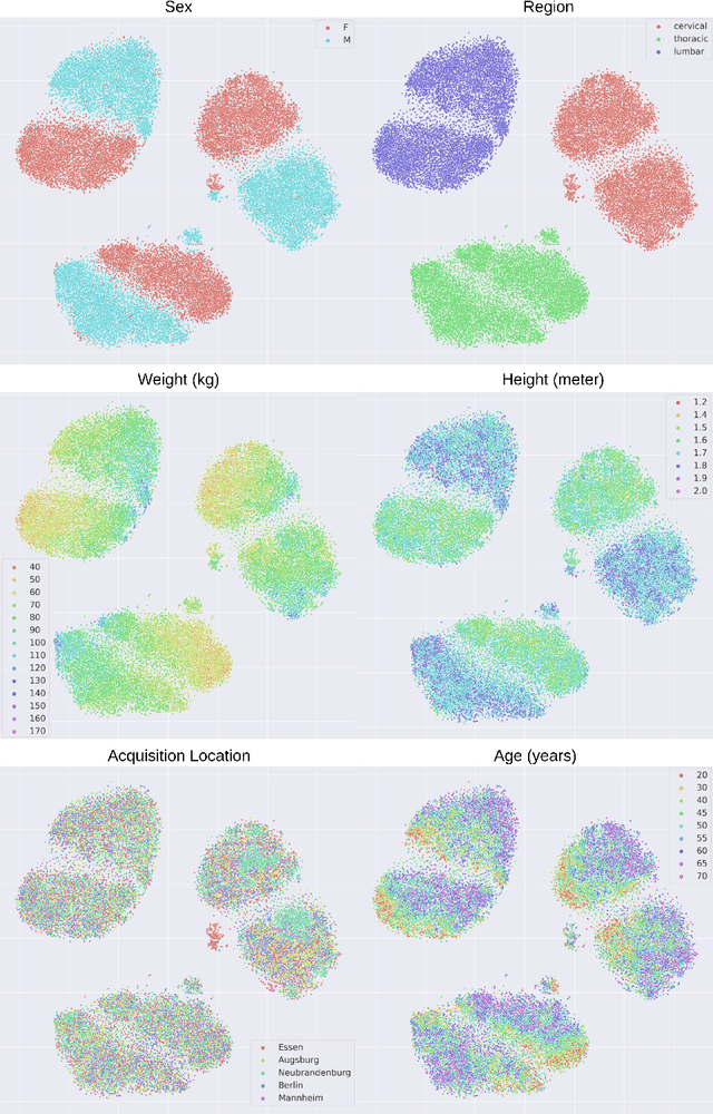

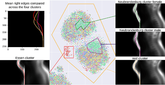
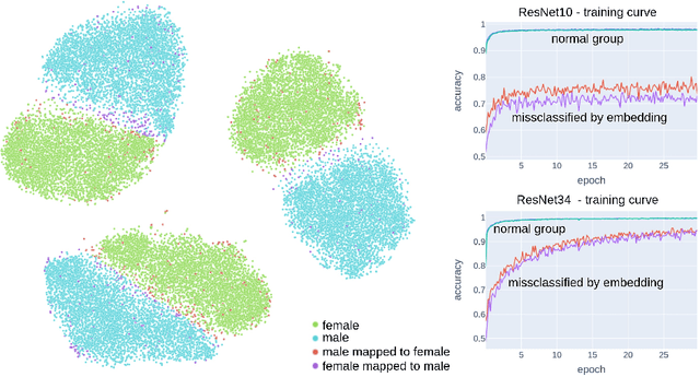
Abstract:Deep learning has made significant strides in medical imaging, leveraging the use of large datasets to improve diagnostics and prognostics. However, large datasets often come with inherent errors through subject selection and acquisition. In this paper, we investigate the use of Diffusion Autoencoder (DAE) embeddings for uncovering and understanding data characteristics and biases, including biases for protected variables like sex and data abnormalities indicative of unwanted protocol variations. We use sagittal T2-weighted magnetic resonance (MR) images of the neck, chest, and lumbar region from 11186 German National Cohort (NAKO) participants. We compare DAE embeddings with existing generative models like StyleGAN and Variational Autoencoder. Evaluations on a large-scale dataset consisting of sagittal T2-weighted MR images of three spine regions show that DAE embeddings effectively separate protected variables such as sex and age. Furthermore, we used t-SNE visualization to identify unwanted variations in imaging protocols, revealing differences in head positioning. Our embedding can identify samples where a sex predictor will have issues learning the correct sex. Our findings highlight the potential of using advanced embedding techniques like DAEs to detect data quality issues and biases in medical imaging datasets. Identifying such hidden relations can enhance the reliability and fairness of deep learning models in healthcare applications, ultimately improving patient care and outcomes.
TotalVibeSegmentator: Full Torso Segmentation for the NAKO and UK Biobank in Volumetric Interpolated Breath-hold Examination Body Images
May 31, 2024


Abstract:Objectives: To present a publicly available torso segmentation network for large epidemiology datasets on volumetric interpolated breath-hold examination (VIBE) images. Materials & Methods: We extracted preliminary segmentations from TotalSegmentator, spine, and body composition networks for VIBE images, then improved them iteratively and retrained a nnUNet network. Using subsets of NAKO (85 subjects) and UK Biobank (16 subjects), we evaluated with Dice-score on a holdout set (12 subjects) and existing organ segmentation approach (1000 subjects), generating 71 semantic segmentation types for VIBE images. We provide an additional network for the vertebra segments 22 individual vertebra types. Results: We achieved an average Dice score of 0.89 +- 0.07 overall 71 segmentation labels. We scored > 0.90 Dice-score on the abdominal organs except for the pancreas with a Dice of 0.70. Conclusion: Our work offers a detailed and refined publicly available full torso segmentation on VIBE images.
FedNorm: Modality-Based Normalization in Federated Learning for Multi-Modal Liver Segmentation
May 23, 2022


Abstract:Given the high incidence and effective treatment options for liver diseases, they are of great socioeconomic importance. One of the most common methods for analyzing CT and MRI images for diagnosis and follow-up treatment is liver segmentation. Recent advances in deep learning have demonstrated encouraging results for automatic liver segmentation. Despite this, their success depends primarily on the availability of an annotated database, which is often not available because of privacy concerns. Federated Learning has been recently proposed as a solution to alleviate these challenges by training a shared global model on distributed clients without access to their local databases. Nevertheless, Federated Learning does not perform well when it is trained on a high degree of heterogeneity of image data due to multi-modal imaging, such as CT and MRI, and multiple scanner types. To this end, we propose Fednorm and its extension \fednormp, two Federated Learning algorithms that use a modality-based normalization technique. Specifically, Fednorm normalizes the features on a client-level, while Fednorm+ employs the modality information of single slices in the feature normalization. Our methods were validated using 428 patients from six publicly available databases and compared to state-of-the-art Federated Learning algorithms and baseline models in heterogeneous settings (multi-institutional, multi-modal data). The experimental results demonstrate that our methods show an overall acceptable performance, achieve Dice per patient scores up to 0.961, consistently outperform locally trained models, and are on par or slightly better than centralized models.
An Uncertainty-Aware, Shareable and Transparent Neural Network Architecture for Brain-Age Modeling
Jul 16, 2021
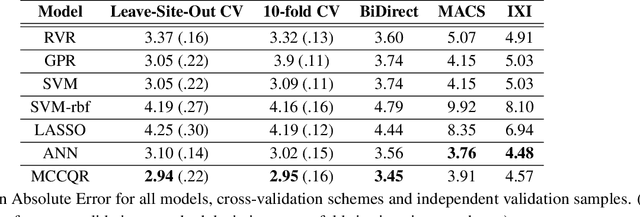
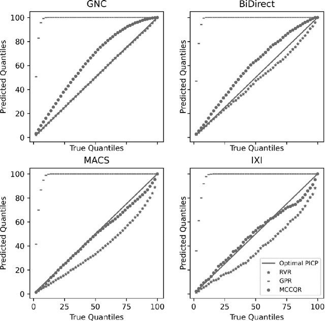
Abstract:The deviation between chronological age and age predicted from neuroimaging data has been identified as a sensitive risk-marker of cross-disorder brain changes, growing into a cornerstone of biological age-research. However, Machine Learning models underlying the field do not consider uncertainty, thereby confounding results with training data density and variability. Also, existing models are commonly based on homogeneous training sets, often not independently validated, and cannot be shared due to data protection issues. Here, we introduce an uncertainty-aware, shareable, and transparent Monte-Carlo Dropout Composite-Quantile-Regression (MCCQR) Neural Network trained on N=10,691 datasets from the German National Cohort. The MCCQR model provides robust, distribution-free uncertainty quantification in high-dimensional neuroimaging data, achieving lower error rates compared to existing models across ten recruitment centers and in three independent validation samples (N=4,004). In two examples, we demonstrate that it prevents spurious associations and increases power to detect accelerated brain-aging. We make the pre-trained model publicly available.
Predicting brain-age from raw T 1 -weighted Magnetic Resonance Imaging data using 3D Convolutional Neural Networks
Mar 22, 2021


Abstract:Age prediction based on Magnetic Resonance Imaging (MRI) data of the brain is a biomarker to quantify the progress of brain diseases and aging. Current approaches rely on preparing the data with multiple preprocessing steps, such as registering voxels to a standardized brain atlas, which yields a significant computational overhead, hampers widespread usage and results in the predicted brain-age to be sensitive to preprocessing parameters. Here we describe a 3D Convolutional Neural Network (CNN) based on the ResNet architecture being trained on raw, non-registered T$_ 1$-weighted MRI data of N=10,691 samples from the German National Cohort and additionally applied and validated in N=2,173 samples from three independent studies using transfer learning. For comparison, state-of-the-art models using preprocessed neuroimaging data are trained and validated on the same samples. The 3D CNN using raw neuroimaging data predicts age with a mean average deviation of 2.84 years, outperforming the state-of-the-art brain-age models using preprocessed data. Since our approach is invariant to preprocessing software and parameter choices, it enables faster, more robust and more accurate brain-age modeling.
Fully Automated and Standardized Segmentation of Adipose Tissue Compartments by Deep Learning in Three-dimensional Whole-body MRI of Epidemiological Cohort Studies
Aug 05, 2020


Abstract:Purpose: To enable fast and reliable assessment of subcutaneous and visceral adipose tissue compartments derived from whole-body MRI. Methods: Quantification and localization of different adipose tissue compartments from whole-body MR images is of high interest to examine metabolic conditions. For correct identification and phenotyping of individuals at increased risk for metabolic diseases, a reliable automatic segmentation of adipose tissue into subcutaneous and visceral adipose tissue is required. In this work we propose a 3D convolutional neural network (DCNet) to provide a robust and objective segmentation. In this retrospective study, we collected 1000 cases (66$\pm$ 13 years; 523 women) from the Tuebingen Family Study and from the German Center for Diabetes research (TUEF/DZD), as well as 300 cases (53$\pm$ 11 years; 152 women) from the German National Cohort (NAKO) database for model training, validation, and testing with a transfer learning between the cohorts. These datasets had variable imaging sequences, imaging contrasts, receiver coil arrangements, scanners and imaging field strengths. The proposed DCNet was compared against a comparable 3D UNet segmentation in terms of sensitivity, specificity, precision, accuracy, and Dice overlap. Results: Fast (5-7seconds) and reliable adipose tissue segmentation can be obtained with high Dice overlap (0.94), sensitivity (96.6%), specificity (95.1%), precision (92.1%) and accuracy (98.4%) from 3D whole-body MR datasets (field of view coverage 450x450x2000mm${}^3$). Segmentation masks and adipose tissue profiles are automatically reported back to the referring physician. Conclusion: Automatic adipose tissue segmentation is feasible in 3D whole-body MR data sets and is generalizable to different epidemiological cohort studies with the proposed DCNet.
A Machine-learning framework for automatic reference-free quality assessment in MRI
Jul 18, 2018


Abstract:Magnetic resonance (MR) imaging offers a wide variety of imaging techniques. A large amount of data is created per examination which needs to be checked for sufficient quality in order to derive a meaningful diagnosis. This is a manual process and therefore time- and cost-intensive. Any imaging artifacts originating from scanner hardware, signal processing or induced by the patient may reduce the image quality and complicate the diagnosis or any image post-processing. Therefore, the assessment or the ensurance of sufficient image quality in an automated manner is of high interest. Usually no reference image is available or difficult to define. Therefore, classical reference-based approaches are not applicable. Model observers mimicking the human observers (HO) can assist in this task. Thus, we propose a new machine-learning-based reference-free MR image quality assessment framework which is trained on HO-derived labels to assess MR image quality immediately after each acquisition. We include the concept of active learning and present an efficient blinded reading platform to reduce the effort in the HO labeling procedure. Derived image features and the applied classifiers (support-vector-machine, deep neural network) are investigated for a cohort of 250 patients. The MR image quality assessment framework can achieve a high test accuracy of 93.7$\%$ for estimating quality classes on a 5-point Likert-scale. The proposed MR image quality assessment framework is able to provide an accurate and efficient quality estimation which can be used as a prospective quality assurance including automatic acquisition adaptation or guided MR scanner operation, and/or as a retrospective quality assessment including support of diagnostic decisions or quality control in cohort studies.
 Add to Chrome
 Add to Firefox
 Add to Edge