U. Rajendra Acharya
SSI-GAN: Semi-Supervised Swin-Inspired Generative Adversarial Networks for Neuronal Spike Classification
Jan 01, 2026Abstract:Mosquitos are the main transmissive agents of arboviral diseases. Manual classification of their neuronal spike patterns is very labor-intensive and expensive. Most available deep learning solutions require fully labeled spike datasets and highly preprocessed neuronal signals. This reduces the feasibility of mass adoption in actual field scenarios. To address the scarcity of labeled data problems, we propose a new Generative Adversarial Network (GAN) architecture that we call the Semi-supervised Swin-Inspired GAN (SSI-GAN). The Swin-inspired, shifted-window discriminator, together with a transformer-based generator, is used to classify neuronal spike trains and, consequently, detect viral neurotropism. We use a multi-head self-attention model in a flat, window-based transformer discriminator that learns to capture sparser high-frequency spike features. Using just 1 to 3% labeled data, SSI-GAN was trained with more than 15 million spike samples collected at five-time post-infection and recording classification into Zika-infected, dengue-infected, or uninfected categories. Hyperparameters were optimized using the Bayesian Optuna framework, and performance for robustness was validated under fivefold Monte Carlo cross-validation. SSI-GAN reached 99.93% classification accuracy on the third day post-infection with only 3% labeled data. It maintained high accuracy across all stages of infection with just 1% supervision. This shows a 97-99% reduction in manual labeling effort relative to standard supervised approaches at the same performance level. The shifted-window transformer design proposed here beat all baselines by a wide margin and set new best marks in spike-based neuronal infection classification.
Uncertainty-Aware Deep Learning for Automated Skin Cancer Classification: A Comprehensive Evaluation
Jun 12, 2025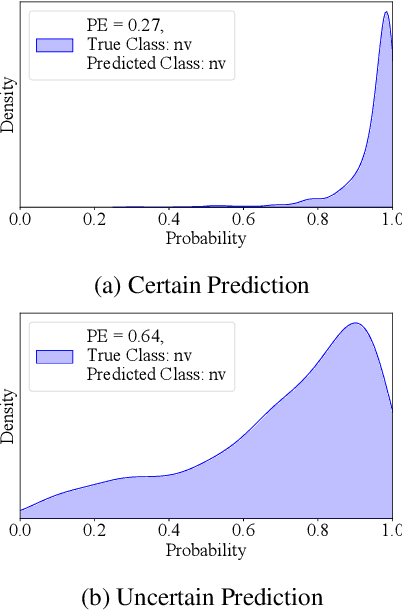
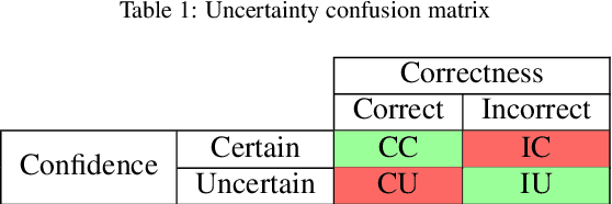
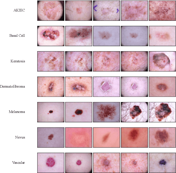

Abstract:Accurate and reliable skin cancer diagnosis is critical for early treatment and improved patient outcomes. Deep learning (DL) models have shown promise in automating skin cancer classification, but their performance can be limited by data scarcity and a lack of uncertainty awareness. In this study, we present a comprehensive evaluation of DL-based skin lesion classification using transfer learning and uncertainty quantification (UQ) on the HAM10000 dataset. In the first phase, we benchmarked several pre-trained feature extractors-including Contrastive Language-Image Pretraining (CLIP) variants, Residual Network-50 (ResNet50), Densely Connected Convolutional Network (DenseNet121), Visual Geometry Group network (VGG16), and EfficientNet-V2-Large-combined with a range of traditional classifiers such as Support Vector Machine (SVM), eXtreme Gradient Boosting (XGBoost), and logistic regression. Our results show that CLIP-based vision transformers, particularly LAION CLIP ViT-H/14 with SVM, deliver the highest classification performance. In the second phase, we incorporated UQ using Monte Carlo Dropout (MCD), Ensemble, and Ensemble Monte Carlo Dropout (EMCD) to assess not only prediction accuracy but also the reliability of model outputs. We evaluated these models using uncertainty-aware metrics such as uncertainty accuracy(UAcc), uncertainty sensitivity(USen), uncertainty specificity(USpe), and uncertainty precision(UPre). The results demonstrate that ensemble methods offer a good trade-off between accuracy and uncertainty handling, while EMCD is more sensitive to uncertain predictions. This study highlights the importance of integrating UQ into DL-based medical diagnosis to enhance both performance and trustworthiness in real-world clinical applications.
Enhancing Osteoporosis Detection: An Explainable Multi-Modal Learning Framework with Feature Fusion and Variable Clustering
Nov 01, 2024


Abstract:Osteoporosis is a common condition that increases fracture risk, especially in older adults. Early diagnosis is vital for preventing fractures, reducing treatment costs, and preserving mobility. However, healthcare providers face challenges like limited labeled data and difficulties in processing medical images. This study presents a novel multi-modal learning framework that integrates clinical and imaging data to improve diagnostic accuracy and model interpretability. The model utilizes three pre-trained networks-VGG19, InceptionV3, and ResNet50-to extract deep features from X-ray images. These features are transformed using PCA to reduce dimensionality and focus on the most relevant components. A clustering-based selection process identifies the most representative components, which are then combined with preprocessed clinical data and processed through a fully connected network (FCN) for final classification. A feature importance plot highlights key variables, showing that Medical History, BMI, and Height were the main contributors, emphasizing the significance of patient-specific data. While imaging features were valuable, they had lower importance, indicating that clinical data are crucial for accurate predictions. This framework promotes precise and interpretable predictions, enhancing transparency and building trust in AI-driven diagnoses for clinical integration.
Functional Classification of Spiking Signal Data Using Artificial Intelligence Techniques: A Review
Sep 26, 2024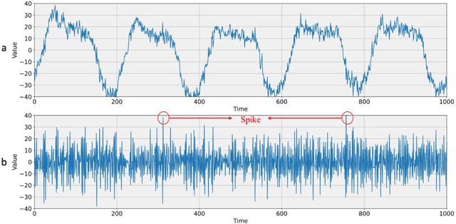
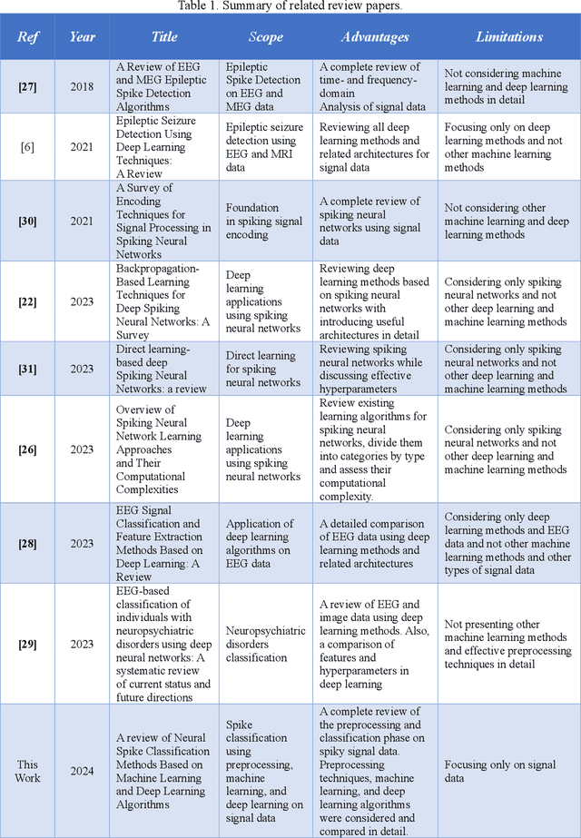
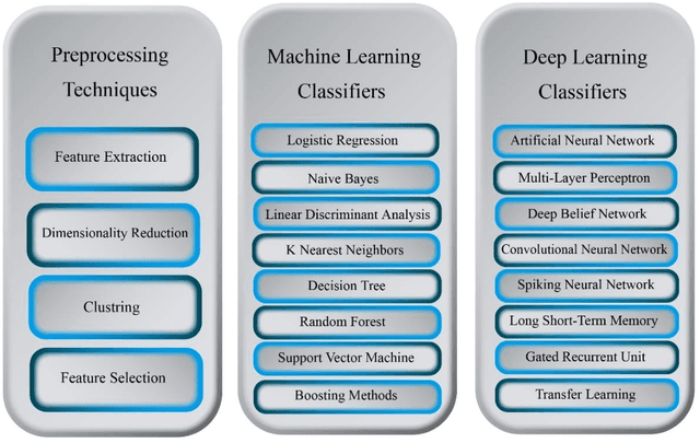
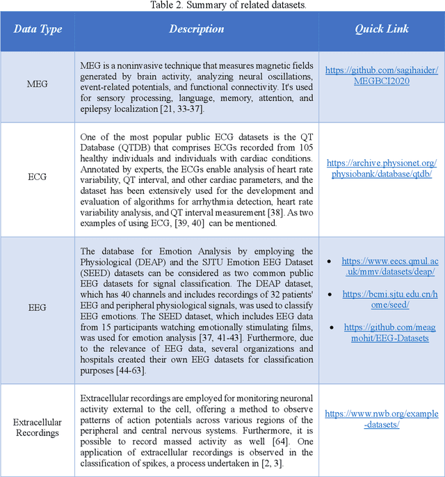
Abstract:Human brain neuron activities are incredibly significant nowadays. Neuronal behavior is assessed by analyzing signal data such as electroencephalography (EEG), which can offer scientists valuable information about diseases and human-computer interaction. One of the difficulties researchers confront while evaluating these signals is the existence of large volumes of spike data. Spikes are some considerable parts of signal data that can happen as a consequence of vital biomarkers or physical issues such as electrode movements. Hence, distinguishing types of spikes is important. From this spot, the spike classification concept commences. Previously, researchers classified spikes manually. The manual classification was not precise enough as it involves extensive analysis. Consequently, Artificial Intelligence (AI) was introduced into neuroscience to assist clinicians in classifying spikes correctly. This review discusses the importance and use of AI in spike classification, focusing on the recognition of neural activity noises. The task is divided into three main components: preprocessing, classification, and evaluation. Existing methods are introduced and their importance is determined. The review also highlights the need for more efficient algorithms. The primary goal is to provide a perspective on spike classification for future research and provide a comprehensive understanding of the methodologies and issues involved. The review organizes materials in the spike classification field for future studies. In this work, numerous studies were extracted from different databases. The PRISMA-related research guidelines were then used to choose papers. Then, research studies based on spike classification using machine learning and deep learning approaches with effective preprocessing were selected.
Artificial Intelligence and Diabetes Mellitus: An Inside Look Through the Retina
Feb 28, 2024


Abstract:Diabetes mellitus (DM) predisposes patients to vascular complications. Retinal images and vasculature reflect the body's micro- and macrovascular health. They can be used to diagnose DM complications, including diabetic retinopathy (DR), neuropathy, nephropathy, and atherosclerotic cardiovascular disease, as well as forecast the risk of cardiovascular events. Artificial intelligence (AI)-enabled systems developed for high-throughput detection of DR using digitized retinal images have become clinically adopted. Beyond DR screening, AI integration also holds immense potential to address challenges associated with the holistic care of the patient with DM. In this work, we aim to comprehensively review the literature for studies on AI applications based on retinal images related to DM diagnosis, prognostication, and management. We will describe the findings of holistic AI-assisted diabetes care, including but not limited to DR screening, and discuss barriers to implementing such systems, including issues concerning ethics, data privacy, equitable access, and explainability. With the ability to evaluate the patient's health status vis a vis DM complication as well as risk prognostication of future cardiovascular complications, AI-assisted retinal image analysis has the potential to become a central tool for modern personalized medicine in patients with DM.
Current and future roles of artificial intelligence in retinopathy of prematurity
Feb 15, 2024


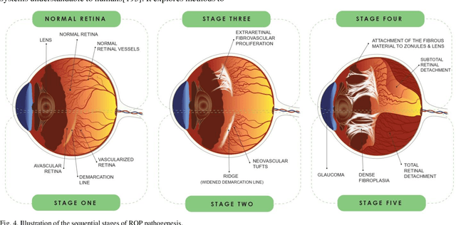
Abstract:Retinopathy of prematurity (ROP) is a severe condition affecting premature infants, leading to abnormal retinal blood vessel growth, retinal detachment, and potential blindness. While semi-automated systems have been used in the past to diagnose ROP-related plus disease by quantifying retinal vessel features, traditional machine learning (ML) models face challenges like accuracy and overfitting. Recent advancements in deep learning (DL), especially convolutional neural networks (CNNs), have significantly improved ROP detection and classification. The i-ROP deep learning (i-ROP-DL) system also shows promise in detecting plus disease, offering reliable ROP diagnosis potential. This research comprehensively examines the contemporary progress and challenges associated with using retinal imaging and artificial intelligence (AI) to detect ROP, offering valuable insights that can guide further investigation in this domain. Based on 89 original studies in this field (out of 1487 studies that were comprehensively reviewed), we concluded that traditional methods for ROP diagnosis suffer from subjectivity and manual analysis, leading to inconsistent clinical decisions. AI holds great promise for improving ROP management. This review explores AI's potential in ROP detection, classification, diagnosis, and prognosis.
Automated detection of Zika and dengue in Aedes aegypti using neural spiking analysis
Dec 14, 2023


Abstract:Mosquito-borne diseases present considerable risks to the health of both animals and humans. Aedes aegypti mosquitoes are the primary vectors for numerous medically important viruses such as dengue, Zika, yellow fever, and chikungunya. To characterize this mosquito neural activity, it is essential to classify the generated electrical spikes. However, no open-source neural spike classification method is currently available for mosquitoes. Our work presented in this paper provides an innovative artificial intelligence-based method to classify the neural spikes in uninfected, dengue-infected, and Zika-infected mosquitoes. Aiming for outstanding performance, the method employs a fusion of normalization, feature importance, and dimension reduction for the preprocessing and combines convolutional neural network and extra gradient boosting (XGBoost) for classification. The method uses the electrical spiking activity data of mosquito neurons recorded by microelectrode array technology. We used data from 0, 1, 2, 3, and 7 days post-infection, containing over 15 million samples, to analyze the method's performance. The performance of the proposed method was evaluated using accuracy, precision, recall, and the F1 scores. The results obtained from the method highlight its remarkable performance in differentiating infected vs uninfected mosquito samples, achieving an average of 98.1%. The performance was also compared with 6 other machine learning algorithms to further assess the method's capability. The method outperformed all other machine learning algorithms' performance. Overall, this research serves as an efficient method to classify the neural spikes of Aedes aegypti mosquitoes and can assist in unraveling the complex interactions between pathogens and mosquitoes.
Artificial Intelligence in Assessing Cardiovascular Diseases and Risk Factors via Retinal Fundus Images: A Review of the Last Decade
Nov 11, 2023


Abstract:Background: Cardiovascular diseases (CVDs) continue to be the leading cause of mortality on a global scale. In recent years, the application of artificial intelligence (AI) techniques, particularly deep learning (DL), has gained considerable popularity for evaluating the various aspects of CVDs. Moreover, using fundus images and optical coherence tomography angiography (OCTA) to diagnose retinal diseases has been extensively studied. To better understand heart function and anticipate changes based on microvascular characteristics and function, researchers are currently exploring the integration of AI with non-invasive retinal scanning. Leveraging AI-assisted early detection and prediction of cardiovascular diseases on a large scale holds excellent potential to mitigate cardiovascular events and alleviate the economic burden on healthcare systems. Method: A comprehensive search was conducted across various databases, including PubMed, Medline, Google Scholar, Scopus, Web of Sciences, IEEE Xplore, and ACM Digital Library, using specific keywords related to cardiovascular diseases and artificial intelligence. Results: A total of 87 English-language publications, selected for relevance were included in the study, and additional references were considered. This study presents an overview of the current advancements and challenges in employing retinal imaging and artificial intelligence to identify cardiovascular disorders and provides insights for further exploration in this field. Conclusion: Researchers aim to develop precise disease prognosis patterns as the aging population and global CVD burden increase. AI and deep learning are transforming healthcare, offering the potential for single retinal image-based diagnosis of various CVDs, albeit with the need for accelerated adoption in healthcare systems.
Solving the multiplication problem of a large language model system using a graph-based method
Oct 18, 2023


Abstract:The generative pre-trained transformer (GPT)-based chatbot software ChatGPT possesses excellent natural language processing capabilities but is inadequate for solving arithmetic problems, especially multiplication. Its GPT structure uses a computational graph for multiplication, which has limited accuracy beyond simple multiplication operations. We developed a graph-based multiplication algorithm that emulated human-like numerical operations by incorporating a 10k operator, where k represents the maximum power to base 10 of the larger of two input numbers. Our proposed algorithm attained 100% accuracy for 1,000,000 large number multiplication tasks, effectively solving the multiplication challenge of GPT-based and other large language models. Our work highlights the importance of blending simple human insights into the design of artificial intelligence algorithms. Keywords: Graph-based multiplication; ChatGPT; Multiplication problem
Empowering Precision Medicine: AI-Driven Schizophrenia Diagnosis via EEG Signals: A Comprehensive Review from 2002-2023
Sep 14, 2023Abstract:Schizophrenia (SZ) is a prevalent mental disorder characterized by cognitive, emotional, and behavioral changes. Symptoms of SZ include hallucinations, illusions, delusions, lack of motivation, and difficulties in concentration. Diagnosing SZ involves employing various tools, including clinical interviews, physical examinations, psychological evaluations, the Diagnostic and Statistical Manual of Mental Disorders (DSM), and neuroimaging techniques. Electroencephalography (EEG) recording is a significant functional neuroimaging modality that provides valuable insights into brain function during SZ. However, EEG signal analysis poses challenges for neurologists and scientists due to the presence of artifacts, long-term recordings, and the utilization of multiple channels. To address these challenges, researchers have introduced artificial intelligence (AI) techniques, encompassing conventional machine learning (ML) and deep learning (DL) methods, to aid in SZ diagnosis. This study reviews papers focused on SZ diagnosis utilizing EEG signals and AI methods. The introduction section provides a comprehensive explanation of SZ diagnosis methods and intervention techniques. Subsequently, review papers in this field are discussed, followed by an introduction to the AI methods employed for SZ diagnosis and a summary of relevant papers presented in tabular form. Additionally, this study reports on the most significant challenges encountered in SZ diagnosis, as identified through a review of papers in this field. Future directions to overcome these challenges are also addressed. The discussion section examines the specific details of each paper, culminating in the presentation of conclusions and findings.
 Add to Chrome
 Add to Firefox
 Add to Edge