Prabal Datta Barua
Solving the multiplication problem of a large language model system using a graph-based method
Oct 18, 2023


Abstract:The generative pre-trained transformer (GPT)-based chatbot software ChatGPT possesses excellent natural language processing capabilities but is inadequate for solving arithmetic problems, especially multiplication. Its GPT structure uses a computational graph for multiplication, which has limited accuracy beyond simple multiplication operations. We developed a graph-based multiplication algorithm that emulated human-like numerical operations by incorporating a 10k operator, where k represents the maximum power to base 10 of the larger of two input numbers. Our proposed algorithm attained 100% accuracy for 1,000,000 large number multiplication tasks, effectively solving the multiplication challenge of GPT-based and other large language models. Our work highlights the importance of blending simple human insights into the design of artificial intelligence algorithms. Keywords: Graph-based multiplication; ChatGPT; Multiplication problem
Full-resolution Lung Nodule Segmentation from Chest X-ray Images using Residual Encoder-Decoder Networks
Jul 13, 2023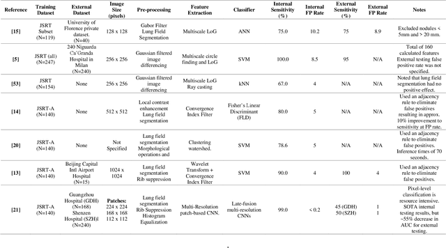

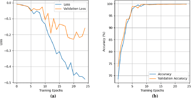

Abstract:Lung cancer is the leading cause of cancer death and early diagnosis is associated with a positive prognosis. Chest X-ray (CXR) provides an inexpensive imaging mode for lung cancer diagnosis. Suspicious nodules are difficult to distinguish from vascular and bone structures using CXR. Computer vision has previously been proposed to assist human radiologists in this task, however, leading studies use down-sampled images and computationally expensive methods with unproven generalization. Instead, this study localizes lung nodules using efficient encoder-decoder neural networks that process full resolution images to avoid any signal loss resulting from down-sampling. Encoder-decoder networks are trained and tested using the JSRT lung nodule dataset. The networks are used to localize lung nodules from an independent external CXR dataset. Sensitivity and false positive rates are measured using an automated framework to eliminate any observer subjectivity. These experiments allow for the determination of the optimal network depth, image resolution and pre-processing pipeline for generalized lung nodule localization. We find that nodule localization is influenced by subtlety, with more subtle nodules being detected in earlier training epochs. Therefore, we propose a novel self-ensemble model from three consecutive epochs centered on the validation optimum. This ensemble achieved a sensitivity of 85% in 10-fold internal testing with false positives of 8 per image. A sensitivity of 81% is achieved at a false positive rate of 6 following morphological false positive reduction. This result is comparable to more computationally complex systems based on linear and spatial filtering, but with a sub-second inference time that is faster than other methods. The proposed algorithm achieved excellent generalization results against an external dataset with sensitivity of 77% at a false positive rate of 7.6.
Natural Language Processing in Electronic Health Records in Relation to Healthcare Decision-making: A Systematic Review
Jun 22, 2023
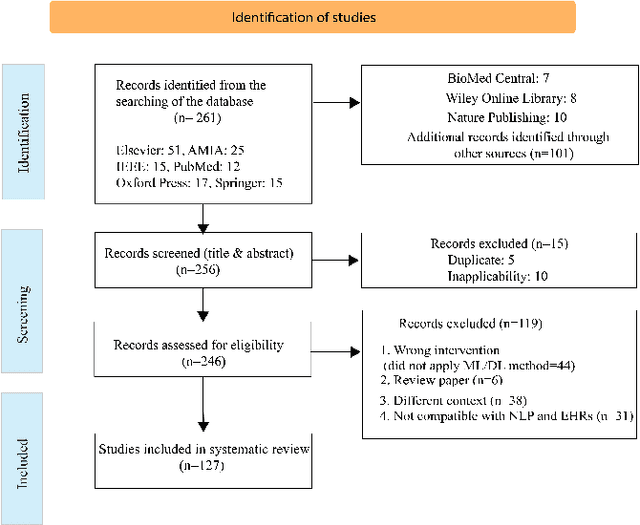
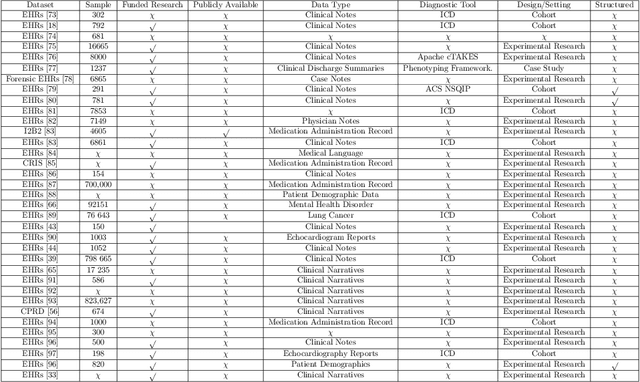
Abstract:Background: Natural Language Processing (NLP) is widely used to extract clinical insights from Electronic Health Records (EHRs). However, the lack of annotated data, automated tools, and other challenges hinder the full utilisation of NLP for EHRs. Various Machine Learning (ML), Deep Learning (DL) and NLP techniques are studied and compared to understand the limitations and opportunities in this space comprehensively. Methodology: After screening 261 articles from 11 databases, we included 127 papers for full-text review covering seven categories of articles: 1) medical note classification, 2) clinical entity recognition, 3) text summarisation, 4) deep learning (DL) and transfer learning architecture, 5) information extraction, 6) Medical language translation and 7) other NLP applications. This study follows the Preferred Reporting Items for Systematic Reviews and Meta-Analyses (PRISMA) guidelines. Result and Discussion: EHR was the most commonly used data type among the selected articles, and the datasets were primarily unstructured. Various ML and DL methods were used, with prediction or classification being the most common application of ML or DL. The most common use cases were: the International Classification of Diseases, Ninth Revision (ICD-9) classification, clinical note analysis, and named entity recognition (NER) for clinical descriptions and research on psychiatric disorders. Conclusion: We find that the adopted ML models were not adequately assessed. In addition, the data imbalance problem is quite important, yet we must find techniques to address this underlining problem. Future studies should address key limitations in studies, primarily identifying Lupus Nephritis, Suicide Attempts, perinatal self-harmed and ICD-9 classification.
NRC-Net: Automated noise robust cardio net for detecting valvular cardiac diseases using optimum transformation method with heart sound signals
Apr 29, 2023Abstract:Cardiovascular diseases (CVDs) can be effectively treated when detected early, reducing mortality rates significantly. Traditionally, phonocardiogram (PCG) signals have been utilized for detecting cardiovascular disease due to their cost-effectiveness and simplicity. Nevertheless, various environmental and physiological noises frequently affect the PCG signals, compromising their essential distinctive characteristics. The prevalence of this issue in overcrowded and resource-constrained hospitals can compromise the accuracy of medical diagnoses. Therefore, this study aims to discover the optimal transformation method for detecting CVDs using noisy heart sound signals and propose a noise robust network to improve the CVDs classification performance.For the identification of the optimal transformation method for noisy heart sound data mel-frequency cepstral coefficients (MFCCs), short-time Fourier transform (STFT), constant-Q nonstationary Gabor transform (CQT) and continuous wavelet transform (CWT) has been used with VGG16. Furthermore, we propose a novel convolutional recurrent neural network (CRNN) architecture called noise robust cardio net (NRC-Net), which is a lightweight model to classify mitral regurgitation, aortic stenosis, mitral stenosis, mitral valve prolapse, and normal heart sounds using PCG signals contaminated with respiratory and random noises. An attention block is included to extract important temporal and spatial features from the noisy corrupted heart sound.The results of this study indicate that,CWT is the optimal transformation method for noisy heart sound signals. When evaluated on the GitHub heart sound dataset, CWT demonstrates an accuracy of 95.69% for VGG16, which is 1.95% better than the second-best CQT transformation technique. Moreover, our proposed NRC-Net with CWT obtained an accuracy of 97.4%, which is 1.71% higher than the VGG16.
Debiasing pipeline improves deep learning model generalization for X-ray based lung nodule detection
Jan 24, 2022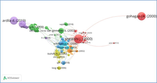
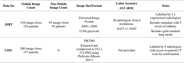
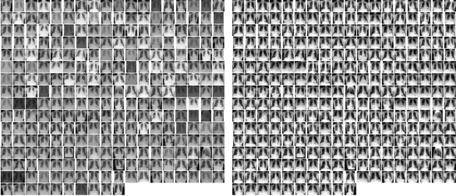
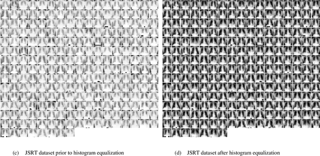
Abstract:Lung cancer is the leading cause of cancer death worldwide and a good prognosis depends on early diagnosis. Unfortunately, screening programs for the early diagnosis of lung cancer are uncommon. This is in-part due to the at-risk groups being located in rural areas far from medical facilities. Reaching these populations would require a scaled approach that combines mobility, low cost, speed, accuracy, and privacy. We can resolve these issues by combining the chest X-ray imaging mode with a federated deep-learning approach, provided that the federated model is trained on homogenous data to ensure that no single data source can adversely bias the model at any point in time. In this study we show that an image pre-processing pipeline that homogenizes and debiases chest X-ray images can improve both internal classification and external generalization, paving the way for a low-cost and accessible deep learning-based clinical system for lung cancer screening. An evolutionary pruning mechanism is used to train a nodule detection deep learning model on the most informative images from a publicly available lung nodule X-ray dataset. Histogram equalization is used to remove systematic differences in image brightness and contrast. Model training is performed using all combinations of lung field segmentation, close cropping, and rib suppression operators. We show that this pre-processing pipeline results in deep learning models that successfully generalize an independent lung nodule dataset using ablation studies to assess the contribution of each operator in this pipeline. In stripping chest X-ray images of known confounding variables by lung field segmentation, along with suppression of signal noise from the bone structure we can train a highly accurate deep learning lung nodule detection algorithm with outstanding generalization accuracy of 89% to nodule samples in unseen data.
 Add to Chrome
 Add to Firefox
 Add to Edge