Mahboobeh Jafari
Empowering Precision Medicine: AI-Driven Schizophrenia Diagnosis via EEG Signals: A Comprehensive Review from 2002-2023
Sep 14, 2023Abstract:Schizophrenia (SZ) is a prevalent mental disorder characterized by cognitive, emotional, and behavioral changes. Symptoms of SZ include hallucinations, illusions, delusions, lack of motivation, and difficulties in concentration. Diagnosing SZ involves employing various tools, including clinical interviews, physical examinations, psychological evaluations, the Diagnostic and Statistical Manual of Mental Disorders (DSM), and neuroimaging techniques. Electroencephalography (EEG) recording is a significant functional neuroimaging modality that provides valuable insights into brain function during SZ. However, EEG signal analysis poses challenges for neurologists and scientists due to the presence of artifacts, long-term recordings, and the utilization of multiple channels. To address these challenges, researchers have introduced artificial intelligence (AI) techniques, encompassing conventional machine learning (ML) and deep learning (DL) methods, to aid in SZ diagnosis. This study reviews papers focused on SZ diagnosis utilizing EEG signals and AI methods. The introduction section provides a comprehensive explanation of SZ diagnosis methods and intervention techniques. Subsequently, review papers in this field are discussed, followed by an introduction to the AI methods employed for SZ diagnosis and a summary of relevant papers presented in tabular form. Additionally, this study reports on the most significant challenges encountered in SZ diagnosis, as identified through a review of papers in this field. Future directions to overcome these challenges are also addressed. The discussion section examines the specific details of each paper, culminating in the presentation of conclusions and findings.
Automated Diagnosis of Cardiovascular Diseases from Cardiac Magnetic Resonance Imaging Using Deep Learning Models: A Review
Oct 26, 2022Abstract:In recent years, cardiovascular diseases (CVDs) have become one of the leading causes of mortality globally. CVDs appear with minor symptoms and progressively get worse. The majority of people experience symptoms such as exhaustion, shortness of breath, ankle swelling, fluid retention, and other symptoms when starting CVD. Coronary artery disease (CAD), arrhythmia, cardiomyopathy, congenital heart defect (CHD), mitral regurgitation, and angina are the most common CVDs. Clinical methods such as blood tests, electrocardiography (ECG) signals, and medical imaging are the most effective methods used for the detection of CVDs. Among the diagnostic methods, cardiac magnetic resonance imaging (CMR) is increasingly used to diagnose, monitor the disease, plan treatment and predict CVDs. Coupled with all the advantages of CMR data, CVDs diagnosis is challenging for physicians due to many slices of data, low contrast, etc. To address these issues, deep learning (DL) techniques have been employed to the diagnosis of CVDs using CMR data, and much research is currently being conducted in this field. This review provides an overview of the studies performed in CVDs detection using CMR images and DL techniques. The introduction section examined CVDs types, diagnostic methods, and the most important medical imaging techniques. In the following, investigations to detect CVDs using CMR images and the most significant DL methods are presented. Another section discussed the challenges in diagnosing CVDs from CMR data. Next, the discussion section discusses the results of this review, and future work in CVDs diagnosis from CMR images and DL techniques are outlined. The most important findings of this study are presented in the conclusion section.
Automatic Diagnosis of Myocarditis Disease in Cardiac MRI Modality using Deep Transformers and Explainable Artificial Intelligence
Oct 26, 2022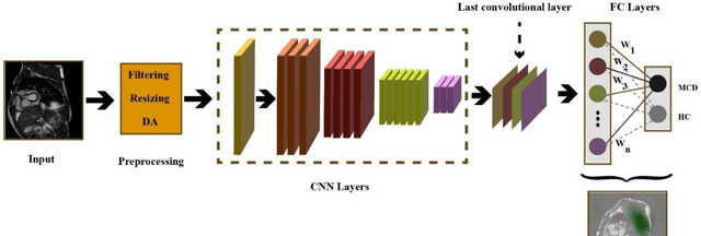

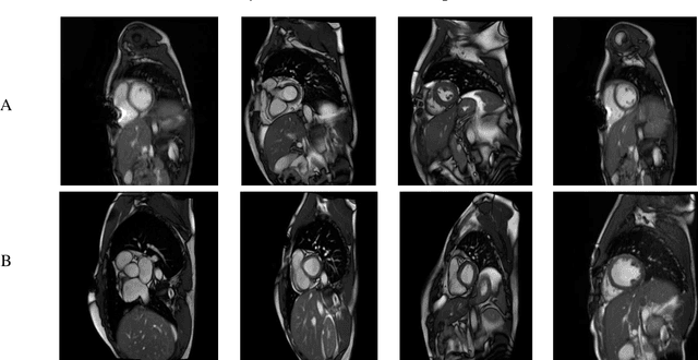

Abstract:Myocarditis is among the most important cardiovascular diseases (CVDs), endangering the health of many individuals by damaging the myocardium. Microbes and viruses, such as HIV, play a vital role in myocarditis disease (MCD) incidence. Lack of MCD diagnosis in the early stages is associated with irreversible complications. Cardiac magnetic resonance imaging (CMRI) is highly popular among cardiologists to diagnose CVDs. In this paper, a deep learning (DL) based computer-aided diagnosis system (CADS) is presented for the diagnosis of MCD using CMRI images. The proposed CADS includes dataset, preprocessing, feature extraction, classification, and post-processing steps. First, the Z-Alizadeh dataset was selected for the experiments. The preprocessing step included noise removal, image resizing, and data augmentation (DA). In this step, CutMix, and MixUp techniques were used for the DA. Then, the most recent pre-trained and transformers models were used for feature extraction and classification using CMRI images. Our results show high performance for the detection of MCD using transformer models compared with the pre-trained architectures. Among the DL architectures, Turbulence Neural Transformer (TNT) architecture achieved an accuracy of 99.73% with 10-fold cross-validation strategy. Explainable-based Grad Cam method is used to visualize the MCD suspected areas in CMRI images.
Automatic Autism Spectrum Disorder Detection Using Artificial Intelligence Methods with MRI Neuroimaging: A Review
Jun 20, 2022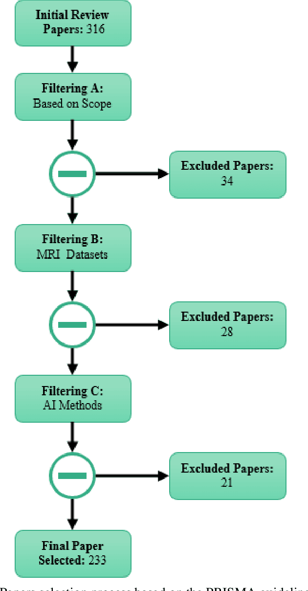

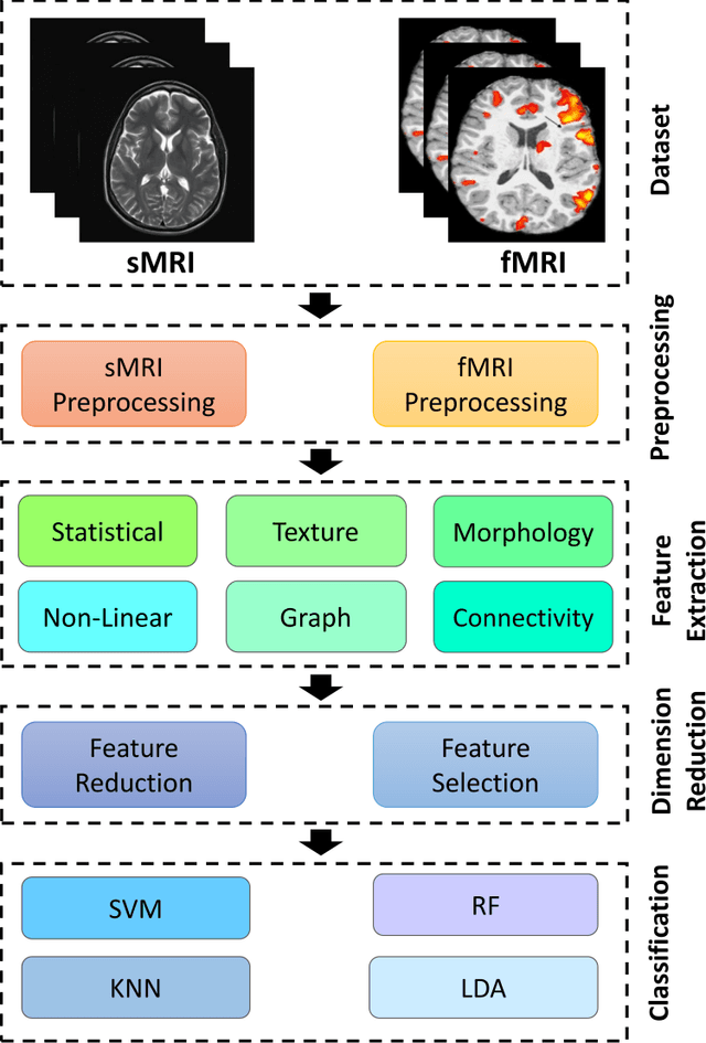
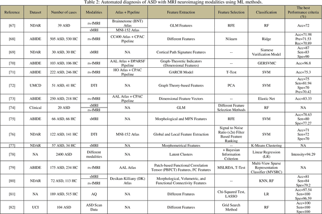
Abstract:Autism spectrum disorder (ASD) is a brain condition characterized by diverse signs and symptoms that appear in early childhood. ASD is also associated with communication deficits and repetitive behavior in affected individuals. Various ASD detection methods have been developed, including neuroimaging modalities and psychological tests. Among these methods, magnetic resonance imaging (MRI) imaging modalities are of paramount importance to physicians. Clinicians rely on MRI modalities to diagnose ASD accurately. The MRI modalities are non-invasive methods that include functional (fMRI) and structural (sMRI) neuroimaging methods. However, the process of diagnosing ASD with fMRI and sMRI for specialists is often laborious and time-consuming; therefore, several computer-aided design systems (CADS) based on artificial intelligence (AI) have been developed to assist the specialist physicians. Conventional machine learning (ML) and deep learning (DL) are the most popular schemes of AI used for diagnosing ASD. This study aims to review the automated detection of ASD using AI. We review several CADS that have been developed using ML techniques for the automated diagnosis of ASD using MRI modalities. There has been very limited work on the use of DL techniques to develop automated diagnostic models for ASD. A summary of the studies developed using DL is provided in the appendix. Then, the challenges encountered during the automated diagnosis of ASD using MRI and AI techniques are described in detail. Additionally, a graphical comparison of studies using ML and DL to diagnose ASD automatically is discussed. We conclude by suggesting future approaches to detecting ASDs using AI techniques and MRI neuroimaging.
Applications of Epileptic Seizures Detection in Neuroimaging Modalities Using Deep Learning Techniques: Methods, Challenges, and Future Works
May 29, 2021
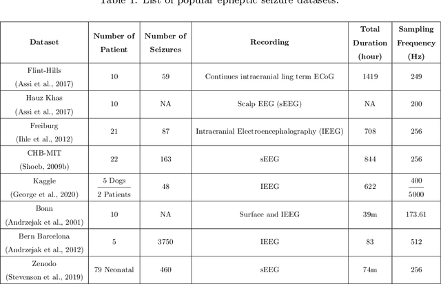
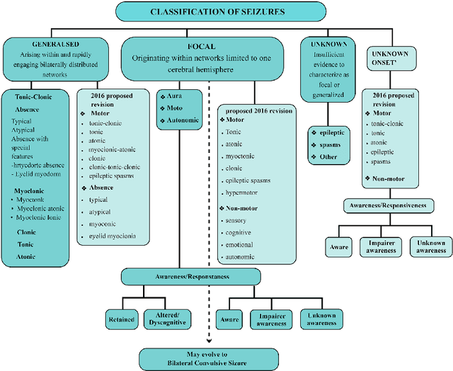
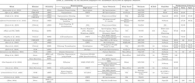
Abstract:Epileptic seizures are a type of neurological disorder that affect many people worldwide. Specialist physicians and neurologists take advantage of structural and functional neuroimaging modalities to diagnose various types of epileptic seizures. Neuroimaging modalities assist specialist physicians considerably in analyzing brain tissue and the changes made in it. One method to accelerate the accurate and fast diagnosis of epileptic seizures is to employ computer aided diagnosis systems (CADS) based on artificial intelligence (AI) and functional and structural neuroimaging modalities. AI encompasses a variety of areas, and one of its branches is deep learning (DL). Not long ago, and before the rise of DL algorithms, feature extraction was an essential part of every conventional machine learning method, yet handcrafting features limit these models' performances to the knowledge of system designers. DL methods resolved this issue entirely by automating the feature extraction and classification process; applications of these methods in many fields of medicine, such as the diagnosis of epileptic seizures, have made notable improvements. In this paper, a comprehensive overview of the types of DL methods exploited to diagnose epileptic seizures from various neuroimaging modalities has been studied. Additionally, rehabilitation systems and cloud computing in epileptic seizures diagnosis applications have been exactly investigated using various modalities.
Applications of Deep Learning Techniques for Automated Multiple Sclerosis Detection Using Magnetic Resonance Imaging: A Review
May 11, 2021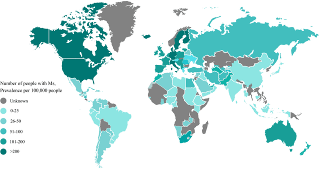

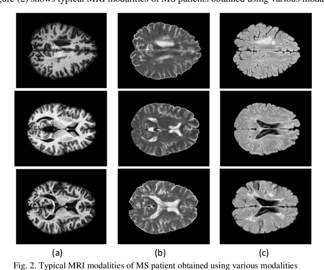
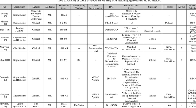
Abstract:Multiple Sclerosis (MS) is a type of brain disease which causes visual, sensory, and motor problems for people with a detrimental effect on the functioning of the nervous system. In order to diagnose MS, multiple screening methods have been proposed so far; among them, magnetic resonance imaging (MRI) has received considerable attention among physicians. MRI modalities provide physicians with fundamental information about the structure and function of the brain, which is crucial for the rapid diagnosis of MS lesions. Diagnosing MS using MRI is time-consuming, tedious, and prone to manual errors. Hence, computer aided diagnosis systems (CADS) based on artificial intelligence (AI) methods have been proposed in recent years for accurate diagnosis of MS using MRI neuroimaging modalities. In the AI field, automated MS diagnosis is being conducted using (i) conventional machine learning and (ii) deep learning (DL) techniques. The conventional machine learning approach is based on feature extraction and selection by trial and error. In DL, these steps are performed by the DL model itself. In this paper, a complete review of automated MS diagnosis methods performed using DL techniques with MRI neuroimaging modalities are discussed. Also, each work is thoroughly reviewed and discussed. Finally, the most important challenges and future directions in the automated MS diagnosis using DL techniques coupled with MRI modalities are presented in detail.
Fusion of convolution neural network, support vector machine and Sobel filter for accurate detection of COVID-19 patients using X-ray images
Feb 13, 2021
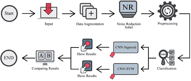

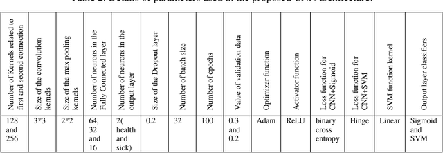
Abstract:The coronavirus (COVID-19) is currently the most common contagious disease which is prevalent all over the world. The main challenge of this disease is the primary diagnosis to prevent secondary infections and its spread from one person to another. Therefore, it is essential to use an automatic diagnosis system along with clinical procedures for the rapid diagnosis of COVID-19 to prevent its spread. Artificial intelligence techniques using computed tomography (CT) images of the lungs and chest radiography have the potential to obtain high diagnostic performance for Covid-19 diagnosis. In this study, a fusion of convolutional neural network (CNN), support vector machine (SVM), and Sobel filter is proposed to detect COVID-19 using X-ray images. A new X-ray image dataset was collected and subjected to high pass filter using a Sobel filter to obtain the edges of the images. Then these images are fed to CNN deep learning model followed by SVM classifier with ten-fold cross validation strategy. This method is designed so that it can learn with not many data. Our results show that the proposed CNN-SVM with Sobel filtering (CNN-SVM+Sobel) achieved the highest classification accuracy of 99.02% in accurate detection of COVID-19. It showed that using Sobel filter can improve the performance of CNN. Unlike most of the other researches, this method does not use a pre-trained network. We have also validated our developed model using six public databases and obtained the highest performance. Hence, our developed model is ready for clinical application
Automated Detection and Forecasting of COVID-19 using Deep Learning Techniques: A Review
Jul 27, 2020


Abstract:Coronavirus, or COVID-19, is a hazardous disease that has endangered the health of many people around the world by directly affecting the lungs. COVID-19 is a medium-sized, coated virus with a single-stranded RNA. This virus has one of the largest RNA genomes and is approximately 120 nm. The X-Ray and computed tomography (CT) imaging modalities are widely used to obtain a fast and accurate medical diagnosis. Identifying COVID-19 from these medical images is extremely challenging as it is time-consuming, demanding, and prone to human errors. Hence, artificial intelligence (AI) methodologies can be used to obtain consistent high performance. Among the AI methodologies, deep learning (DL) networks have gained much popularity compared to traditional machine learning (ML) methods. Unlike ML techniques, all stages of feature extraction, feature selection, and classification are accomplished automatically in DL models. In this paper, a complete survey of studies on the application of DL techniques for COVID-19 diagnostic and automated segmentation of lungs is discussed, concentrating on works that used X-Ray and CT images. Additionally, a review of papers on the forecasting of coronavirus prevalence in different parts of the world with DL techniques is presented. Lastly, the challenges faced in the automated detection of COVID-19 using DL techniques and directions for future research are discussed.
Deep Learning for Neuroimaging-based Diagnosis and Rehabilitation of Autism Spectrum Disorder: A Review
Jul 11, 2020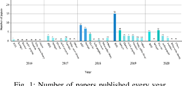
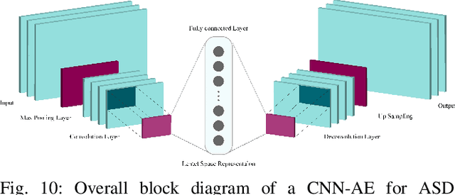


Abstract:Accurate diagnosis of Autism Spectrum Disorder (ASD) is essential for its management and rehabilitation. Neuroimaging techniques that are non-invasive are disease markers and may be leveraged to aid ASD diagnosis. Structural and functional neuroimaging techniques provide physicians substantial information about the structure (anatomy and structural connectivity) and function (activity and functional connectivity) of the brain. Due to the intricate structure and function of the brain, diagnosing ASD with neuroimaging data without exploiting artificial intelligence (AI) techniques is extremely challenging. AI techniques comprise traditional machine learning (ML) approaches and deep learning (DL) techniques. Conventional ML methods employ various feature extraction and classification techniques, but in DL, the process of feature extraction and classification is accomplished intelligently and integrally. In this paper, studies conducted with the aid of DL networks to distinguish ASD were investigated. Rehabilitation tools provided by supporting ASD patients utilizing DL networks were also assessed. Finally, we presented important challenges in this automated detection and rehabilitation of ASD.
Epileptic seizure detection using deep learning techniques: A Review
Jul 02, 2020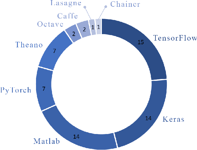
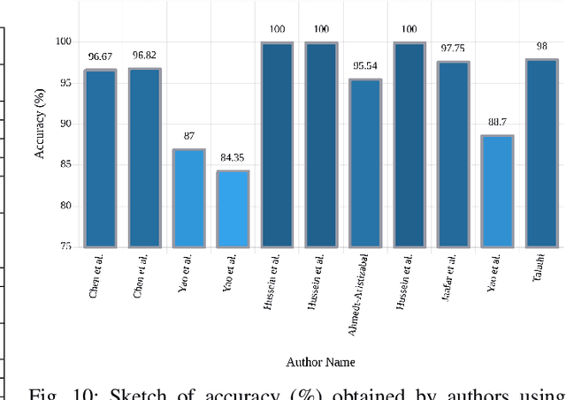
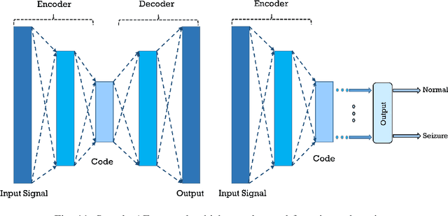
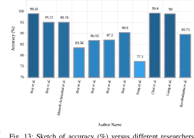
Abstract:A variety of screening approaches have been proposed to diagnose epileptic seizures, using Electroencephalography (EEG) and Magnetic Resonance Imaging (MRI) modalities. Artificial intelligence encompasses a variety of areas, and one of its branches is deep learning. Before the rise of deep learning, conventional machine learning algorithms involving feature extraction were performed. This limited their performance to the ability of those handcrafting the features. However, in deep learning, the extraction of features and classification is entirely automated. The advent of these techniques in many areas of medicine such as diagnosis of epileptic seizures, has made significant advances. In this study, a comprehensive overview of the types of deep learning methods exploited to diagnose epileptic seizures from various modalities has been studied. Additionally, hardware implementation and cloud-based works are discussed as they are most suited for applied medicine.
 Add to Chrome
 Add to Firefox
 Add to Edge