Zahra Alizadeh Sani
FCM-DNN: diagnosing coronary artery disease by deep accuracy Fuzzy C-Means clustering model
Feb 28, 2022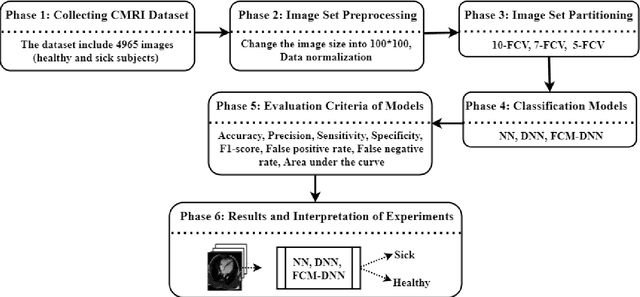

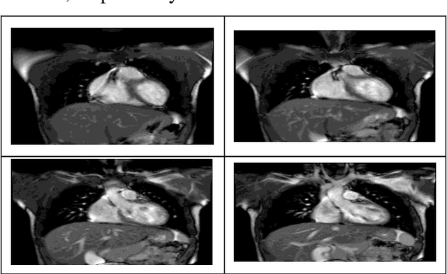

Abstract:Cardiovascular disease is one of the most challenging diseases in middle-aged and older people, which causes high mortality. Coronary artery disease (CAD) is known as a common cardiovascular disease. A standard clinical tool for diagnosing CAD is angiography. The main challenges are dangerous side effects and high angiography costs. Today, the development of artificial intelligence-based methods is a valuable achievement for diagnosing disease. Hence, in this paper, artificial intelligence methods such as neural network (NN), deep neural network (DNN), and Fuzzy C-Means clustering combined with deep neural network (FCM-DNN) are developed for diagnosing CAD on a cardiac magnetic resonance imaging (CMRI) dataset. The original dataset is used in two different approaches. First, the labeled dataset is applied to the NN and DNN to create the NN and DNN models. Second, the labels are removed, and the unlabeled dataset is clustered via the FCM method, and then, the clustered dataset is fed to the DNN to create the FCM-DNN model. By utilizing the second clustering and modeling, the training process is improved, and consequently, the accuracy is increased. As a result, the proposed FCM-DNN model achieves the best performance with a 99.91% accuracy specifying 10 clusters, i.e., 5 clusters for healthy subjects and 5 clusters for sick subjects, through the 10-fold cross-validation technique compared to the NN and DNN models reaching the accuracies of 92.18% and 99.63%, respectively. To the best of our knowledge, no study has been conducted for CAD diagnosis on the CMRI dataset using artificial intelligence methods. The results confirm that the proposed FCM-DNN model can be helpful for scientific and research centers.
CNN AE: Convolution Neural Network combined with Autoencoder approach to detect survival chance of COVID 19 patients
Apr 18, 2021


Abstract:In this paper, we propose a novel method named CNN-AE to predict survival chance of COVID-19 patients using a CNN trained on clinical information. To further increase the prediction accuracy, we use the CNN in combination with an autoencoder. Our method is one of the first that aims to predict survival chance of already infected patients. We rely on clinical data to carry out the prediction. The motivation is that the required resources to prepare CT images are expensive and limited compared to the resources required to collect clinical data such as blood pressure, liver disease, etc. We evaluate our method on a publicly available clinical dataset of deceased and recovered patients which we have collected. Careful analysis of the dataset properties is also presented which consists of important features extraction and correlation computation between features. Since most of COVID-19 patients are usually recovered, the number of deceased samples of our dataset is low leading to data imbalance. To remedy this issue, a data augmentation procedure based on autoencoders is proposed. To demonstrate the generality of our augmentation method, we train random forest and Na\"ive Bayes on our dataset with and without augmentation and compare their performance. We also evaluate our method on another dataset for further generality verification. Experimental results reveal the superiority of CNN-AE method compared to the standard CNN as well as other methods such as random forest and Na\"ive Bayes. COVID-19 detection average accuracy of CNN-AE is 96.05% which is higher than CNN average accuracy of 92.49%. To show that clinical data can be used as a reliable dataset for COVID-19 survival chance prediction, CNN-AE is compared with a standard CNN which is trained on CT images.
Fusion of convolution neural network, support vector machine and Sobel filter for accurate detection of COVID-19 patients using X-ray images
Feb 13, 2021
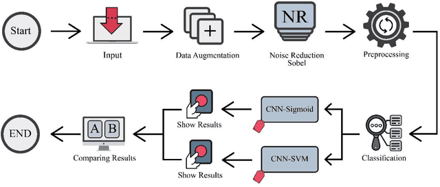

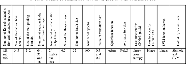
Abstract:The coronavirus (COVID-19) is currently the most common contagious disease which is prevalent all over the world. The main challenge of this disease is the primary diagnosis to prevent secondary infections and its spread from one person to another. Therefore, it is essential to use an automatic diagnosis system along with clinical procedures for the rapid diagnosis of COVID-19 to prevent its spread. Artificial intelligence techniques using computed tomography (CT) images of the lungs and chest radiography have the potential to obtain high diagnostic performance for Covid-19 diagnosis. In this study, a fusion of convolutional neural network (CNN), support vector machine (SVM), and Sobel filter is proposed to detect COVID-19 using X-ray images. A new X-ray image dataset was collected and subjected to high pass filter using a Sobel filter to obtain the edges of the images. Then these images are fed to CNN deep learning model followed by SVM classifier with ten-fold cross validation strategy. This method is designed so that it can learn with not many data. Our results show that the proposed CNN-SVM with Sobel filtering (CNN-SVM+Sobel) achieved the highest classification accuracy of 99.02% in accurate detection of COVID-19. It showed that using Sobel filter can improve the performance of CNN. Unlike most of the other researches, this method does not use a pre-trained network. We have also validated our developed model using six public databases and obtained the highest performance. Hence, our developed model is ready for clinical application
Uncertainty-Aware Semi-supervised Method using Large Unlabelled and Limited Labeled COVID-19 Data
Feb 12, 2021
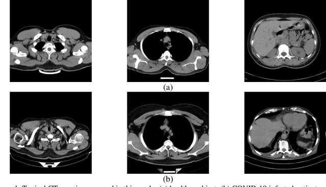
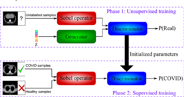
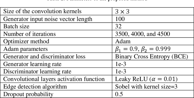
Abstract:The new coronavirus has caused more than 1 million deaths and continues to spread rapidly. This virus targets the lungs, causing respiratory distress which can be mild or severe. The X-ray or computed tomography (CT) images of lungs can reveal whether the patient is infected with COVID-19 or not. Many researchers are trying to improve COVID-19 detection using artificial intelligence. In this paper, relying on Generative Adversarial Networks (GAN), we propose a Semi-supervised Classification using Limited Labelled Data (SCLLD) for automated COVID-19 detection. Our motivation is to develop learning method which can cope with scenarios that preparing labelled data is time consuming or expensive. We further improved the detection accuracy of the proposed method by applying Sobel edge detection. The GAN discriminator output is a probability value which is used for classification in this work. The proposed system is trained using 10,000 CT scans collected from Omid hospital. Also, we validate our system using the public dataset. The proposed method is compared with other state of the art supervised methods such as Gaussian processes. To the best of our knowledge, this is the first time a COVID-19 semi-supervised detection method is presented. Our method is capable of learning from a mixture of limited labelled and unlabelled data where supervised learners fail due to lack of sufficient amount of labelled data. Our semi-supervised training method significantly outperforms the supervised training of Convolutional Neural Network (CNN) in case labelled training data is scarce. Our method has achieved an accuracy of 99.60%, sensitivity of 99.39%, and specificity of 99.80% where CNN (trained supervised) has achieved an accuracy of 69.87%, sensitivity of 94%, and specificity of 46.40%.
Objective Evaluation of Deep Uncertainty Predictions for COVID-19 Detection
Dec 22, 2020

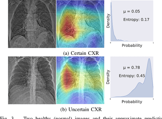

Abstract:Deep neural networks (DNNs) have been widely applied for detecting COVID-19 in medical images. Existing studies mainly apply transfer learning and other data representation strategies to generate accurate point estimates. The generalization power of these networks is always questionable due to being developed using small datasets and failing to report their predictive confidence. Quantifying uncertainties associated with DNN predictions is a prerequisite for their trusted deployment in medical settings. Here we apply and evaluate three uncertainty quantification techniques for COVID-19 detection using chest X-Ray (CXR) images. The novel concept of uncertainty confusion matrix is proposed and new performance metrics for the objective evaluation of uncertainty estimates are introduced. Through comprehensive experiments, it is shown that networks pertained on CXR images outperform networks pretrained on natural image datasets such as ImageNet. Qualitatively and quantitatively evaluations also reveal that the predictive uncertainty estimates are statistically higher for erroneous predictions than correct predictions. Accordingly, uncertainty quantification methods are capable of flagging risky predictions with high uncertainty estimates. We also observe that ensemble methods more reliably capture uncertainties during the inference.
Automated Detection and Forecasting of COVID-19 using Deep Learning Techniques: A Review
Jul 27, 2020


Abstract:Coronavirus, or COVID-19, is a hazardous disease that has endangered the health of many people around the world by directly affecting the lungs. COVID-19 is a medium-sized, coated virus with a single-stranded RNA. This virus has one of the largest RNA genomes and is approximately 120 nm. The X-Ray and computed tomography (CT) imaging modalities are widely used to obtain a fast and accurate medical diagnosis. Identifying COVID-19 from these medical images is extremely challenging as it is time-consuming, demanding, and prone to human errors. Hence, artificial intelligence (AI) methodologies can be used to obtain consistent high performance. Among the AI methodologies, deep learning (DL) networks have gained much popularity compared to traditional machine learning (ML) methods. Unlike ML techniques, all stages of feature extraction, feature selection, and classification are accomplished automatically in DL models. In this paper, a complete survey of studies on the application of DL techniques for COVID-19 diagnostic and automated segmentation of lungs is discussed, concentrating on works that used X-Ray and CT images. Additionally, a review of papers on the forecasting of coronavirus prevalence in different parts of the world with DL techniques is presented. Lastly, the challenges faced in the automated detection of COVID-19 using DL techniques and directions for future research are discussed.
 Add to Chrome
 Add to Firefox
 Add to Edge