Navid Ghassemi
Moo-ving Beyond Tradition: Revolutionizing Cattle Behavioural Phenotyping with Pose Estimation Techniques
Aug 12, 2024Abstract:The cattle industry has been a major contributor to the economy of many countries, including the US and Canada. The integration of Artificial Intelligence (AI) has revolutionized this sector, mirroring its transformative impact across all industries by enabling scalable and automated monitoring and intervention practices. AI has also introduced tools and methods that automate many tasks previously performed by human labor with the help of computer vision, including health inspections. Among these methods, pose estimation has a special place; pose estimation is the process of finding the position of joints in an image of animals. Analyzing the pose of animal subjects enables precise identification and tracking of the animal's movement and the movements of its body parts. By summarizing the video and imagery data into movement and joint location using pose estimation and then analyzing this information, we can address the scalability challenge in cattle management, focusing on health monitoring, behavioural phenotyping and welfare concerns. Our study reviews recent advancements in pose estimation methodologies, their applicability in improving the cattle industry, existing challenges, and gaps in this field. Furthermore, we propose an initiative to enhance open science frameworks within this field of study by launching a platform designed to connect industry and academia.
Automated Diagnosis of Cardiovascular Diseases from Cardiac Magnetic Resonance Imaging Using Deep Learning Models: A Review
Oct 26, 2022Abstract:In recent years, cardiovascular diseases (CVDs) have become one of the leading causes of mortality globally. CVDs appear with minor symptoms and progressively get worse. The majority of people experience symptoms such as exhaustion, shortness of breath, ankle swelling, fluid retention, and other symptoms when starting CVD. Coronary artery disease (CAD), arrhythmia, cardiomyopathy, congenital heart defect (CHD), mitral regurgitation, and angina are the most common CVDs. Clinical methods such as blood tests, electrocardiography (ECG) signals, and medical imaging are the most effective methods used for the detection of CVDs. Among the diagnostic methods, cardiac magnetic resonance imaging (CMR) is increasingly used to diagnose, monitor the disease, plan treatment and predict CVDs. Coupled with all the advantages of CMR data, CVDs diagnosis is challenging for physicians due to many slices of data, low contrast, etc. To address these issues, deep learning (DL) techniques have been employed to the diagnosis of CVDs using CMR data, and much research is currently being conducted in this field. This review provides an overview of the studies performed in CVDs detection using CMR images and DL techniques. The introduction section examined CVDs types, diagnostic methods, and the most important medical imaging techniques. In the following, investigations to detect CVDs using CMR images and the most significant DL methods are presented. Another section discussed the challenges in diagnosing CVDs from CMR data. Next, the discussion section discusses the results of this review, and future work in CVDs diagnosis from CMR images and DL techniques are outlined. The most important findings of this study are presented in the conclusion section.
Automatic Diagnosis of Myocarditis Disease in Cardiac MRI Modality using Deep Transformers and Explainable Artificial Intelligence
Oct 26, 2022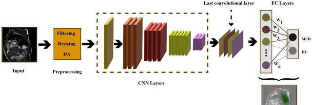

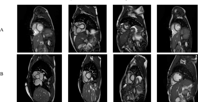

Abstract:Myocarditis is among the most important cardiovascular diseases (CVDs), endangering the health of many individuals by damaging the myocardium. Microbes and viruses, such as HIV, play a vital role in myocarditis disease (MCD) incidence. Lack of MCD diagnosis in the early stages is associated with irreversible complications. Cardiac magnetic resonance imaging (CMRI) is highly popular among cardiologists to diagnose CVDs. In this paper, a deep learning (DL) based computer-aided diagnosis system (CADS) is presented for the diagnosis of MCD using CMRI images. The proposed CADS includes dataset, preprocessing, feature extraction, classification, and post-processing steps. First, the Z-Alizadeh dataset was selected for the experiments. The preprocessing step included noise removal, image resizing, and data augmentation (DA). In this step, CutMix, and MixUp techniques were used for the DA. Then, the most recent pre-trained and transformers models were used for feature extraction and classification using CMRI images. Our results show high performance for the detection of MCD using transformer models compared with the pre-trained architectures. Among the DL architectures, Turbulence Neural Transformer (TNT) architecture achieved an accuracy of 99.73% with 10-fold cross-validation strategy. Explainable-based Grad Cam method is used to visualize the MCD suspected areas in CMRI images.
A Comprehensive Review of Trends, Applications and Challenges In Out-of-Distribution Detection
Sep 26, 2022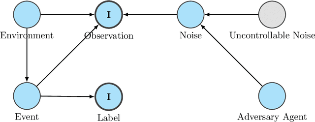
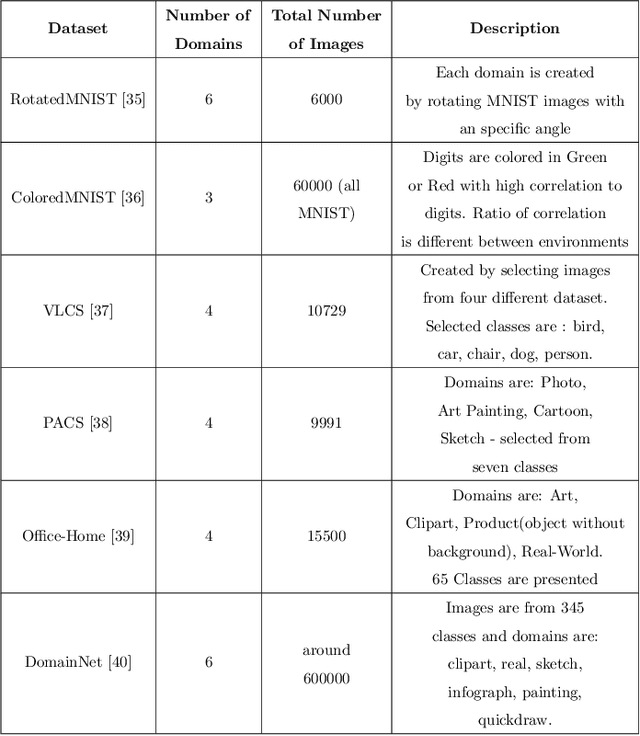
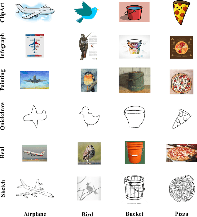
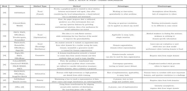
Abstract:With recent advancements in artificial intelligence, its applications can be seen in every aspect of humans' daily life. From voice assistants to mobile healthcare and autonomous driving, we rely on the performance of AI methods for many critical tasks; therefore, it is essential to assert the performance of models in proper means to prevent damage. One of the shortfalls of AI models in general, and deep machine learning in particular, is a drop in performance when faced with shifts in the distribution of data. Nonetheless, these shifts are always expected in real-world applications; thus, a field of study has emerged, focusing on detecting out-of-distribution data subsets and enabling a more comprehensive generalization. Furthermore, as many deep learning based models have achieved near-perfect results on benchmark datasets, the need to evaluate these models' reliability and trustworthiness for pushing towards real-world applications is felt more strongly than ever. This has given rise to a growing number of studies in the field of out-of-distribution detection and domain generalization, which begs the need for surveys that compare these studies from various perspectives and highlight their straightens and weaknesses. This paper presents a survey that, in addition to reviewing more than 70 papers in this field, presents challenges and directions for future works and offers a unifying look into various types of data shifts and solutions for better generalization.
Automatic Autism Spectrum Disorder Detection Using Artificial Intelligence Methods with MRI Neuroimaging: A Review
Jun 20, 2022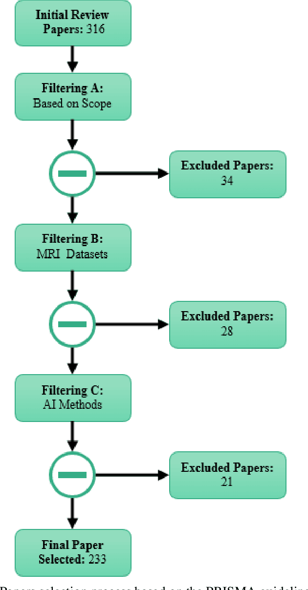

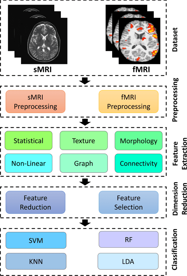
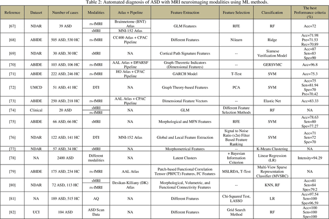
Abstract:Autism spectrum disorder (ASD) is a brain condition characterized by diverse signs and symptoms that appear in early childhood. ASD is also associated with communication deficits and repetitive behavior in affected individuals. Various ASD detection methods have been developed, including neuroimaging modalities and psychological tests. Among these methods, magnetic resonance imaging (MRI) imaging modalities are of paramount importance to physicians. Clinicians rely on MRI modalities to diagnose ASD accurately. The MRI modalities are non-invasive methods that include functional (fMRI) and structural (sMRI) neuroimaging methods. However, the process of diagnosing ASD with fMRI and sMRI for specialists is often laborious and time-consuming; therefore, several computer-aided design systems (CADS) based on artificial intelligence (AI) have been developed to assist the specialist physicians. Conventional machine learning (ML) and deep learning (DL) are the most popular schemes of AI used for diagnosing ASD. This study aims to review the automated detection of ASD using AI. We review several CADS that have been developed using ML techniques for the automated diagnosis of ASD using MRI modalities. There has been very limited work on the use of DL techniques to develop automated diagnostic models for ASD. A summary of the studies developed using DL is provided in the appendix. Then, the challenges encountered during the automated diagnosis of ASD using MRI and AI techniques are described in detail. Additionally, a graphical comparison of studies using ML and DL to diagnose ASD automatically is discussed. We conclude by suggesting future approaches to detecting ASDs using AI techniques and MRI neuroimaging.
Automatic Diagnosis of Schizophrenia and Attention Deficit Hyperactivity Disorder in rs-fMRI Modality using Convolutional Autoencoder Model and Interval Type-2 Fuzzy Regression
May 31, 2022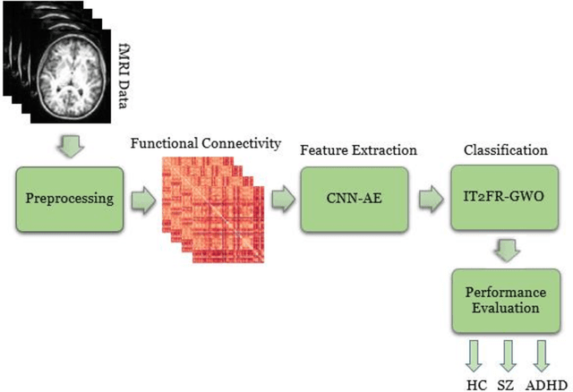
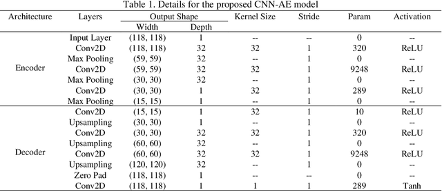
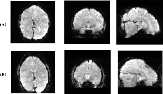
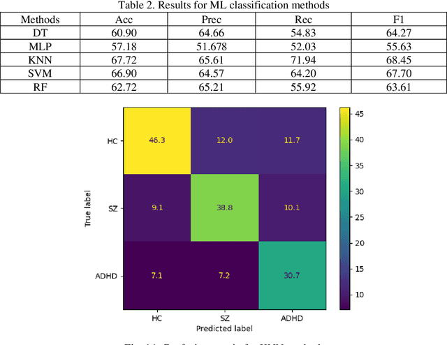
Abstract:Nowadays, many people worldwide suffer from brain disorders, and their health is in danger. So far, numerous methods have been proposed for the diagnosis of Schizophrenia (SZ) and attention deficit hyperactivity disorder (ADHD), among which functional magnetic resonance imaging (fMRI) modalities are known as a popular method among physicians. This paper presents an SZ and ADHD intelligent detection method of resting-state fMRI (rs-fMRI) modality using a new deep learning (DL) method. The University of California Los Angeles (UCLA) dataset, which contains the rs-fMRI modalities of SZ and ADHD patients, has been used for experiments. The FMRIB software library (FSL) toolbox first performed preprocessing on rs-fMRI data. Then, a convolutional Autoencoder (CNN-AE) model with the proposed number of layers is used to extract features from rs-fMRI data. In the classification step, a new fuzzy method called interval type-2 fuzzy regression (IT2FR) is introduced and then optimized by genetic algorithm (GA), particle swarm optimization (PSO), and gray wolf optimization (GWO) techniques. Also, the results of IT2FR methods are compared with multilayer perceptron (MLP), k-nearest neighbors (KNN), support vector machine (SVM), random forest (RF), decision tree (DT), and adaptive neuro-fuzzy inference system (ANFIS) methods. The experiment results show that the IT2FR method with the GWO optimization algorithm has achieved satisfactory results compared to other classifier methods. Finally, the proposed classification technique was able to provide 72.71% accuracy.
Detection of Epileptic Seizures on EEG Signals Using ANFIS Classifier, Autoencoders and Fuzzy Entropies
Sep 06, 2021
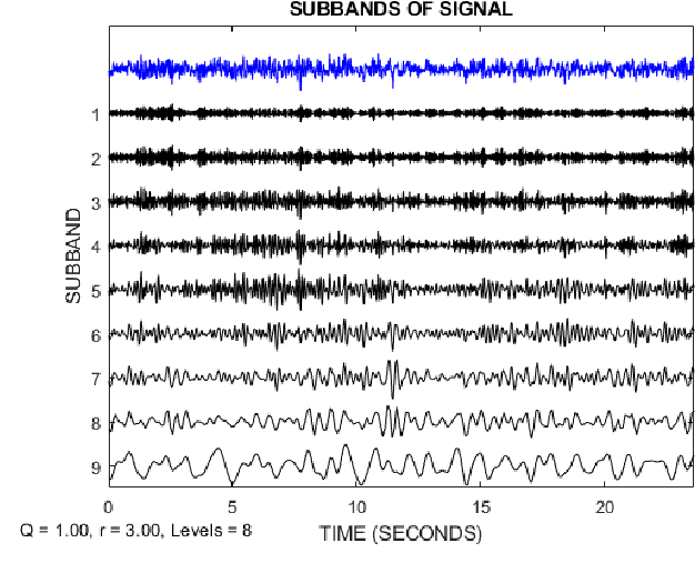
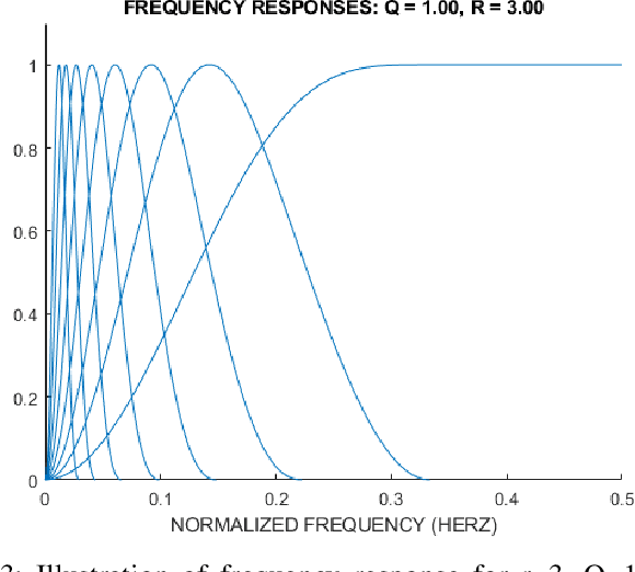
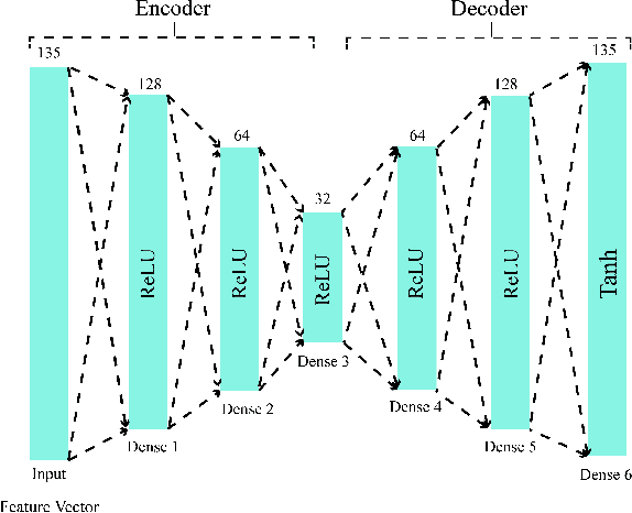
Abstract:Epilepsy is one of the most crucial neurological disorders, and its early diagnosis will help the clinicians to provide accurate treatment for the patients. The electroencephalogram (EEG) signals are widely used for epileptic seizures detection, which provides specialists with substantial information about the functioning of the brain. In this paper, a novel diagnostic procedure using fuzzy theory and deep learning techniques are introduced. The proposed method is evaluated on the Bonn University dataset with six classification combinations and also on the Freiburg dataset. The tunable-Q wavelet transform (TQWT) is employed to decompose the EEG signals into different sub-bands. In the feature extraction step, 13 different fuzzy entropies are calculated from different sub-bands of TQWT, and their computational complexities are calculated to help researchers choose the best feature sets. In the following, an autoencoder (AE) with six layers is employed for dimensionality reduction. Finally, the standard adaptive neuro-fuzzy inference system (ANFIS), and also its variants with grasshopper optimization algorithm (ANFIS-GOA), particle swarm optimization (ANFIS-PSO), and breeding swarm optimization (ANFIS-BS) methods are used for classification. Using our proposed method, ANFIS-BS method has obtained an accuracy of 99.74% in classifying into two classes and an accuracy of 99.46% in ternary classification on the Bonn dataset and 99.28% on the Freiburg dataset, reaching state-of-the-art performances on both of them.
Automatic Diagnosis of Schizophrenia using EEG Signals and CNN-LSTM Models
Sep 02, 2021
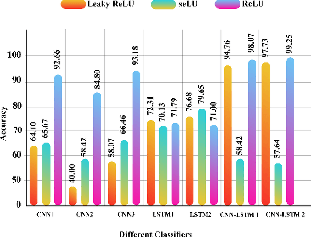
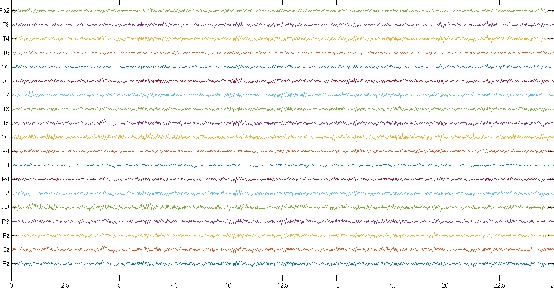
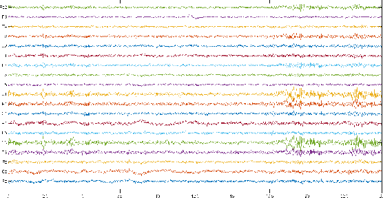
Abstract:Schizophrenia (SZ) is a mental disorder whereby due to the secretion of specific chemicals in the brain, the function of some brain regions is out of balance, leading to the lack of coordination between thoughts, actions, and emotions. This study provides various intelligent Deep Learning (DL)-based methods for automated SZ diagnosis via EEG signals. The obtained results are compared with those of conventional intelligent methods. In order to implement the proposed methods, the dataset of the Institute of Psychiatry and Neurology in Warsaw, Poland, has been used. First, EEG signals are divided into 25-seconds time frames and then were normalized by z-score or norm L2. In the classification step, two different approaches are considered for SZ diagnosis via EEG signals. In this step, the classification of EEG signals is first carried out by conventional DL methods, e.g., KNN, DT, SVM, Bayes, bagging, RF, and ET. Various proposed DL models, including LSTMs, 1D-CNNs, and 1D-CNN-LSTMs, are used in the following. In this step, the DL models were implemented and compared with different activation functions. Among the proposed DL models, the CNN-LSTM architecture has had the best performance. In this architecture, the ReLU activation function and the z-score and L2 combined normalization are used. The proposed CNN-LSTM model has achieved an accuracy percentage of 99.25\%, better than the results of most former studies in this field. It is worth mentioning that in order to perform all simulations, the k-fold cross-validation method with k=5 has been used.
Applications of Epileptic Seizures Detection in Neuroimaging Modalities Using Deep Learning Techniques: Methods, Challenges, and Future Works
May 29, 2021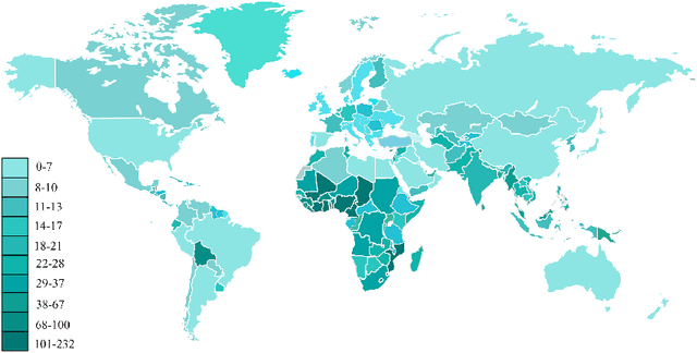
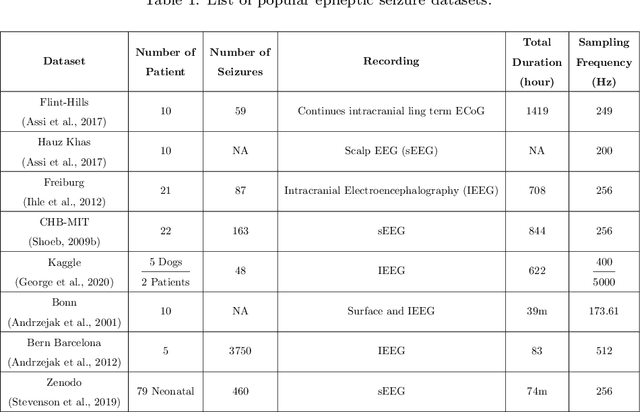
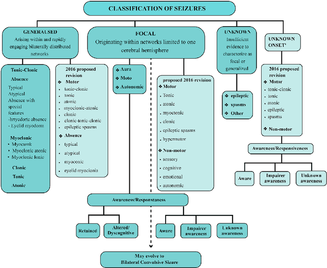
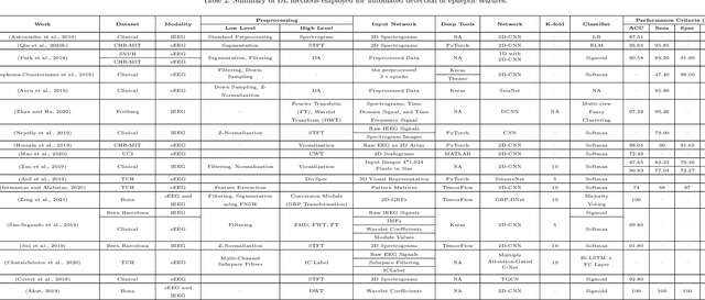
Abstract:Epileptic seizures are a type of neurological disorder that affect many people worldwide. Specialist physicians and neurologists take advantage of structural and functional neuroimaging modalities to diagnose various types of epileptic seizures. Neuroimaging modalities assist specialist physicians considerably in analyzing brain tissue and the changes made in it. One method to accelerate the accurate and fast diagnosis of epileptic seizures is to employ computer aided diagnosis systems (CADS) based on artificial intelligence (AI) and functional and structural neuroimaging modalities. AI encompasses a variety of areas, and one of its branches is deep learning (DL). Not long ago, and before the rise of DL algorithms, feature extraction was an essential part of every conventional machine learning method, yet handcrafting features limit these models' performances to the knowledge of system designers. DL methods resolved this issue entirely by automating the feature extraction and classification process; applications of these methods in many fields of medicine, such as the diagnosis of epileptic seizures, have made notable improvements. In this paper, a comprehensive overview of the types of DL methods exploited to diagnose epileptic seizures from various neuroimaging modalities has been studied. Additionally, rehabilitation systems and cloud computing in epileptic seizures diagnosis applications have been exactly investigated using various modalities.
Automatic Diagnosis of COVID-19 from CT Images using CycleGAN and Transfer Learning
Apr 24, 2021
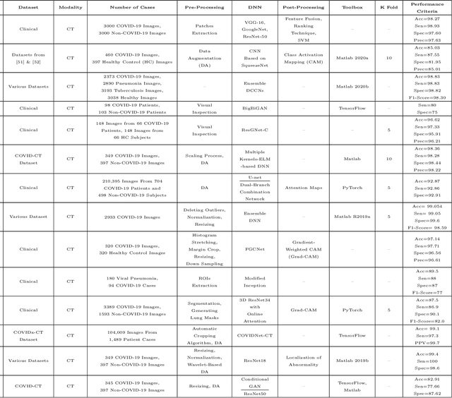
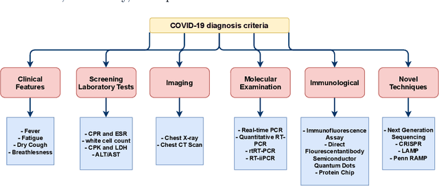
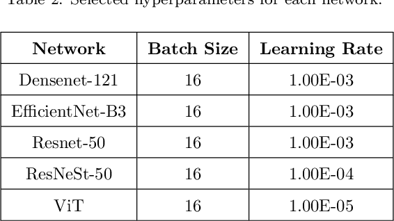
Abstract:The outbreak of the corona virus disease (COVID-19) has changed the lives of most people on Earth. Given the high prevalence of this disease, its correct diagnosis in order to quarantine patients is of the utmost importance in steps of fighting this pandemic. Among the various modalities used for diagnosis, medical imaging, especially computed tomography (CT) imaging, has been the focus of many previous studies due to its accuracy and availability. In addition, automation of diagnostic methods can be of great help to physicians. In this paper, a method based on pre-trained deep neural networks is presented, which, by taking advantage of a cyclic generative adversarial net (CycleGAN) model for data augmentation, has reached state-of-the-art performance for the task at hand, i.e., 99.60% accuracy. Also, in order to evaluate the method, a dataset containing 3163 images from 189 patients has been collected and labeled by physicians. Unlike prior datasets, normal data have been collected from people suspected of having COVID-19 disease and not from data from other diseases, and this database is made available publicly.
 Add to Chrome
 Add to Firefox
 Add to Edge