 Add to Chrome
 Add to Firefox
 Add to Edge
Improving Deep Learning-Based Target Volume Auto-Delineation for Adaptive MR-Guided Radiotherapy in Head and Neck Cancer: Impact of a Volume-Aware Dice Loss
Apr 11, 2026Background: Manual delineation of target volumes in head and neck cancer (HNC) remains a significant bottleneck in radiotherapy planning, characterized by high inter-observer variability and time consumption. This study evaluates the integration of a Volume-Aware (VA) Dice loss function into a self-configuring deep learning framework to enhance the auto-segmentation of primary tumors (PT) and metastatic lymph nodes (LN) for adaptive MR-guided radiotherapy. We investigate how volume-sensitive weighting affects the detection of small, anatomically complex nodal metastases compared to conventional loss functions. Methods: Utilizing the HNTS-MRG 2024 dataset, we implemented an nnU-Net ResEnc M architecture. We conducted a multi-label segmentation task, comparing a standard Dice loss baseline against two Volume-Aware configurations: a "Dual Mask" setup (VA loss on both PT and LN) and a "Selective LN Mask" setup (VA loss on LN only). Evaluation metrics included volumetric Dice scores, surface-based metrics (SDS, MSD, HD95), and lesion-wise binary detection sensitivity and precision. Results: The Selective LN Mask configuration achieved the highest LN Volumetric Dice Score (0.758 vs. 0.734 baseline) and significantly improved LN Lesion-Wise Detection Sensitivity (84.93% vs. 81.80%). However, a critical trade-off was observed; PT detection precision declined significantly in the selective setup (63.65% vs. 81.27%). The Dual Mask configuration provided the most balanced performance across both targets, maintaining primary tumor precision at 82.04% while improving LN sensitivity to 83.46%. Conclusions: A volume-sensitive loss function mitigated the under-representation of small metastatic lesions in HNC. While selective weighting yielded the best nodal detection, a dual-mask approach is required in multi-label tasks to maintain segmentation accuracy for larger primary tumor volumes.
Improving Generalization of Deep Learning for Brain Metastases Segmentation Across Institutions
Apr 01, 2026Background: Deep learning has demonstrated significant potential for automated brain metastases (BM) segmentation; however, models trained at a singular institution often exhibit suboptimal performance at various sites due to disparities in scanner hardware, imaging protocols, and patient demographics. The goal of this work is to create a domain adaptation framework that will allow for BM segmentation to be used across multiple institutions. Methods: We propose a VAE-MMD preprocessing pipeline that combines variational autoencoders (VAE) with maximum mean discrepancy (MMD) loss, incorporating skip connections and self-attention mechanisms alongside nnU-Net segmentation. The method was tested on 740 patients from four public databases: Stanford, UCSF, UCLM, and PKG, evaluated by domain classifier's accuracy, sensitivity, precision, F1/F2 scores, surface Dice (sDice), and 95th percentile Hausdorff distance (HD95). Results: VAE-MMD reduced domain classifier accuracy from 0.91 to 0.50, indicating successful feature alignment across institutions. Reconstructed volumes attained a PSNR greater than 36 dB, maintaining anatomical accuracy. The combined method raised the mean F1 by 11.1% (0.700 to 0.778), the mean sDice by 7.93% (0.7121 to 0.7686), and reduced the mean HD95 by 65.5% (11.33 to 3.91 mm) across all four centers compared to the baseline nnU-Net. Conclusions: VAE-MMD effectively diminishes cross-institutional data heterogeneity and enhances BM segmentation generalization across volumetric, detection, and boundary-level metrics without necessitating target-domain labels, thereby overcoming a significant obstacle to the clinical implementation of AI-assisted segmentation.
A Self-supervised Multimodal Deep Learning Approach to Differentiate Post-radiotherapy Progression from Pseudoprogression in Glioblastoma
Feb 06, 2025


Accurate differentiation of pseudoprogression (PsP) from True Progression (TP) following radiotherapy (RT) in glioblastoma (GBM) patients is crucial for optimal treatment planning. However, this task remains challenging due to the overlapping imaging characteristics of PsP and TP. This study therefore proposes a multimodal deep-learning approach utilizing complementary information from routine anatomical MR images, clinical parameters, and RT treatment planning information for improved predictive accuracy. The approach utilizes a self-supervised Vision Transformer (ViT) to encode multi-sequence MR brain volumes to effectively capture both global and local context from the high dimensional input. The encoder is trained in a self-supervised upstream task on unlabeled glioma MRI datasets from the open BraTS2021, UPenn-GBM, and UCSF-PDGM datasets to generate compact, clinically relevant representations from FLAIR and T1 post-contrast sequences. These encoded MR inputs are then integrated with clinical data and RT treatment planning information through guided cross-modal attention, improving progression classification accuracy. This work was developed using two datasets from different centers: the Burdenko Glioblastoma Progression Dataset (n = 59) for training and validation, and the GlioCMV progression dataset from the University Hospital Erlangen (UKER) (n = 20) for testing. The proposed method achieved an AUC of 75.3%, outperforming the current state-of-the-art data-driven approaches. Importantly, the proposed approach relies on readily available anatomical MRI sequences, clinical data, and RT treatment planning information, enhancing its clinical feasibility. The proposed approach addresses the challenge of limited data availability for PsP and TP differentiation and could allow for improved clinical decision-making and optimized treatment plans for GBM patients.
Exploring the Capabilities and Limitations of Large Language Models for Radiation Oncology Decision Support
Jan 04, 2025


Thanks to the rapidly evolving integration of LLMs into decision-support tools, a significant transformation is happening across large-scale systems. Like other medical fields, the use of LLMs such as GPT-4 is gaining increasing interest in radiation oncology as well. An attempt to assess GPT-4's performance in radiation oncology was made via a dedicated 100-question examination on the highly specialized topic of radiation oncology physics, revealing GPT-4's superiority over other LLMs. GPT-4's performance on a broader field of clinical radiation oncology is further benchmarked by the ACR Radiation Oncology In-Training (TXIT) exam where GPT-4 achieved a high accuracy of 74.57%. Its performance on re-labelling structure names in accordance with the AAPM TG-263 report has also been benchmarked, achieving above 96% accuracies. Such studies shed light on the potential of LLMs in radiation oncology. As interest in the potential and constraints of LLMs in general healthcare applications continues to rise5, the capabilities and limitations of LLMs in radiation oncology decision support have not yet been fully explored.
* Officially published in the Red Journal
Task-Specific Data Preparation for Deep Learning to Reconstruct Structures of Interest from Severely Truncated CBCT Data
Sep 13, 2024
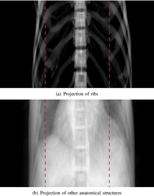
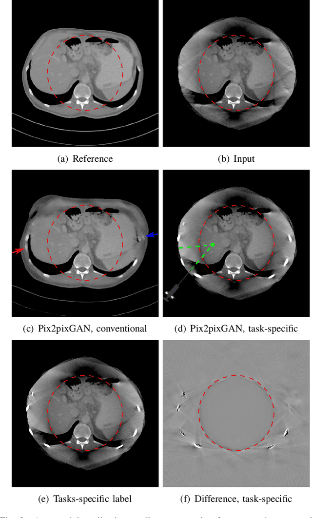
Cone-beam computed tomography (CBCT) is widely used in interventional surgeries and radiation oncology. Due to the limited size of flat-panel detectors, anatomical structures might be missing outside the limited field-of-view (FOV), which restricts the clinical applications of CBCT systems. Recently, deep learning methods have been proposed to extend the FOV for multi-slice CT systems. However, in mobile CBCT system with a smaller FOV size, projection data is severely truncated and it is challenging for a network to restore all missing structures outside the FOV. In some applications, only certain structures outside the FOV are of interest, e.g., ribs in needle path planning for liver/lung cancer diagnosis. Therefore, a task-specific data preparation method is proposed in this work, which automatically let the network focus on structures of interest instead of all the structures. Our preliminary experiment shows that Pix2pixGAN with a conventional training has the risk to reconstruct false positive and false negative rib structures from severely truncated CBCT data, whereas Pix2pixGAN with the proposed task-specific training can reconstruct all the ribs reliably. The proposed method is promising to empower CBCT with more clinical applications.
Enhancing Cross-Modality Synthesis: Subvolume Merging for MRI-to-CT Conversion
Sep 09, 2024


Providing more precise tissue attenuation information, synthetic computed tomography (sCT) generated from magnetic resonance imaging (MRI) contributes to improved radiation therapy treatment planning. In our study, we employ the advanced SwinUNETR framework for synthesizing CT from MRI images. Additionally, we introduce a three-dimensional subvolume merging technique in the prediction process. By selecting an optimal overlap percentage for adjacent subvolumes, stitching artifacts are effectively mitigated, leading to a decrease in the mean absolute error (MAE) between sCT and the labels from 52.65 HU to 47.75 HU. Furthermore, implementing a weight function with a gamma value of 0.9 results in the lowest MAE within the same overlap area. By setting the overlap percentage between 50% and 70%, we achieve a balance between image quality and computational efficiency.
Fine-Tuning a Local LLaMA-3 Large Language Model for Automated Privacy-Preserving Physician Letter Generation in Radiation Oncology
Aug 20, 2024


Generating physician letters is a time-consuming task in daily clinical practice. This study investigates local fine-tuning of large language models (LLMs), specifically LLaMA models, for physician letter generation in a privacy-preserving manner within the field of radiation oncology. Our findings demonstrate that base LLaMA models, without fine-tuning, are inadequate for effectively generating physician letters. The QLoRA algorithm provides an efficient method for local intra-institutional fine-tuning of LLMs with limited computational resources (i.e., a single 48 GB GPU workstation within the hospital). The fine-tuned LLM successfully learns radiation oncology-specific information and generates physician letters in an institution-specific style. ROUGE scores of the generated summary reports highlight the superiority of the 8B LLaMA-3 model over the 13B LLaMA-2 model. Further multidimensional physician evaluations of 10 cases reveal that, although the fine-tuned LLaMA-3 model has limited capacity to generate content beyond the provided input data, it successfully generates salutations, diagnoses and treatment histories, recommendations for further treatment, and planned schedules. Overall, clinical benefit was rated highly by the clinical experts (average score of 3.44 on a 4-point scale). With careful physician review and correction, automated LLM-based physician letter generation has significant practical value.
Comprehensive Multimodal Deep Learning Survival Prediction Enabled by a Transformer Architecture: A Multicenter Study in Glioblastoma
May 21, 2024

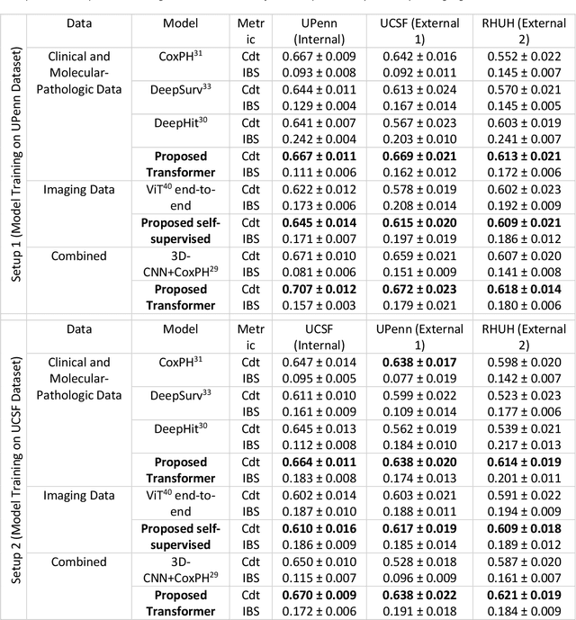

Background: This research aims to improve glioblastoma survival prediction by integrating MR images, clinical and molecular-pathologic data in a transformer-based deep learning model, addressing data heterogeneity and performance generalizability. Method: We propose and evaluate a transformer-based non-linear and non-proportional survival prediction model. The model employs self-supervised learning techniques to effectively encode the high-dimensional MRI input for integration with non-imaging data using cross-attention. To demonstrate model generalizability, the model is assessed with the time-dependent concordance index (Cdt) in two training setups using three independent public test sets: UPenn-GBM, UCSF-PDGM, and RHUH-GBM, each comprising 378, 366, and 36 cases, respectively. Results: The proposed transformer model achieved promising performance for imaging as well as non-imaging data, effectively integrating both modalities for enhanced performance (UPenn-GBM test-set, imaging Cdt 0.645, multimodal Cdt 0.707) while outperforming state-of-the-art late-fusion 3D-CNN-based models. Consistent performance was observed across the three independent multicenter test sets with Cdt values of 0.707 (UPenn-GBM, internal test set), 0.672 (UCSF-PDGM, first external test set) and 0.618 (RHUH-GBM, second external test set). The model achieved significant discrimination between patients with favorable and unfavorable survival for all three datasets (logrank p 1.9\times{10}^{-8}, 9.7\times{10}^{-3}, and 1.2\times{10}^{-2}). Conclusions: The proposed transformer-based survival prediction model integrates complementary information from diverse input modalities, contributing to improved glioblastoma survival prediction compared to state-of-the-art methods. Consistent performance was observed across institutions supporting model generalizability.
Multicenter Privacy-Preserving Model Training for Deep Learning Brain Metastases Autosegmentation
May 17, 2024Objectives: This work aims to explore the impact of multicenter data heterogeneity on deep learning brain metastases (BM) autosegmentation performance, and assess the efficacy of an incremental transfer learning technique, namely learning without forgetting (LWF), to improve model generalizability without sharing raw data. Materials and methods: A total of six BM datasets from University Hospital Erlangen (UKER), University Hospital Zurich (USZ), Stanford, UCSF, NYU and BraTS Challenge 2023 on BM segmentation were used for this evaluation. First, the multicenter performance of a convolutional neural network (DeepMedic) for BM autosegmentation was established for exclusive single-center training and for training on pooled data, respectively. Subsequently bilateral collaboration was evaluated, where a UKER pretrained model is shared to another center for further training using transfer learning (TL) either with or without LWF. Results: For single-center training, average F1 scores of BM detection range from 0.625 (NYU) to 0.876 (UKER) on respective single-center test data. Mixed multicenter training notably improves F1 scores at Stanford and NYU, with negligible improvement at other centers. When the UKER pretrained model is applied to USZ, LWF achieves a higher average F1 score (0.839) than naive TL (0.570) and single-center training (0.688) on combined UKER and USZ test data. Naive TL improves sensitivity and contouring accuracy, but compromises precision. Conversely, LWF demonstrates commendable sensitivity, precision and contouring accuracy. When applied to Stanford, similar performance was observed. Conclusion: Data heterogeneity results in varying performance in BM autosegmentation, posing challenges to model generalizability. LWF is a promising approach to peer-to-peer privacy-preserving model training.
Reference-Free Multi-Modality Volume Registration of X-Ray Microscopy and Light-Sheet Fluorescence Microscopy
Apr 23, 2024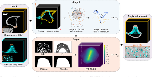
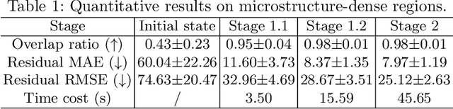
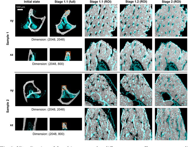
Recently, X-ray microscopy (XRM) and light-sheet fluorescence microscopy (LSFM) have emerged as two pivotal imaging tools in preclinical research on bone remodeling diseases, offering micrometer-level resolution. Integrating these complementary modalities provides a holistic view of bone microstructures, facilitating function-oriented volume analysis across different disease cycles. However, registering such independently acquired large-scale volumes is extremely challenging under real and reference-free scenarios. This paper presents a fast two-stage pipeline for volume registration of XRM and LSFM. The first stage extracts the surface features and employs two successive point cloud-based methods for coarse alignment. The second stage fine-tunes the initial alignment using a modified cross-correlation method, ensuring precise volumetric registration. Moreover, we propose residual similarity as a novel metric to assess the alignment of two complementary modalities. The results imply robust gradual improvement across the stages. In the end, all correlating microstructures, particularly lacunae in XRM and bone cells in LSFM, are precisely matched, enabling new insights into bone diseases like osteoporosis which are a substantial burden in aging societies.