Xilin Yang
Automated HER2 scoring with uncertainty quantification using lensfree holography and deep learning
Jan 26, 2026Abstract:Accurate assessment of human epidermal growth factor receptor 2 (HER2) expression is critical for breast cancer diagnosis, prognosis, and therapy selection; yet, most existing digital HER2 scoring methods rely on bulky and expensive optical systems. Here, we present a compact and cost-effective lensfree holography platform integrated with deep learning for automated HER2 scoring of immunohistochemically stained breast tissue sections. The system captures lensfree diffraction patterns of stained HER2 tissue sections under RGB laser illumination and acquires complex field information over a sample area of ~1,250 mm^2 at an effective throughput of ~84 mm^2 per minute. To enhance diagnostic reliability, we incorporated an uncertainty quantification strategy based on Bayesian Monte Carlo dropout, which provides autonomous uncertainty estimates for each prediction and supports reliable, robust HER2 scoring, with an overall correction rate of 30.4%. Using a blinded test set of 412 unique tissue samples, our approach achieved a testing accuracy of 84.9% for 4-class (0, 1+, 2+, 3+) HER2 classification and 94.8% for binary (0/1+ vs. 2+/3+) HER2 scoring with uncertainty quantification. Overall, this lensfree holography approach provides a practical pathway toward portable, high-throughput, and cost-effective HER2 scoring, particularly suited for resource-limited settings, where traditional digital pathology infrastructure is unavailable.
Snapshot multi-spectral imaging through defocusing and a Fourier imager network
Jan 24, 2025


Abstract:Multi-spectral imaging, which simultaneously captures the spatial and spectral information of a scene, is widely used across diverse fields, including remote sensing, biomedical imaging, and agricultural monitoring. Here, we introduce a snapshot multi-spectral imaging approach employing a standard monochrome image sensor with no additional spectral filters or customized components. Our system leverages the inherent chromatic aberration of wavelength-dependent defocusing as a natural source of physical encoding of multi-spectral information; this encoded image information is rapidly decoded via a deep learning-based multi-spectral Fourier Imager Network (mFIN). We experimentally tested our method with six illumination bands and demonstrated an overall accuracy of 92.98% for predicting the illumination channels at the input and achieved a robust multi-spectral image reconstruction on various test objects. This deep learning-powered framework achieves high-quality multi-spectral image reconstruction using snapshot image acquisition with a monochrome image sensor and could be useful for applications in biomedicine, industrial quality control, and agriculture, among others.
Diagnosing Hate Speech Classification: Where Do Humans and Machines Disagree, and Why?
Oct 14, 2024


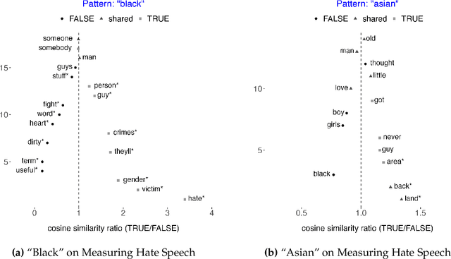
Abstract:This study uses the cosine similarity ratio, embedding regression, and manual re-annotation to diagnose hate speech classification. We begin by computing cosine similarity ratio on a dataset "Measuring Hate Speech" that contains 135,556 annotated comments on social media. This way, we show a basic use of cosine similarity as a description of hate speech content. We then diagnose hate speech classification starting from understanding the inconsistency of human annotation from the dataset. Using embedding regression as a basic diagnostic, we found that female annotators are more sensitive to racial slurs that target the black population. We perform with a more complicated diagnostic by training a hate speech classifier using a SoTA pre-trained large language model, NV-Embed-v2, to convert texts to embeddings and run a logistic regression. This classifier achieves a testing accuracy of 94%. In diagnosing where machines disagree with human annotators, we found that machines make fewer mistakes than humans despite the fact that human annotations are treated as ground truth in the training set. Machines perform better in correctly labeling long statements of facts, but perform worse in labeling short instances of swear words. We hypothesize that this is due to model alignment - while curating models at their creation prevents the models from producing obvious hate speech, it also reduces the model's ability to detect such content.
Label-free evaluation of lung and heart transplant biopsies using virtual staining
Sep 09, 2024


Abstract:Organ transplantation serves as the primary therapeutic strategy for end-stage organ failures. However, allograft rejection is a common complication of organ transplantation. Histological assessment is essential for the timely detection and diagnosis of transplant rejection and remains the gold standard. Nevertheless, the traditional histochemical staining process is time-consuming, costly, and labor-intensive. Here, we present a panel of virtual staining neural networks for lung and heart transplant biopsies, which digitally convert autofluorescence microscopic images of label-free tissue sections into their brightfield histologically stained counterparts, bypassing the traditional histochemical staining process. Specifically, we virtually generated Hematoxylin and Eosin (H&E), Masson's Trichrome (MT), and Elastic Verhoeff-Van Gieson (EVG) stains for label-free transplant lung tissue, along with H&E and MT stains for label-free transplant heart tissue. Subsequent blind evaluations conducted by three board-certified pathologists have confirmed that the virtual staining networks consistently produce high-quality histology images with high color uniformity, closely resembling their well-stained histochemical counterparts across various tissue features. The use of virtually stained images for the evaluation of transplant biopsies achieved comparable diagnostic outcomes to those obtained via traditional histochemical staining, with a concordance rate of 82.4% for lung samples and 91.7% for heart samples. Moreover, virtual staining models create multiple stains from the same autofluorescence input, eliminating structural mismatches observed between adjacent sections stained in the traditional workflow, while also saving tissue, expert time, and staining costs.
Unidirectional imaging with partially coherent light
Aug 10, 2024


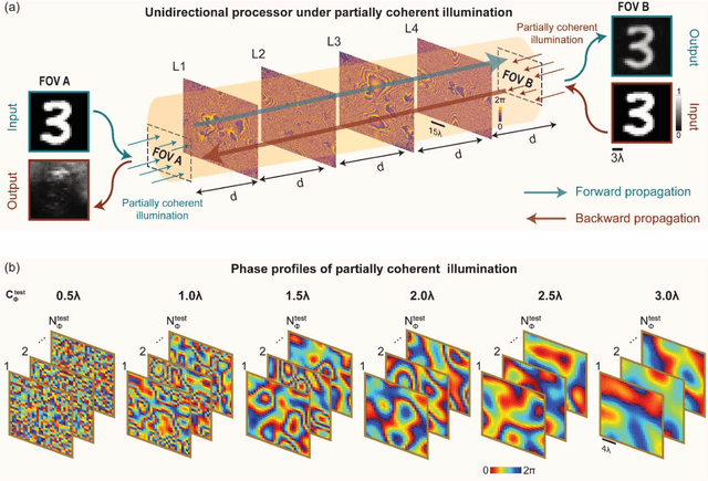
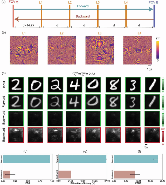
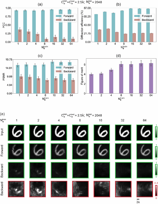
Abstract:Unidirectional imagers form images of input objects only in one direction, e.g., from field-of-view (FOV) A to FOV B, while blocking the image formation in the reverse direction, from FOV B to FOV A. Here, we report unidirectional imaging under spatially partially coherent light and demonstrate high-quality imaging only in the forward direction (A->B) with high power efficiency while distorting the image formation in the backward direction (B->A) along with low power efficiency. Our reciprocal design features a set of spatially engineered linear diffractive layers that are statistically optimized for partially coherent illumination with a given phase correlation length. Our analyses reveal that when illuminated by a partially coherent beam with a correlation length of ~1.5 w or larger, where w is the wavelength of light, diffractive unidirectional imagers achieve robust performance, exhibiting asymmetric imaging performance between the forward and backward directions - as desired. A partially coherent unidirectional imager designed with a smaller correlation length of less than 1.5 w still supports unidirectional image transmission, but with a reduced figure of merit. These partially coherent diffractive unidirectional imagers are compact (axially spanning less than 75 w), polarization-independent, and compatible with various types of illumination sources, making them well-suited for applications in asymmetric visual information processing and communication.
An insertable glucose sensor using a compact and cost-effective phosphorescence lifetime imager and machine learning
Jun 12, 2024Abstract:Optical continuous glucose monitoring (CGM) systems are emerging for personalized glucose management owing to their lower cost and prolonged durability compared to conventional electrochemical CGMs. Here, we report a computational CGM system, which integrates a biocompatible phosphorescence-based insertable biosensor and a custom-designed phosphorescence lifetime imager (PLI). This compact and cost-effective PLI is designed to capture phosphorescence lifetime images of an insertable sensor through the skin, where the lifetime of the emitted phosphorescence signal is modulated by the local concentration of glucose. Because this phosphorescence signal has a very long lifetime compared to tissue autofluorescence or excitation leakage processes, it completely bypasses these noise sources by measuring the sensor emission over several tens of microseconds after the excitation light is turned off. The lifetime images acquired through the skin are processed by neural network-based models for misalignment-tolerant inference of glucose levels, accurately revealing normal, low (hypoglycemia) and high (hyperglycemia) concentration ranges. Using a 1-mm thick skin phantom mimicking the optical properties of human skin, we performed in vitro testing of the PLI using glucose-spiked samples, yielding 88.8% inference accuracy, also showing resilience to random and unknown misalignments within a lateral distance of ~4.7 mm with respect to the position of the insertable sensor underneath the skin phantom. Furthermore, the PLI accurately identified larger lateral misalignments beyond 5 mm, prompting user intervention for re-alignment. The misalignment-resilient glucose concentration inference capability of this compact and cost-effective phosphorescence lifetime imager makes it an appealing wearable diagnostics tool for real-time tracking of glucose and other biomarkers.
Automated HER2 Scoring in Breast Cancer Images Using Deep Learning and Pyramid Sampling
Apr 01, 2024Abstract:Human epidermal growth factor receptor 2 (HER2) is a critical protein in cancer cell growth that signifies the aggressiveness of breast cancer (BC) and helps predict its prognosis. Accurate assessment of immunohistochemically (IHC) stained tissue slides for HER2 expression levels is essential for both treatment guidance and understanding of cancer mechanisms. Nevertheless, the traditional workflow of manual examination by board-certified pathologists encounters challenges, including inter- and intra-observer inconsistency and extended turnaround times. Here, we introduce a deep learning-based approach utilizing pyramid sampling for the automated classification of HER2 status in IHC-stained BC tissue images. Our approach analyzes morphological features at various spatial scales, efficiently managing the computational load and facilitating a detailed examination of cellular and larger-scale tissue-level details. This method addresses the tissue heterogeneity of HER2 expression by providing a comprehensive view, leading to a blind testing classification accuracy of 84.70%, on a dataset of 523 core images from tissue microarrays. Our automated system, proving reliable as an adjunct pathology tool, has the potential to enhance diagnostic precision and evaluation speed, and might significantly impact cancer treatment planning.
Virtual birefringence imaging and histological staining of amyloid deposits in label-free tissue using autofluorescence microscopy and deep learning
Mar 14, 2024Abstract:Systemic amyloidosis is a group of diseases characterized by the deposition of misfolded proteins in various organs and tissues, leading to progressive organ dysfunction and failure. Congo red stain is the gold standard chemical stain for the visualization of amyloid deposits in tissue sections, as it forms complexes with the misfolded proteins and shows a birefringence pattern under polarized light microscopy. However, Congo red staining is tedious and costly to perform, and prone to false diagnoses due to variations in the amount of amyloid, staining quality and expert interpretation through manual examination of tissue under a polarization microscope. Here, we report the first demonstration of virtual birefringence imaging and virtual Congo red staining of label-free human tissue to show that a single trained neural network can rapidly transform autofluorescence images of label-free tissue sections into brightfield and polarized light microscopy equivalent images, matching the histochemically stained versions of the same samples. We demonstrate the efficacy of our method with blind testing and pathologist evaluations on cardiac tissue where the virtually stained images agreed well with the histochemically stained ground truth images. Our virtually stained polarization and brightfield images highlight amyloid birefringence patterns in a consistent, reproducible manner while mitigating diagnostic challenges due to variations in the quality of chemical staining and manual imaging processes as part of the clinical workflow.
Multiplexed all-optical permutation operations using a reconfigurable diffractive optical network
Feb 04, 2024


Abstract:Large-scale and high-dimensional permutation operations are important for various applications in e.g., telecommunications and encryption. Here, we demonstrate the use of all-optical diffractive computing to execute a set of high-dimensional permutation operations between an input and output field-of-view through layer rotations in a diffractive optical network. In this reconfigurable multiplexed material designed by deep learning, every diffractive layer has four orientations: 0, 90, 180, and 270 degrees. Each unique combination of these rotatable layers represents a distinct rotation state of the diffractive design tailored for a specific permutation operation. Therefore, a K-layer rotatable diffractive material is capable of all-optically performing up to 4^K independent permutation operations. The original input information can be decrypted by applying the specific inverse permutation matrix to output patterns, while applying other inverse operations will lead to loss of information. We demonstrated the feasibility of this reconfigurable multiplexed diffractive design by approximating 256 randomly selected permutation matrices using K=4 rotatable diffractive layers. We also experimentally validated this reconfigurable diffractive network using terahertz radiation and 3D-printed diffractive layers, providing a decent match to our numerical results. The presented rotation-multiplexed diffractive processor design is particularly useful due to its mechanical reconfigurability, offering multifunctional representation through a single fabrication process.
Subwavelength Imaging using a Solid-Immersion Diffractive Optical Processor
Jan 17, 2024Abstract:Phase imaging is widely used in biomedical imaging, sensing, and material characterization, among other fields. However, direct imaging of phase objects with subwavelength resolution remains a challenge. Here, we demonstrate subwavelength imaging of phase and amplitude objects based on all-optical diffractive encoding and decoding. To resolve subwavelength features of an object, the diffractive imager uses a thin, high-index solid-immersion layer to transmit high-frequency information of the object to a spatially-optimized diffractive encoder, which converts/encodes high-frequency information of the input into low-frequency spatial modes for transmission through air. The subsequent diffractive decoder layers (in air) are jointly designed with the encoder using deep-learning-based optimization, and communicate with the encoder layer to create magnified images of input objects at its output, revealing subwavelength features that would otherwise be washed away due to diffraction limit. We demonstrate that this all-optical collaboration between a diffractive solid-immersion encoder and the following decoder layers in air can resolve subwavelength phase and amplitude features of input objects in a highly compact design. To experimentally demonstrate its proof-of-concept, we used terahertz radiation and developed a fabrication method for creating monolithic multi-layer diffractive processors. Through these monolithically fabricated diffractive encoder-decoder pairs, we demonstrated phase-to-intensity transformations and all-optically reconstructed subwavelength phase features of input objects by directly transforming them into magnified intensity features at the output. This solid-immersion-based diffractive imager, with its compact and cost-effective design, can find wide-ranging applications in bioimaging, endoscopy, sensing and materials characterization.
 Add to Chrome
 Add to Firefox
 Add to Edge