Lanzhuju Mei
SDF-Net: A Hybrid Detection Network for Mediastinal Lymph Node Detection on Contrast CT Images
Sep 10, 2024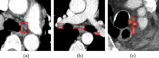
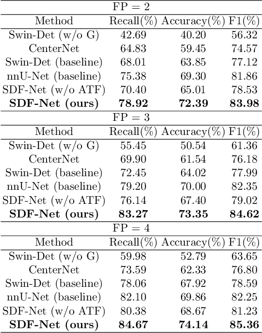
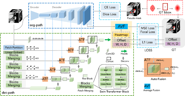
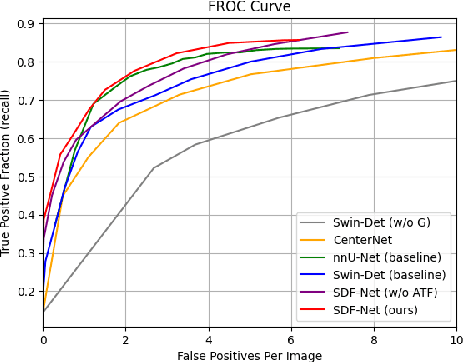
Abstract:Accurate lymph node detection and quantification are crucial for cancer diagnosis and staging on contrast-enhanced CT images, as they impact treatment planning and prognosis. However, detecting lymph nodes in the mediastinal area poses challenges due to their low contrast, irregular shapes and dispersed distribution. In this paper, we propose a Swin-Det Fusion Network (SDF-Net) to effectively detect lymph nodes. SDF-Net integrates features from both segmentation and detection to enhance the detection capability of lymph nodes with various shapes and sizes. Specifically, an auto-fusion module is designed to merge the feature maps of segmentation and detection networks at different levels. To facilitate effective learning without mask annotations, we introduce a shape-adaptive Gaussian kernel to represent lymph node in the training stage and provide more anatomical information for effective learning. Comparative results demonstrate promising performance in addressing the complex lymph node detection problem.
Cephalometric Landmark Detection across Ages with Prototypical Network
Jun 18, 2024


Abstract:Automated cephalometric landmark detection is crucial in real-world orthodontic diagnosis. Current studies mainly focus on only adult subjects, neglecting the clinically crucial scenario presented by adolescents whose landmarks often exhibit significantly different appearances compared to adults. Hence, an open question arises about how to develop a unified and effective detection algorithm across various age groups, including adolescents and adults. In this paper, we propose CeLDA, the first work for Cephalometric Landmark Detection across Ages. Our method leverages a prototypical network for landmark detection by comparing image features with landmark prototypes. To tackle the appearance discrepancy of landmarks between age groups, we design new strategies for CeLDA to improve prototype alignment and obtain a holistic estimation of landmark prototypes from a large set of training images. Moreover, a novel prototype relation mining paradigm is introduced to exploit the anatomical relations between the landmark prototypes. Extensive experiments validate the superiority of CeLDA in detecting cephalometric landmarks on both adult and adolescent subjects. To our knowledge, this is the first effort toward developing a unified solution and dataset for cephalometric landmark detection across age groups. Our code and dataset will be made public on https://github.com/ShanghaiTech-IMPACT/Cephalometric-Landmark-Detection-across-Ages-with-Prototypical-Network
3D Structure-guided Network for Tooth Alignment in 2D Photograph
Oct 17, 2023Abstract:Orthodontics focuses on rectifying misaligned teeth (i.e., malocclusions), affecting both masticatory function and aesthetics. However, orthodontic treatment often involves complex, lengthy procedures. As such, generating a 2D photograph depicting aligned teeth prior to orthodontic treatment is crucial for effective dentist-patient communication and, more importantly, for encouraging patients to accept orthodontic intervention. In this paper, we propose a 3D structure-guided tooth alignment network that takes 2D photographs as input (e.g., photos captured by smartphones) and aligns the teeth within the 2D image space to generate an orthodontic comparison photograph featuring aesthetically pleasing, aligned teeth. Notably, while the process operates within a 2D image space, our method employs 3D intra-oral scanning models collected in clinics to learn about orthodontic treatment, i.e., projecting the pre- and post-orthodontic 3D tooth structures onto 2D tooth contours, followed by a diffusion model to learn the mapping relationship. Ultimately, the aligned tooth contours are leveraged to guide the generation of a 2D photograph with aesthetically pleasing, aligned teeth and realistic textures. We evaluate our network on various facial photographs, demonstrating its exceptional performance and strong applicability within the orthodontic industry.
ChatCAD+: Towards a Universal and Reliable Interactive CAD using LLMs
May 26, 2023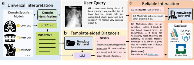
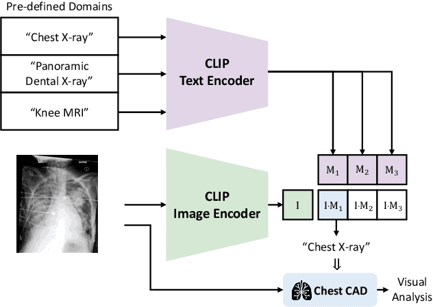
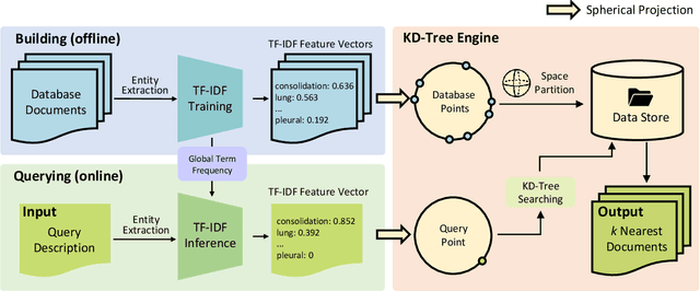
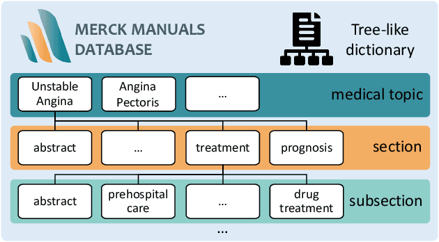
Abstract:The potential of integrating Computer-Assisted Diagnosis (CAD) with Large Language Models (LLMs) in clinical applications, particularly in digital family doctor and clinic assistant roles, shows promise. However, existing works have limitations in terms of reliability, effectiveness, and their narrow applicability to specific image domains, which restricts their overall processing capabilities. Moreover, the mismatch in writing style between LLMs and radiologists undermines their practical utility. To address these challenges, we present ChatCAD+, an interactive CAD system that is universal, reliable, and capable of handling medical images from diverse domains. ChatCAD+ utilizes current information obtained from reputable medical websites to offer precise medical advice. Additionally, it incorporates a template retrieval system that emulates real-world diagnostic reporting, thereby improving its seamless integration into existing clinical workflows. The source code is available at https://github.com/zhaozh10/ChatCAD. The online demo will be available soon.
SNAF: Sparse-view CBCT Reconstruction with Neural Attenuation Fields
Nov 30, 2022


Abstract:Cone beam computed tomography (CBCT) has been widely used in clinical practice, especially in dental clinics, while the radiation dose of X-rays when capturing has been a long concern in CBCT imaging. Several research works have been proposed to reconstruct high-quality CBCT images from sparse-view 2D projections, but the current state-of-the-arts suffer from artifacts and the lack of fine details. In this paper, we propose SNAF for sparse-view CBCT reconstruction by learning the neural attenuation fields, where we have invented a novel view augmentation strategy to overcome the challenges introduced by insufficient data from sparse input views. Our approach achieves superior performance in terms of high reconstruction quality (30+ PSNR) with only 20 input views (25 times fewer than clinical collections), which outperforms the state-of-the-arts. We have further conducted comprehensive experiments and ablation analysis to validate the effectiveness of our approach.
 Add to Chrome
 Add to Firefox
 Add to Edge