Lukas Folle
Differentiable Score-Based Likelihoods: Learning CT Motion Compensation From Clean Images
Apr 23, 2024


Abstract:Motion artifacts can compromise the diagnostic value of computed tomography (CT) images. Motion correction approaches require a per-scan estimation of patient-specific motion patterns. In this work, we train a score-based model to act as a probability density estimator for clean head CT images. Given the trained model, we quantify the deviation of a given motion-affected CT image from the ideal distribution through likelihood computation. We demonstrate that the likelihood can be utilized as a surrogate metric for motion artifact severity in the CT image facilitating the application of an iterative, gradient-based motion compensation algorithm. By optimizing the underlying motion parameters to maximize likelihood, our method effectively reduces motion artifacts, bringing the image closer to the distribution of motion-free scans. Our approach achieves comparable performance to state-of-the-art methods while eliminating the need for a representative data set of motion-affected samples. This is particularly advantageous in real-world applications, where patient motion patterns may exhibit unforeseen variability, ensuring robustness without implicit assumptions about recoverable motion types.
A gradient-based approach to fast and accurate head motion compensation in cone-beam CT
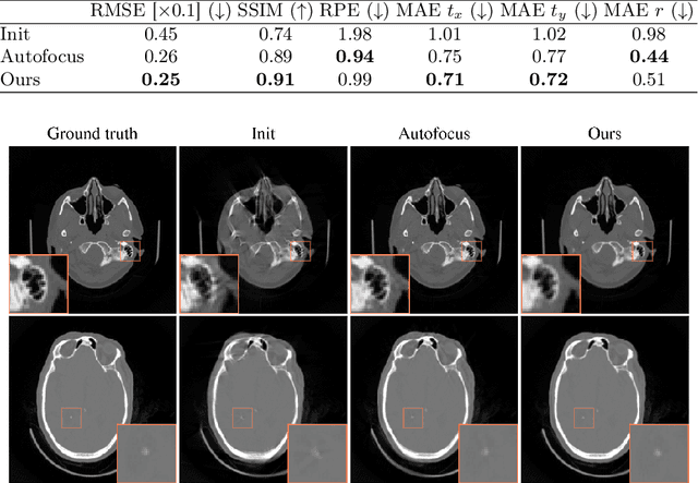
Jan 17, 2024Abstract:Cone-beam computed tomography (CBCT) systems, with their portability, present a promising avenue for direct point-of-care medical imaging, particularly in critical scenarios such as acute stroke assessment. However, the integration of CBCT into clinical workflows faces challenges, primarily linked to long scan duration resulting in patient motion during scanning and leading to image quality degradation in the reconstructed volumes. This paper introduces a novel approach to CBCT motion estimation using a gradient-based optimization algorithm, which leverages generalized derivatives of the backprojection operator for cone-beam CT geometries. Building on that, a fully differentiable target function is formulated which grades the quality of the current motion estimate in reconstruction space. We drastically accelerate motion estimation yielding a 19-fold speed-up compared to existing methods. Additionally, we investigate the architecture of networks used for quality metric regression and propose predicting voxel-wise quality maps, favoring autoencoder-like architectures over contracting ones. This modification improves gradient flow, leading to more accurate motion estimation. The presented method is evaluated through realistic experiments on head anatomy. It achieves a reduction in reprojection error from an initial average of 3mm to 0.61mm after motion compensation and consistently demonstrates superior performance compared to existing approaches. The analytic Jacobian for the backprojection operation, which is at the core of the proposed method, is made publicly available. In summary, this paper contributes to the advancement of CBCT integration into clinical workflows by proposing a robust motion estimation approach that enhances efficiency and accuracy, addressing critical challenges in time-sensitive scenarios.
Gradient-Based Geometry Learning for Fan-Beam CT Reconstruction
Dec 05, 2022Abstract:Incorporating computed tomography (CT) reconstruction operators into differentiable pipelines has proven beneficial in many applications. Such approaches usually focus on the projection data and keep the acquisition geometry fixed. However, precise knowledge of the acquisition geometry is essential for high quality reconstruction results. In this paper, the differentiable formulation of fan-beam CT reconstruction is extended to the acquisition geometry. This allows to propagate gradient information from a loss function on the reconstructed image into the geometry parameters. As a proof-of-concept experiment, this idea is applied to rigid motion compensation. The cost function is parameterized by a trained neural network which regresses an image quality metric from the motion affected reconstruction alone. Using the proposed method, we are the first to optimize such an autofocus-inspired algorithm based on analytical gradients. The algorithm achieves a reduction in MSE by 35.5 % and an improvement in SSIM by 12.6 % over the motion affected reconstruction. Next to motion compensation, we see further use cases of our differentiable method for scanner calibration or hybrid techniques employing deep models.
Generation of Anonymous Chest Radiographs Using Latent Diffusion Models for Training Thoracic Abnormality Classification Systems
Nov 04, 2022Abstract:The availability of large-scale chest X-ray datasets is a requirement for developing well-performing deep learning-based algorithms in thoracic abnormality detection and classification. However, biometric identifiers in chest radiographs hinder the public sharing of such data for research purposes due to the risk of patient re-identification. To counteract this issue, synthetic data generation offers a solution for anonymizing medical images. This work employs a latent diffusion model to synthesize an anonymous chest X-ray dataset of high-quality class-conditional images. We propose a privacy-enhancing sampling strategy to ensure the non-transference of biometric information during the image generation process. The quality of the generated images and the feasibility of serving as exclusive training data are evaluated on a thoracic abnormality classification task. Compared to a real classifier, we achieve competitive results with a performance gap of only 3.5% in the area under the receiver operating characteristic curve.
Learned Cone-Beam CT Reconstruction Using Neural Ordinary Differential Equations
Jan 19, 2022


Abstract:Learned iterative reconstruction algorithms for inverse problems offer the flexibility to combine analytical knowledge about the problem with modules learned from data. This way, they achieve high reconstruction performance while ensuring consistency with the measured data. In computed tomography, extending such approaches from 2D fan-beam to 3D cone-beam data is challenging due to the prohibitively high GPU memory that would be needed to train such models. This paper proposes to use neural ordinary differential equations to solve the reconstruction problem in a residual formulation via numerical integration. For training, there is no need to backpropagate through several unrolled network blocks nor through the internals of the solver. Instead, the gradients are obtained very memory-efficiently in the neural ODE setting allowing for training on a single consumer graphics card. The method is able to reduce the root mean squared error by over 30% compared to the best performing classical iterative reconstruction algorithm and produces high quality cone-beam reconstructions even in a sparse view scenario.
Towards Super-Resolution CEST MRI for Visualization of Small Structures
Dec 03, 2021

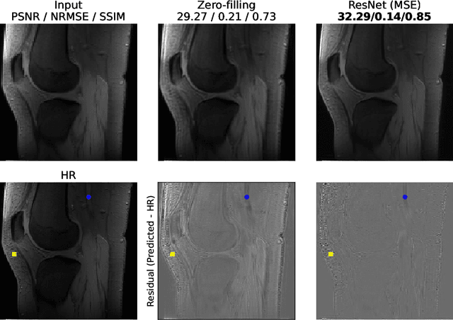
Abstract:The onset of rheumatic diseases such as rheumatoid arthritis is typically subclinical, which results in challenging early detection of the disease. However, characteristic changes in the anatomy can be detected using imaging techniques such as MRI or CT. Modern imaging techniques such as chemical exchange saturation transfer (CEST) MRI drive the hope to improve early detection even further through the imaging of metabolites in the body. To image small structures in the joints of patients, typically one of the first regions where changes due to the disease occur, a high resolution for the CEST MR imaging is necessary. Currently, however, CEST MR suffers from an inherently low resolution due to the underlying physical constraints of the acquisition. In this work we compared established up-sampling techniques to neural network-based super-resolution approaches. We could show, that neural networks are able to learn the mapping from low-resolution to high-resolution unsaturated CEST images considerably better than present methods. On the test set a PSNR of 32.29dB (+10%), a NRMSE of 0.14 (+28%), and a SSIM of 0.85 (+15%) could be achieved using a ResNet neural network, improving the baseline considerably. This work paves the way for the prospective investigation of neural networks for super-resolution CEST MRI and, followingly, might lead to a earlier detection of the onset of rheumatic diseases.
Dilated deeply supervised networks for hippocampus segmentation in MRI
Mar 20, 2019


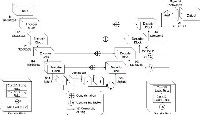
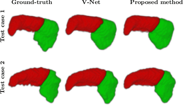
Abstract:Tissue loss in the hippocampi has been heavily correlated with the progression of Alzheimer's Disease (AD). The shape and structure of the hippocampus are important factors in terms of early AD diagnosis and prognosis by clinicians. However, manual segmentation of such subcortical structures in MR studies is a challenging and subjective task. In this paper, we investigate variants of the well known 3D U-Net, a type of convolution neural network (CNN) for semantic segmentation tasks. We propose an alternative form of the 3D U-Net, which uses dilated convolutions and deep supervision to incorporate multi-scale information into the model. The proposed method is evaluated on the task of hippocampus head and body segmentation in an MRI dataset, provided as part of the MICCAI 2018 segmentation decathlon challenge. The experimental results show that our approach outperforms other conventional methods in terms of different segmentation accuracy metrics.
 Add to Chrome
 Add to Firefox
 Add to Edge