 Add to Chrome
 Add to Firefox
 Add to Edge
Automated Report-Derived Oncology VQA Benchmark for Evaluating Vision-Language Models on 3D Medical Imaging
Jun 01, 2026Evaluating vision-language models (VLMs) on medical images requires benchmarks that are clinically grounded, scalable, and controlled for evaluation confounds. Existing public benchmarks are limited in scale, manually annotated, or potentially leaked into VLM pretraining corpora. We present an automated agent-driven pipeline that generates multiple-choice VQA datasets directly from paired private radiology reports and 3D oncology imaging, producing two complementary question types: RADS-style questions deterministically derived from clinician-defined reporting schemas, and radiology report-derived questions generated by an LLM from radiologist findings and verified against the source report. Applied to four in-house cancer cohorts, the pipeline yields an instance-contamination-controlled benchmark without per-question human annotation. Zero-shot evaluation of six VLMs reveals no dominant model and substantial headroom across all cells. A blind ablation reveals that visual reliance is highly dataset-specific: liver Report-derived questions genuinely require the image, while Lung CT is essentially solvable without it - the leading closed model exceeds its sighted accuracy on Lung CT when blinded - indicating that even private clinical data does not guarantee a contamination-controlled read of visual capability. The pipeline is released as an open agent skill for in-house redeployment.
LegSegNet: A Public Deep Learning System for Lower Extremity CT Tissue Segmentation and Quantification
May 29, 2026Lower extremity computed tomography (CT) contains clinically relevant information for body composition analysis, sarcopenia assessment, and musculoskeletal disease monitoring, but extracting these measurements at scale requires accurate tissue segmentation and an automated quantification workflow. Existing public segmentation tools are not designed for comprehensive lower extremity CT analysis, particularly for clinically important inter/intramuscular adipose tissue, and most public methods only provide mask prediction rather than an end-to-end quantification system. To address this problem, we present LegSegNet, a deep learning system for lower extremity CT tissue segmentation and body composition quantification. Given an input CT scan, LegSegNet segments bone, skeletal muscle, subcutaneous adipose tissue, and inter/intramuscular adipose tissue. It then computes quantitative tissue measurements for downstream analysis. We developed the segmentation model using 1,302 manually annotated CT slices and evaluated it on 900 held-out test slices, with all annotations reviewed by radiologists. We benchmark LegSegNet against a broad set of 2D segmentation methods, including CNN-based models, transformer-based models, and finetuned foundation models, and further evaluate its generalization on an external public CT dataset. LegSegNet achieves the best overall segmentation performance, with an average Dice score of 89.31 on the held-out test set. To our knowledge, LegSegNet is the first publicly available end-to-end system for lower extremity CT tissue segmentation and quantification, providing a practical evaluation tool for future computer vision research in medical image analysis. The code and model weights are available at: https://github.com/mazurowski-lab/LegSegNet
Automated glenoid bone loss measurement and segmentation in CT scans for pre-operative planning in shoulder instability
Nov 18, 2025Reliable measurement of glenoid bone loss is essential for operative planning in shoulder instability, but current manual and semi-automated methods are time-consuming and often subject to interreader variability. We developed and validated a fully automated deep learning pipeline for measuring glenoid bone loss on three-dimensional computed tomography (CT) scans using a linear-based, en-face view, best-circle method. Shoulder CT images of 91 patients (average age, 40 years; range, 14-89 years; 65 men) were retrospectively collected along with manual labels including glenoid segmentation, landmarks, and bone loss measurements. The multi-stage algorithm has three main stages: (1) segmentation, where we developed a U-Net to automatically segment the glenoid and humerus; (2) anatomical landmark detection, where a second network predicts glenoid rim points; and (3) geometric fitting, where we applied principal component analysis (PCA), projection, and circle fitting to compute the percentage of bone loss. The automated measurements showed strong agreement with consensus readings and exceeded surgeon-to-surgeon consistency (intraclass correlation coefficient (ICC) 0.84 vs 0.78), including in low- and high-bone-loss subgroups (ICC 0.71 vs 0.63 and 0.83 vs 0.21, respectively; P < 0.001). For classifying patients into low, medium, and high bone-loss categories, the pipeline achieved a recall of 0.714 for low and 0.857 for high severity, with no low cases misclassified as high or vice versa. These results suggest that our method is a time-efficient and clinically reliable tool for preoperative planning in shoulder instability and for screening patients with substantial glenoid bone loss. Code and dataset are available at https://github.com/Edenliu1/Auto-Glenoid-Measurement-DL-Pipeline.
SAMora: Enhancing SAM through Hierarchical Self-Supervised Pre-Training for Medical Images
Nov 09, 2025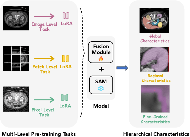
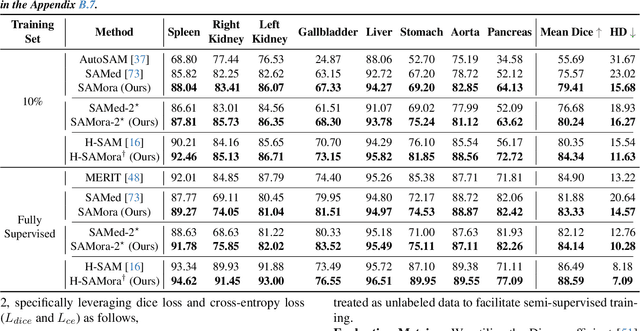
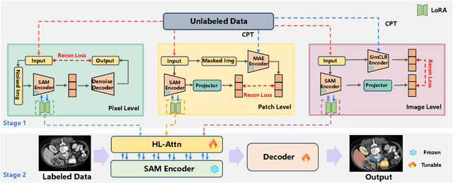
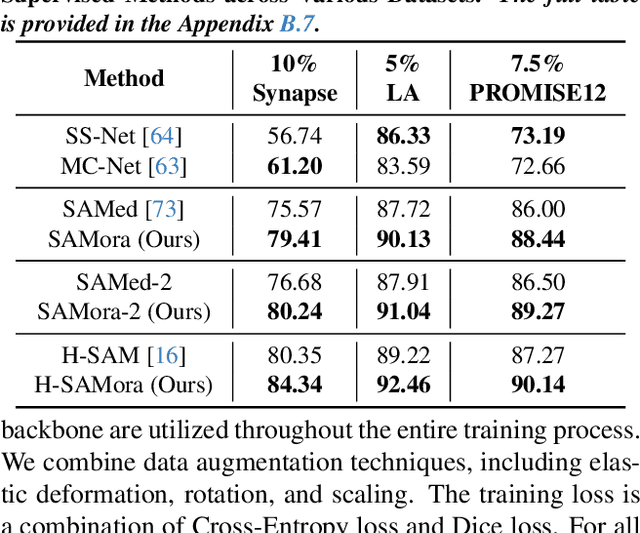
The Segment Anything Model (SAM) has demonstrated significant potential in medical image segmentation. Yet, its performance is limited when only a small amount of labeled data is available, while there is abundant valuable yet often overlooked hierarchical information in medical data. To address this limitation, we draw inspiration from self-supervised learning and propose SAMora, an innovative framework that captures hierarchical medical knowledge by applying complementary self-supervised learning objectives at the image, patch, and pixel levels. To fully exploit the complementarity of hierarchical knowledge within LoRAs, we introduce HL-Attn, a hierarchical fusion module that integrates multi-scale features while maintaining their distinct characteristics. SAMora is compatible with various SAM variants, including SAM2, SAMed, and H-SAM. Experimental results on the Synapse, LA, and PROMISE12 datasets demonstrate that SAMora outperforms existing SAM variants. It achieves state-of-the-art performance in both few-shot and fully supervised settings while reducing fine-tuning epochs by 90%. The code is available at https://github.com/ShChen233/SAMora.
Transplant-Ready? Evaluating AI Lung Segmentation Models in Candidates with Severe Lung Disease
Sep 18, 2025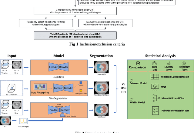
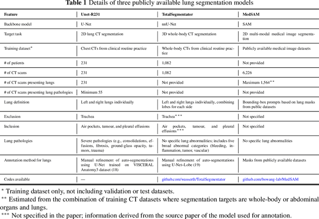
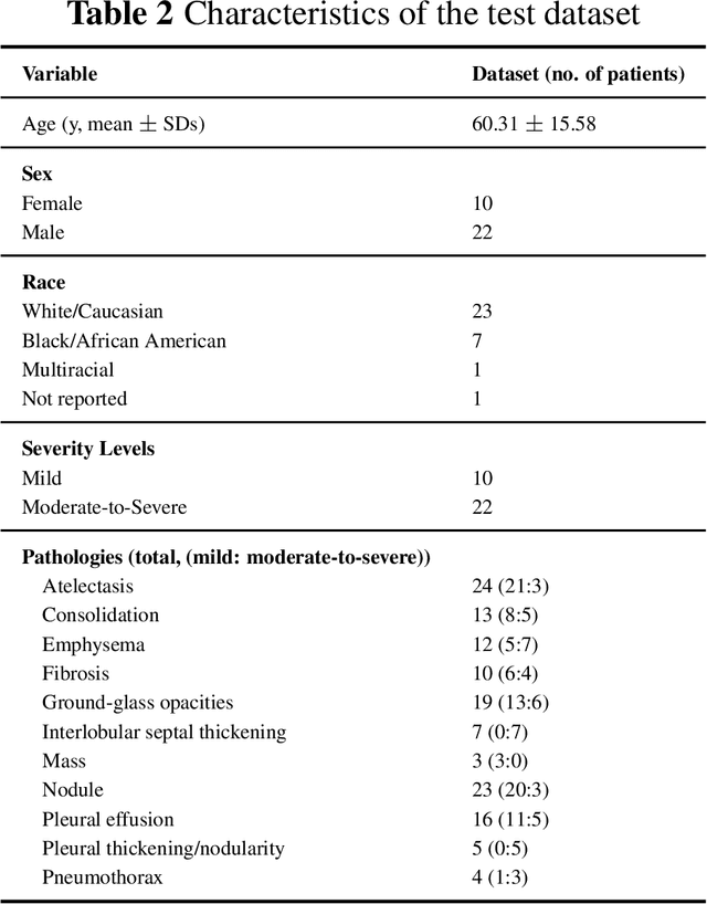
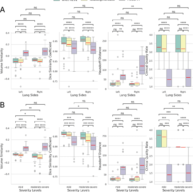
This study evaluates publicly available deep-learning based lung segmentation models in transplant-eligible patients to determine their performance across disease severity levels, pathology categories, and lung sides, and to identify limitations impacting their use in preoperative planning in lung transplantation. This retrospective study included 32 patients who underwent chest CT scans at Duke University Health System between 2017 and 2019 (total of 3,645 2D axial slices). Patients with standard axial CT scans were selected based on the presence of two or more lung pathologies of varying severity. Lung segmentation was performed using three previously developed deep learning models: Unet-R231, TotalSegmentator, MedSAM. Performance was assessed using quantitative metrics (volumetric similarity, Dice similarity coefficient, Hausdorff distance) and a qualitative measure (four-point clinical acceptability scale). Unet-R231 consistently outperformed TotalSegmentator and MedSAM in general, for different severity levels, and pathology categories (p<0.05). All models showed significant performance declines from mild to moderate-to-severe cases, particularly in volumetric similarity (p<0.05), without significant differences among lung sides or pathology types. Unet-R231 provided the most accurate automated lung segmentation among evaluated models with TotalSegmentator being a close second, though their performance declined significantly in moderate-to-severe cases, emphasizing the need for specialized model fine-tuning in severe pathology contexts.
BreastSegNet: Multi-label Segmentation of Breast MRI
Jul 18, 2025Breast MRI provides high-resolution imaging critical for breast cancer screening and preoperative staging. However, existing segmentation methods for breast MRI remain limited in scope, often focusing on only a few anatomical structures, such as fibroglandular tissue or tumors, and do not cover the full range of tissues seen in scans. This narrows their utility for quantitative analysis. In this study, we present BreastSegNet, a multi-label segmentation algorithm for breast MRI that covers nine anatomical labels: fibroglandular tissue (FGT), vessel, muscle, bone, lesion, lymph node, heart, liver, and implant. We manually annotated a large set of 1123 MRI slices capturing these structures with detailed review and correction from an expert radiologist. Additionally, we benchmark nine segmentation models, including U-Net, SwinUNet, UNet++, SAM, MedSAM, and nnU-Net with multiple ResNet-based encoders. Among them, nnU-Net ResEncM achieves the highest average Dice scores of 0.694 across all labels. It performs especially well on heart, liver, muscle, FGT, and bone, with Dice scores exceeding 0.73, and approaching 0.90 for heart and liver. All model code and weights are publicly available, and we plan to release the data at a later date.
Improving Surgical Risk Prediction Through Integrating Automated Body Composition Analysis: a Retrospective Trial on Colectomy Surgery
Jun 16, 2025Objective: To evaluate whether preoperative body composition metrics automatically extracted from CT scans can predict postoperative outcomes after colectomy, either alone or combined with clinical variables or existing risk predictors. Main outcomes and measures: The primary outcome was the predictive performance for 1-year all-cause mortality following colectomy. A Cox proportional hazards model with 1-year follow-up was used, and performance was evaluated using the concordance index (C-index) and Integrated Brier Score (IBS). Secondary outcomes included postoperative complications, unplanned readmission, blood transfusion, and severe infection, assessed using AUC and Brier Score from logistic regression. Odds ratios (OR) described associations between individual CT-derived body composition metrics and outcomes. Over 300 features were extracted from preoperative CTs across multiple vertebral levels, including skeletal muscle area, density, fat areas, and inter-tissue metrics. NSQIP scores were available for all surgeries after 2012.
MRI-CORE: A Foundation Model for Magnetic Resonance Imaging
Jun 13, 2025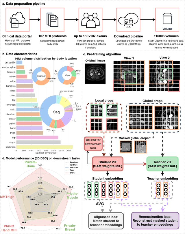
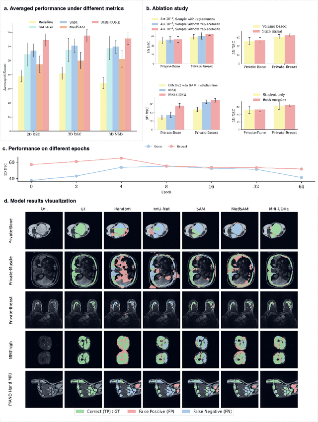
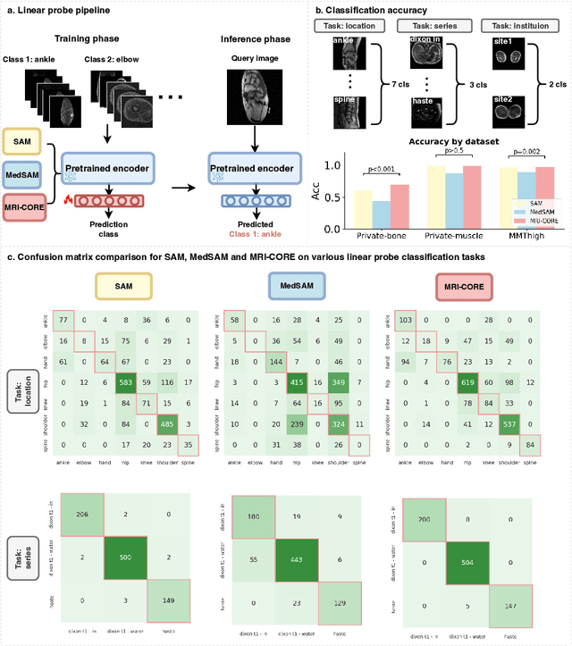
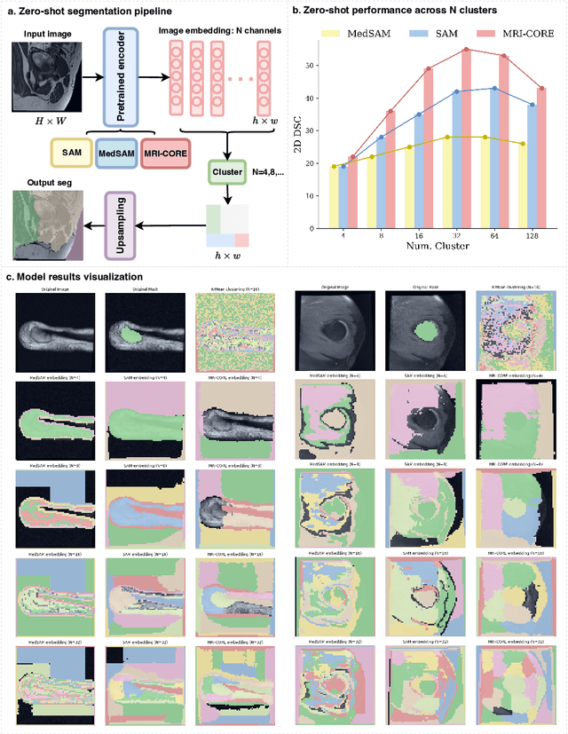
The widespread use of Magnetic Resonance Imaging (MRI) and the rise of deep learning have enabled the development of powerful predictive models for a wide range of diagnostic tasks in MRI, such as image classification or object segmentation. However, training models for specific new tasks often requires large amounts of labeled data, which is difficult to obtain due to high annotation costs and data privacy concerns. To circumvent this issue, we introduce MRI-CORE (MRI COmprehensive Representation Encoder), a vision foundation model pre-trained using more than 6 million slices from over 110,000 MRI volumes across 18 main body locations. Experiments on five diverse object segmentation tasks in MRI demonstrate that MRI-CORE can significantly improve segmentation performance in realistic scenarios with limited labeled data availability, achieving an average gain of 6.97% 3D Dice Coefficient using only 10 annotated slices per task. We further demonstrate new model capabilities in MRI such as classification of image properties including body location, sequence type and institution, and zero-shot segmentation. These results highlight the value of MRI-CORE as a generalist vision foundation model for MRI, potentially lowering the data annotation resource barriers for many applications.
GuidedMorph: Two-Stage Deformable Registration for Breast MRI
May 19, 2025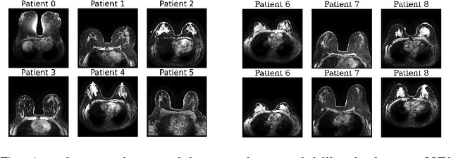
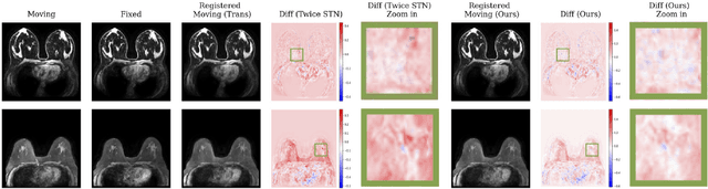
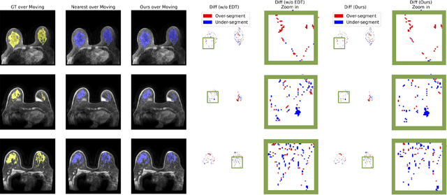
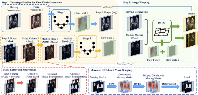
Accurately registering breast MR images from different time points enables the alignment of anatomical structures and tracking of tumor progression, supporting more effective breast cancer detection, diagnosis, and treatment planning. However, the complexity of dense tissue and its highly non-rigid nature pose challenges for conventional registration methods, which primarily focus on aligning general structures while overlooking intricate internal details. To address this, we propose \textbf{GuidedMorph}, a novel two-stage registration framework designed to better align dense tissue. In addition to a single-scale network for global structure alignment, we introduce a framework that utilizes dense tissue information to track breast movement. The learned transformation fields are fused by introducing the Dual Spatial Transformer Network (DSTN), improving overall alignment accuracy. A novel warping method based on the Euclidean distance transform (EDT) is also proposed to accurately warp the registered dense tissue and breast masks, preserving fine structural details during deformation. The framework supports paradigms that require external segmentation models and with image data only. It also operates effectively with the VoxelMorph and TransMorph backbones, offering a versatile solution for breast registration. We validate our method on ISPY2 and internal dataset, demonstrating superior performance in dense tissue, overall breast alignment, and breast structural similarity index measure (SSIM), with notable improvements by over 13.01% in dense tissue Dice, 3.13% in breast Dice, and 1.21% in breast SSIM compared to the best learning-based baseline.
Accelerating Volumetric Medical Image Annotation via Short-Long Memory SAM 2
May 03, 2025Manual annotation of volumetric medical images, such as magnetic resonance imaging (MRI) and computed tomography (CT), is a labor-intensive and time-consuming process. Recent advancements in foundation models for video object segmentation, such as Segment Anything Model 2 (SAM 2), offer a potential opportunity to significantly speed up the annotation process by manually annotating one or a few slices and then propagating target masks across the entire volume. However, the performance of SAM 2 in this context varies. Our experiments show that relying on a single memory bank and attention module is prone to error propagation, particularly at boundary regions where the target is present in the previous slice but absent in the current one. To address this problem, we propose Short-Long Memory SAM 2 (SLM-SAM 2), a novel architecture that integrates distinct short-term and long-term memory banks with separate attention modules to improve segmentation accuracy. We evaluate SLM-SAM 2 on three public datasets covering organs, bones, and muscles across MRI and CT modalities. We show that the proposed method markedly outperforms the default SAM 2, achieving average Dice Similarity Coefficient improvement of 0.14 and 0.11 in the scenarios when 5 volumes and 1 volume are available for the initial adaptation, respectively. SLM-SAM 2 also exhibits stronger resistance to over-propagation, making a notable step toward more accurate automated annotation of medical images for segmentation model development.