Chubin Ou
Patch Progression Masked Autoencoder with Fusion CNN Network for Classifying Evolution Between Two Pairs of 2D OCT Slices
Aug 27, 2025Abstract:Age-related Macular Degeneration (AMD) is a prevalent eye condition affecting visual acuity. Anti-vascular endothelial growth factor (anti-VEGF) treatments have been effective in slowing the progression of neovascular AMD, with better outcomes achieved through timely diagnosis and consistent monitoring. Tracking the progression of neovascular activity in OCT scans of patients with exudative AMD allows for the development of more personalized and effective treatment plans. This was the focus of the Monitoring Age-related Macular Degeneration Progression in Optical Coherence Tomography (MARIO) challenge, in which we participated. In Task 1, which involved classifying the evolution between two pairs of 2D slices from consecutive OCT acquisitions, we employed a fusion CNN network with model ensembling to further enhance the model's performance. For Task 2, which focused on predicting progression over the next three months based on current exam data, we proposed the Patch Progression Masked Autoencoder that generates an OCT for the next exam and then classifies the evolution between the current OCT and the one generated using our solution from Task 1. The results we achieved allowed us to place in the Top 10 for both tasks. Some team members are part of the same organization as the challenge organizers; therefore, we are not eligible to compete for the prize.
* 10 pages, 5 figures, 3 tables, challenge/conference paper
MuTri: Multi-view Tri-alignment for OCT to OCTA 3D Image Translation
Apr 02, 2025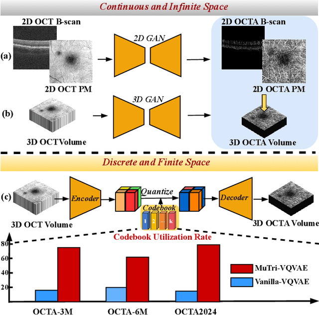

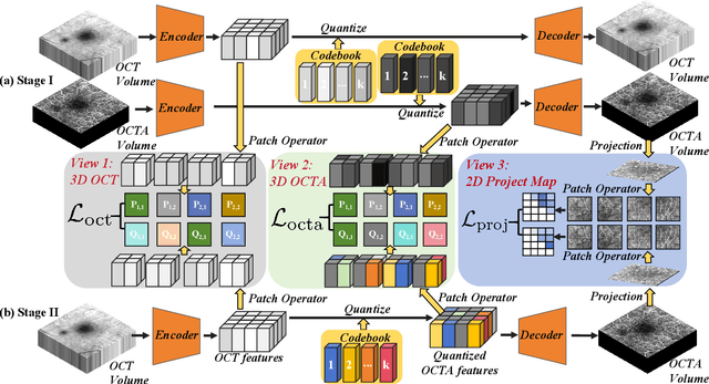

Abstract:Optical coherence tomography angiography (OCTA) shows its great importance in imaging microvascular networks by providing accurate 3D imaging of blood vessels, but it relies upon specialized sensors and expensive devices. For this reason, previous works show the potential to translate the readily available 3D Optical Coherence Tomography (OCT) images into 3D OCTA images. However, existing OCTA translation methods directly learn the mapping from the OCT domain to the OCTA domain in continuous and infinite space with guidance from only a single view, i.e., the OCTA project map, resulting in suboptimal results. To this end, we propose the multi-view Tri-alignment framework for OCT to OCTA 3D image translation in discrete and finite space, named MuTri. In the first stage, we pre-train two vector-quantized variational auto-encoder (VQ- VAE) by reconstructing 3D OCT and 3D OCTA data, providing semantic prior for subsequent multi-view guidances. In the second stage, our multi-view tri-alignment facilitates another VQVAE model to learn the mapping from the OCT domain to the OCTA domain in discrete and finite space. Specifically, a contrastive-inspired semantic alignment is proposed to maximize the mutual information with the pre-trained models from OCT and OCTA views, to facilitate codebook learning. Meanwhile, a vessel structure alignment is proposed to minimize the structure discrepancy with the pre-trained models from the OCTA project map view, benefiting from learning the detailed vessel structure information. We also collect the first large-scale dataset, namely, OCTA2024, which contains a pair of OCT and OCTA volumes from 846 subjects.
MultiEYE: Dataset and Benchmark for OCT-Enhanced Retinal Disease Recognition from Fundus Images
Dec 12, 2024


Abstract:Existing multi-modal learning methods on fundus and OCT images mostly require both modalities to be available and strictly paired for training and testing, which appears less practical in clinical scenarios. To expand the scope of clinical applications, we formulate a novel setting, "OCT-enhanced disease recognition from fundus images", that allows for the use of unpaired multi-modal data during the training phase and relies on the widespread fundus photographs for testing. To benchmark this setting, we present the first large multi-modal multi-class dataset for eye disease diagnosis, MultiEYE, and propose an OCT-assisted Conceptual Distillation Approach (OCT-CoDA), which employs semantically rich concepts to extract disease-related knowledge from OCT images and leverage them into the fundus model. Specifically, we regard the image-concept relation as a link to distill useful knowledge from the OCT teacher model to the fundus student model, which considerably improves the diagnostic performance based on fundus images and formulates the cross-modal knowledge transfer into an explainable process. Through extensive experiments on the multi-disease classification task, our proposed OCT-CoDA demonstrates remarkable results and interpretability, showing great potential for clinical application. Our dataset and code are available at https://github.com/xmed-lab/MultiEYE.
Fundus-Enhanced Disease-Aware Distillation Model for Retinal Disease Classification from OCT Images
Aug 01, 2023

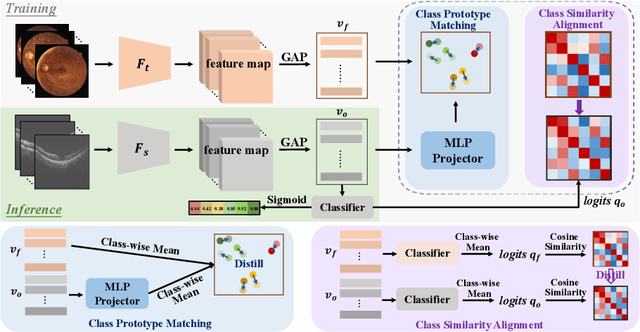
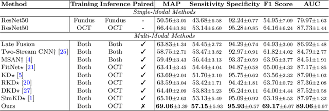
Abstract:Optical Coherence Tomography (OCT) is a novel and effective screening tool for ophthalmic examination. Since collecting OCT images is relatively more expensive than fundus photographs, existing methods use multi-modal learning to complement limited OCT data with additional context from fundus images. However, the multi-modal framework requires eye-paired datasets of both modalities, which is impractical for clinical use. To address this problem, we propose a novel fundus-enhanced disease-aware distillation model (FDDM), for retinal disease classification from OCT images. Our framework enhances the OCT model during training by utilizing unpaired fundus images and does not require the use of fundus images during testing, which greatly improves the practicality and efficiency of our method for clinical use. Specifically, we propose a novel class prototype matching to distill disease-related information from the fundus model to the OCT model and a novel class similarity alignment to enforce consistency between disease distribution of both modalities. Experimental results show that our proposed approach outperforms single-modal, multi-modal, and state-of-the-art distillation methods for retinal disease classification. Code is available at https://github.com/xmed-lab/FDDM.
Vessel-Promoted OCT to OCTA Image Translation by Heuristic Contextual Constraints
Mar 13, 2023Abstract:Optical Coherence Tomography Angiography (OCTA) has become increasingly vital in the clinical screening of fundus diseases due to its ability to capture accurate 3D imaging of blood vessels in a non-contact scanning manner. However, the acquisition of OCTA images remains challenging due to the requirement of exclusive sensors and expensive devices. In this paper, we propose a novel framework, TransPro, that translates 3D Optical Coherence Tomography (OCT) images into exclusive 3D OCTA images using an image translation pattern. Our main objective is to address two issues in existing image translation baselines, namely, the aimlessness in the translation process and incompleteness of the translated object. The former refers to the overall quality of the translated OCTA images being satisfactory, but the retinal vascular quality being low. The latter refers to incomplete objects in translated OCTA images due to the lack of global contexts. TransPro merges a 2D retinal vascular segmentation model and a 2D OCTA image translation model into a 3D image translation baseline for the 2D projection map projected by the translated OCTA images. The 2D retinal vascular segmentation model can improve attention to the retinal vascular, while the 2D OCTA image translation model introduces beneficial heuristic contextual information. Extensive experimental results on two challenging datasets demonstrate that TransPro can consistently outperform existing approaches with minimal computational overhead during training and none during testing.
GAMMA Challenge:Glaucoma grAding from Multi-Modality imAges
Feb 16, 2022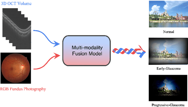
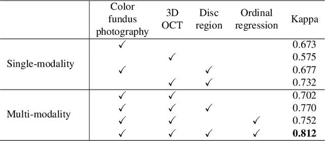
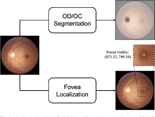
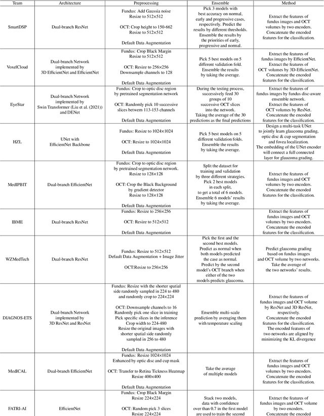
Abstract:Color fundus photography and Optical Coherence Tomography (OCT) are the two most cost-effective tools for glaucoma screening. Both two modalities of images have prominent biomarkers to indicate glaucoma suspected. Clinically, it is often recommended to take both of the screenings for a more accurate and reliable diagnosis. However, although numerous algorithms are proposed based on fundus images or OCT volumes in computer-aided diagnosis, there are still few methods leveraging both of the modalities for the glaucoma assessment. Inspired by the success of Retinal Fundus Glaucoma Challenge (REFUGE) we held previously, we set up the Glaucoma grAding from Multi-Modality imAges (GAMMA) Challenge to encourage the development of fundus \& OCT-based glaucoma grading. The primary task of the challenge is to grade glaucoma from both the 2D fundus images and 3D OCT scanning volumes. As part of GAMMA, we have publicly released a glaucoma annotated dataset with both 2D fundus color photography and 3D OCT volumes, which is the first multi-modality dataset for glaucoma grading. In addition, an evaluation framework is also established to evaluate the performance of the submitted methods. During the challenge, 1272 results were submitted, and finally, top-10 teams were selected to the final stage. We analysis their results and summarize their methods in the paper. Since all these teams submitted their source code in the challenge, a detailed ablation study is also conducted to verify the effectiveness of the particular modules proposed. We find many of the proposed techniques are practical for the clinical diagnosis of glaucoma. As the first in-depth study of fundus \& OCT multi-modality glaucoma grading, we believe the GAMMA Challenge will be an essential starting point for future research.
 Add to Chrome
 Add to Firefox
 Add to Edge