Anas Tahir
Blind ECG Restoration by Operational Cycle-GANs
Jan 29, 2022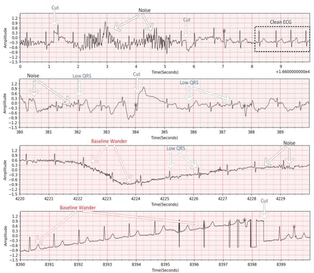
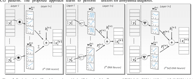

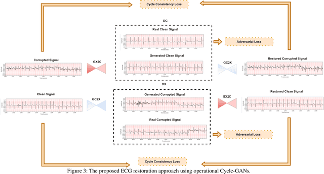
Abstract:Continuous long-term monitoring of electrocardiography (ECG) signals is crucial for the early detection of cardiac abnormalities such as arrhythmia. Non-clinical ECG recordings acquired by Holter and wearable ECG sensors often suffer from severe artifacts such as baseline wander, signal cuts, motion artifacts, variations on QRS amplitude, noise, and other interferences. Usually, a set of such artifacts occur on the same ECG signal with varying severity and duration, and this makes an accurate diagnosis by machines or medical doctors extremely difficult. Despite numerous studies that have attempted ECG denoising, they naturally fail to restore the actual ECG signal corrupted with such artifacts due to their simple and naive noise model. In this study, we propose a novel approach for blind ECG restoration using cycle-consistent generative adversarial networks (Cycle-GANs) where the quality of the signal can be improved to a clinical level ECG regardless of the type and severity of the artifacts corrupting the signal. To further boost the restoration performance, we propose 1D operational Cycle-GANs with the generative neuron model. The proposed approach has been evaluated extensively using one of the largest benchmark ECG datasets from the China Physiological Signal Challenge (CPSC-2020) with more than one million beats. Besides the quantitative and qualitative evaluations, a group of cardiologists performed medical evaluations to validate the quality and usability of the restored ECG, especially for an accurate arrhythmia diagnosis.
A Shallow U-Net Architecture for Reliably Predicting Blood Pressure from Photoplethysmogram and Electrocardiogram Signals
Nov 12, 2021
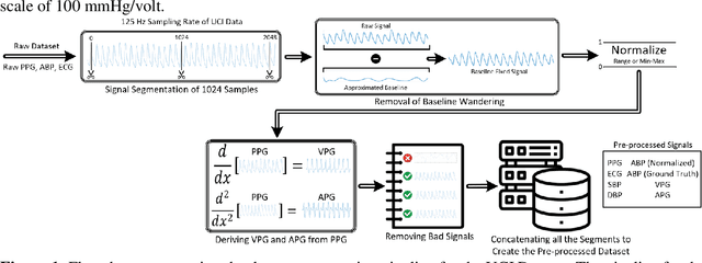
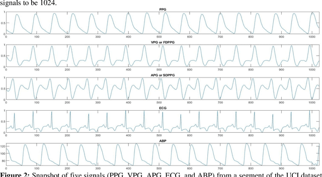

Abstract:Cardiovascular diseases are the most common causes of death around the world. To detect and treat heart-related diseases, continuous Blood Pressure (BP) monitoring along with many other parameters are required. Several invasive and non-invasive methods have been developed for this purpose. Most existing methods used in the hospitals for continuous monitoring of BP are invasive. On the contrary, cuff-based BP monitoring methods, which can predict Systolic Blood Pressure (SBP) and Diastolic Blood Pressure (DBP), cannot be used for continuous monitoring. Several studies attempted to predict BP from non-invasively collectible signals such as Photoplethysmogram (PPG) and Electrocardiogram (ECG), which can be used for continuous monitoring. In this study, we explored the applicability of autoencoders in predicting BP from PPG and ECG signals. The investigation was carried out on 12,000 instances of 942 patients of the MIMIC-II dataset and it was found that a very shallow, one-dimensional autoencoder can extract the relevant features to predict the SBP and DBP with the state-of-the-art performance on a very large dataset. Independent test set from a portion of the MIMIC-II dataset provides an MAE of 2.333 and 0.713 for SBP and DBP, respectively. On an external dataset of forty subjects, the model trained on the MIMIC-II dataset, provides an MAE of 2.728 and 1.166 for SBP and DBP, respectively. For both the cases, the results met British Hypertension Society (BHS) Grade A and surpassed the studies from the current literature.
Robust Peak Detection for Holter ECGs by Self-Organized Operational Neural Networks
Sep 30, 2021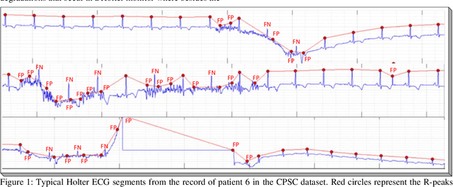
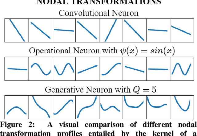
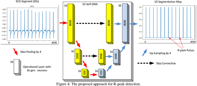
Abstract:Although numerous R-peak detectors have been proposed in the literature, their robustness and performance levels may significantly deteriorate in low quality and noisy signals acquired from mobile ECG sensors such as Holter monitors. Recently, this issue has been addressed by deep 1D Convolutional Neural Networks (CNNs) that have achieved state-of-the-art performance levels in Holter monitors; however, they pose a high complexity level that requires special parallelized hardware setup for real-time processing. On the other hand, their performance deteriorates when a compact network configuration is used instead. This is an expected outcome as recent studies have demonstrated that the learning performance of CNNs is limited due to their strictly homogenous configuration with the sole linear neuron model. This has been addressed by Operational Neural Networks (ONNs) with their heterogenous network configuration encapsulating neurons with various non-linear operators. In this study, to further boost the peak detection performance along with an elegant computational efficiency, we propose 1D Self-Organized Operational Neural Networks (Self-ONNs) with generative neurons. The most crucial advantage of 1D Self-ONNs over the ONNs is their self-organization capability that voids the need to search for the best operator set per neuron since each generative neuron has the ability to create the optimal operator during training. The experimental results over the China Physiological Signal Challenge-2020 (CPSC) dataset with more than one million ECG beats show that the proposed 1D Self-ONNs can significantly surpass the state-of-the-art deep CNN with less computational complexity. Results demonstrate that the proposed solution achieves 99.10% F1-score, 99.79% sensitivity, and 98.42% positive predictivity in the CPSC dataset which is the best R-peak detection performance ever achieved.
EDITH :ECG biometrics aided by Deep learning for reliable Individual auTHentication
Feb 16, 2021

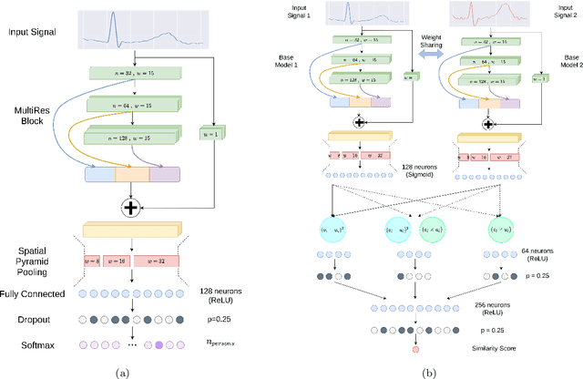

Abstract:In recent years, physiological signal based authentication has shown great promises,for its inherent robustness against forgery. Electrocardiogram (ECG) signal, being the most widely studied biosignal, has also received the highest level of attention in this regard. It has been proven with numerous studies that by analyzing ECG signals from different persons, it is possible to identify them, with acceptable accuracy. In this work, we present, EDITH, a deep learning-based framework for ECG biometrics authentication system. Moreover, we hypothesize and demonstrate that Siamese architectures can be used over typical distance metrics for improved performance. We have evaluated EDITH using 4 commonly used datasets and outperformed the prior works using less number of beats. EDITH performs competitively using just a single heartbeat (96-99.75% accuracy) and can be further enhanced by fusing multiple beats (100% accuracy from 3 to 6 beats). Furthermore, the proposed Siamese architecture manages to reduce the identity verification Equal Error Rate (EER) to 1.29%. A limited case study of EDITH with real-world experimental data also suggests its potential as a practical authentication system.
Detection and severity classification of COVID-19 in CT images using deep learning
Feb 15, 2021
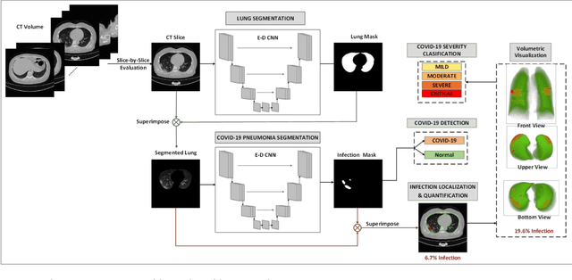

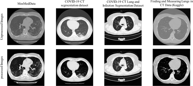
Abstract:Since the breakout of coronavirus disease (COVID-19), the computer-aided diagnosis has become a necessity to prevent the spread of the virus. Detecting COVID-19 at an early stage is essential to reduce the mortality risk of the patients. In this study, a cascaded system is proposed to segment the lung, detect, localize, and quantify COVID-19 infections from computed tomography (CT) images Furthermore, the system classifies the severity of COVID-19 as mild, moderate, severe, or critical based on the percentage of infected lungs. An extensive set of experiments were performed using state-of-the-art deep Encoder-Decoder Convolutional Neural Networks (ED-CNNs), UNet, and Feature Pyramid Network (FPN), with different backbone (encoder) structures using the variants of DenseNet and ResNet. The conducted experiments showed the best performance for lung region segmentation with Dice Similarity Coefficient (DSC) of 97.19% and Intersection over Union (IoU) of 95.10% using U-Net model with the DenseNet 161 encoder. Furthermore, the proposed system achieved an elegant performance for COVID-19 infection segmentation with a DSC of 94.13% and IoU of 91.85% using the FPN model with the DenseNet201 encoder. The achieved performance is significantly superior to previous methods for COVID-19 lesion localization. Besides, the proposed system can reliably localize infection of various shapes and sizes, especially small infection regions, which are rarely considered in recent studies. Moreover, the proposed system achieved high COVID-19 detection performance with 99.64% sensitivity and 98.72% specificity. Finally, the system was able to discriminate between different severity levels of COVID-19 infection over a dataset of 1,110 subjects with sensitivity values of 98.3%, 71.2%, 77.8%, and 100% for mild, moderate, severe, and critical infections, respectively.
Robust R-Peak Detection in Low-Quality Holter ECGs using 1D Convolutional Neural Network
Dec 29, 2020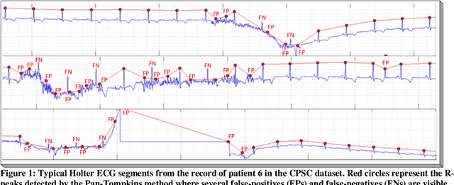
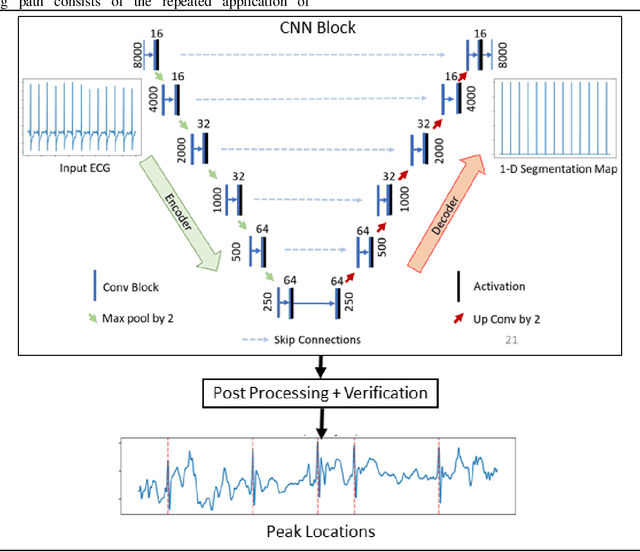
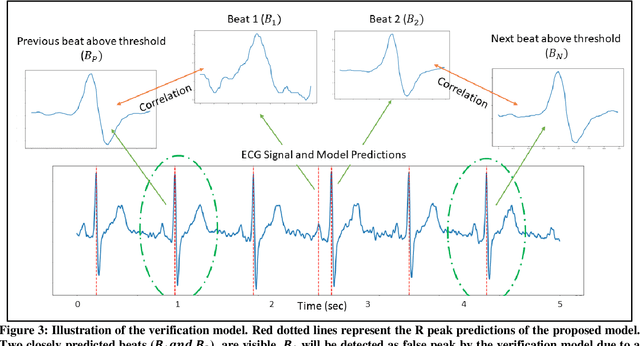
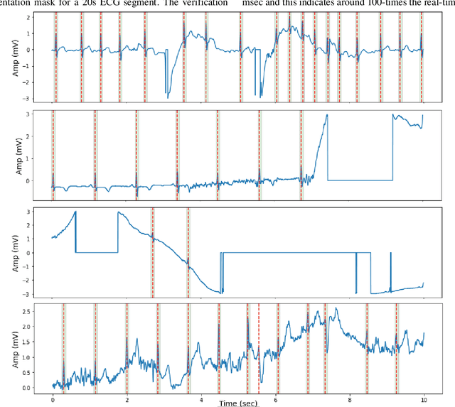
Abstract:Noise and low quality of ECG signals acquired from Holter or wearable devices deteriorate the accuracy and robustness of R-peak detection algorithms. This paper presents a generic and robust system for R-peak detection in Holter ECG signals. While many proposed algorithms have successfully addressed the problem of ECG R-peak detection, there is still a notable gap in the performance of these detectors on such low-quality ECG records. Therefore, in this study, a novel implementation of the 1D Convolutional Neural Network (CNN) is used integrated with a verification model to reduce the number of false alarms. This CNN architecture consists of an encoder block and a corresponding decoder block followed by a sample-wise classification layer to construct the 1D segmentation map of R- peaks from the input ECG signal. Once the proposed model has been trained, it can solely be used to detect R-peaks possibly in a single channel ECG data stream quickly and accurately, or alternatively, such a solution can be conveniently employed for real-time monitoring on a lightweight portable device. The model is tested on two open-access ECG databases: The China Physiological Signal Challenge (2020) database (CPSC-DB) with more than one million beats, and the commonly used MIT-BIH Arrhythmia Database (MIT-DB). Experimental results demonstrate that the proposed systematic approach achieves 99.30% F1-score, 99.69% recall, and 98.91% precision in CPSC-DB, which is the best R-peak detection performance ever achieved. Compared to all competing methods, the proposed approach can reduce the false-positives and false-negatives in Holter ECG signals by more than 54% and 82%, respectively. Results also demonstrate similar or better performance than most competing algorithms on MIT-DB with 99.83% F1-score, 99.85% recall, and 99.82% precision.
Exploring the Effect of Image Enhancement Techniques on COVID-19 Detection using Chest X-rays Images
Nov 25, 2020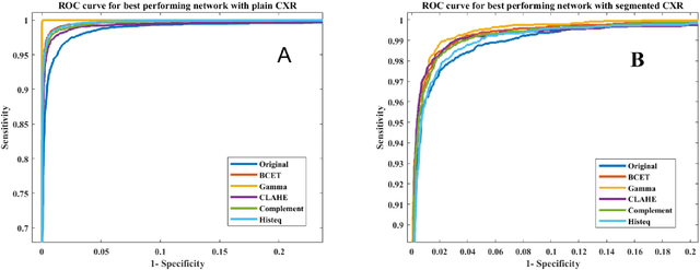
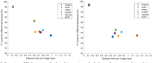
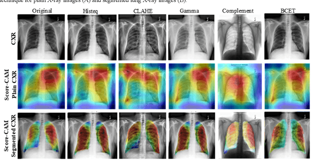
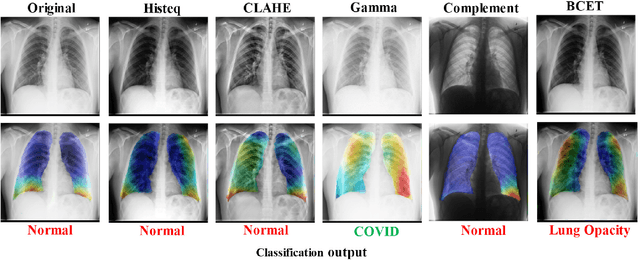
Abstract:The use of computer-aided diagnosis in the reliable and fast detection of coronavirus disease (COVID-19) has become a necessity to prevent the spread of the virus during the pandemic to ease the burden on the medical infrastructure. Chest X-ray (CXR) imaging has several advantages over other imaging techniques as it is cheap, easily accessible, fast and portable. This paper explores the effect of various popular image enhancement techniques and states the effect of each of them on the detection performance. We have compiled the largest X-ray dataset called COVQU-20, consisting of 18,479 normal, non-COVID lung opacity and COVID-19 CXR images. To the best of our knowledge, this is the largest public COVID positive database. Ground glass opacity is the common symptom reported in COVID-19 pneumonia patients and so a mixture of 3616 COVID-19, 6012 non-COVID lung opacity, and 8851 normal chest X-ray images were used to create this dataset. Five different image enhancement techniques: histogram equalization, contrast limited adaptive histogram equalization, image complement, gamma correction, and Balance Contrast Enhancement Technique were used to improve COVID-19 detection accuracy. Six different Convolutional Neural Networks (CNNs) were investigated in this study. Gamma correction technique outperforms other enhancement techniques in detecting COVID-19 from standard and segmented lung CXR images. The accuracy, precision, sensitivity, f1-score, and specificity in the detection of COVID-19 with gamma correction on CXR images were 96.29%, 96.28%, 96.29%, 96.28% and 96.27% respectively. The accuracy, precision, sensitivity, F1-score, and specificity were 95.11 %, 94.55 %, 94.56 %, 94.53 % and 95.59 % respectively for segmented lung images. The proposed approach with very high and comparable performance will boost the fast and robust COVID-19 detection using chest X-ray images.
Coronavirus: Comparing COVID-19, SARS and MERS in the eyes of AI
Jun 08, 2020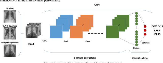


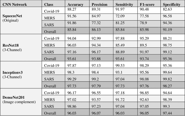
Abstract:Novel Coronavirus disease (COVID-19) is an extremely contagious and quickly spreading Coronavirus disease. Severe Acute Respiratory Syndrome (SARS)-CoV, Middle East Respiratory Syndrome (MERS)-CoV outbreak in 2002 and 2011 and current COVID-19 pandemic all from the same family of Coronavirus. The fatality rate due to SARS and MERS were higher than COVID-19 however, the spread of those were limited to few countries while COVID-19 affected more than two-hundred countries of the world. In this work, authors used deep machine learning algorithms along with innovative image pre-processing techniques to distinguish COVID-19 images from SARS and MERS images. Several deep learning algorithms were trained, and tested and four outperforming algorithms were reported: SqueezeNet, ResNet18, Inceptionv3 and DenseNet201. Original, Contrast limited adaptive histogram equalized and complemented image were used individually and in concatenation as the inputs to the networks. It was observed that inceptionv3 outperforms all networks for 3-channel concatenation technique and provide an excellent sensitivity of 99.5%, 93.1% and 97% for classifying COVID-19, MERS and SARS images respectively. Investigating deep layer activation mapping of the correctly classified images and miss-classified images, it was observed that some overlapping features between COVID-19 and MERS images were identified by the deep layer network. Interestingly these features were present in MERS images and 10 out of 144 images were miss-classified as COVID while only one out of 423 COVID-19 images was miss-classified as MERS. None of the MERS images was miss-classified to SARS and only one COVID-19 image was miss-classified as SARS. Therefore, it can be summarized that SARS images are significantly different from MERS and COVID-19 in the eyes of AI while there are some overlapping feature available between MERS and COVID-19.
 Add to Chrome
 Add to Firefox
 Add to Edge