Chuanbo Wang
Department of Computer Science, University of Wisconsin-Milwaukee, Milwaukee, WI, USA
Wound Tissue Segmentation in Diabetic Foot Ulcer Images Using Deep Learning: A Pilot Study
Jun 23, 2024Abstract:Identifying individual tissues, so-called tissue segmentation, in diabetic foot ulcer (DFU) images is a challenging task and little work has been published, largely due to the limited availability of a clinical image dataset. To address this gap, we have created a DFUTissue dataset for the research community to evaluate wound tissue segmentation algorithms. The dataset contains 110 images with tissues labeled by wound experts and 600 unlabeled images. Additionally, we conducted a pilot study on segmenting wound characteristics including fibrin, granulation, and callus using deep learning. Due to the limited amount of annotated data, our framework consists of both supervised learning (SL) and semi-supervised learning (SSL) phases. In the SL phase, we propose a hybrid model featuring a Mix Transformer (MiT-b3) in the encoder and a CNN in the decoder, enhanced by the integration of a parallel spatial and channel squeeze-and-excitation (P-scSE) module known for its efficacy in improving boundary accuracy. The SSL phase employs a pseudo-labeling-based approach, iteratively identifying and incorporating valuable unlabeled images to enhance overall segmentation performance. Comparative evaluations with state-of-the-art methods are conducted for both SL and SSL phases. The SL achieves a Dice Similarity Coefficient (DSC) of 84.89%, which has been improved to 87.64% in the SSL phase. Furthermore, the results are benchmarked against two widely used SSL approaches: Generative Adversarial Networks and Cross-Consistency Training. Additionally, our hybrid model outperforms the state-of-the-art methods with a 92.99% DSC in performing binary segmentation of DFU wound areas when tested on the Chronic Wound dataset. Codes and data are available at https://github.com/uwm-bigdata/DFUTissueSegNet.
Biomedical image analysis competitions: The state of current participation practice
Dec 16, 2022Abstract:The number of international benchmarking competitions is steadily increasing in various fields of machine learning (ML) research and practice. So far, however, little is known about the common practice as well as bottlenecks faced by the community in tackling the research questions posed. To shed light on the status quo of algorithm development in the specific field of biomedical imaging analysis, we designed an international survey that was issued to all participants of challenges conducted in conjunction with the IEEE ISBI 2021 and MICCAI 2021 conferences (80 competitions in total). The survey covered participants' expertise and working environments, their chosen strategies, as well as algorithm characteristics. A median of 72% challenge participants took part in the survey. According to our results, knowledge exchange was the primary incentive (70%) for participation, while the reception of prize money played only a minor role (16%). While a median of 80 working hours was spent on method development, a large portion of participants stated that they did not have enough time for method development (32%). 25% perceived the infrastructure to be a bottleneck. Overall, 94% of all solutions were deep learning-based. Of these, 84% were based on standard architectures. 43% of the respondents reported that the data samples (e.g., images) were too large to be processed at once. This was most commonly addressed by patch-based training (69%), downsampling (37%), and solving 3D analysis tasks as a series of 2D tasks. K-fold cross-validation on the training set was performed by only 37% of the participants and only 50% of the participants performed ensembling based on multiple identical models (61%) or heterogeneous models (39%). 48% of the respondents applied postprocessing steps.
FUSeg: The Foot Ulcer Segmentation Challenge
Jan 02, 2022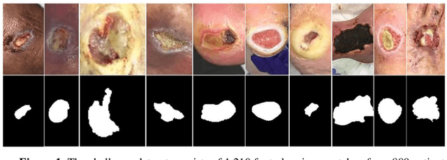
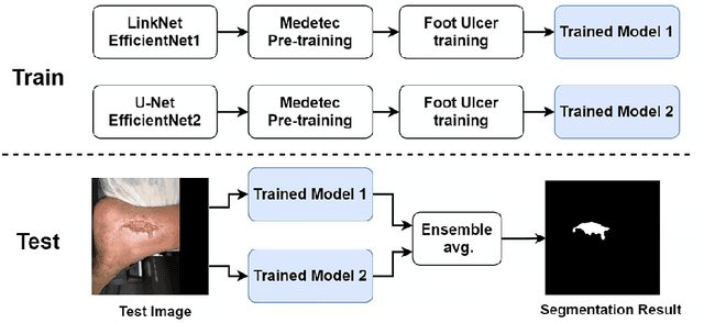
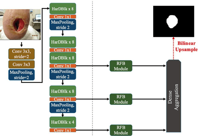

Abstract:Acute and chronic wounds with varying etiologies burden the healthcare systems economically. The advanced wound care market is estimated to reach $22 billion by 2024. Wound care professionals provide proper diagnosis and treatment with heavy reliance on images and image documentation. Segmentation of wound boundaries in images is a key component of the care and diagnosis protocol since it is important to estimate the area of the wound and provide quantitative measurement for the treatment. Unfortunately, this process is very time-consuming and requires a high level of expertise. Recently automatic wound segmentation methods based on deep learning have shown promising performance but require large datasets for training and it is unclear which methods perform better. To address these issues, we propose the Foot Ulcer Segmentation challenge (FUSeg) organized in conjunction with the 2021 International Conference on Medical Image Computing and Computer Assisted Intervention (MICCAI). We built a wound image dataset containing 1,210 foot ulcer images collected over 2 years from 889 patients. It is pixel-wise annotated by wound care experts and split into a training set with 1010 images and a testing set with 200 images for evaluation. Teams around the world developed automated methods to predict wound segmentations on the testing set of which annotations were kept private. The predictions were evaluated and ranked based on the average Dice coefficient. The FUSeg challenge remains an open challenge as a benchmark for wound segmentation after the conference.
Multiclass Wound Image Classification using an Ensemble Deep CNN-based Classifier
Oct 19, 2020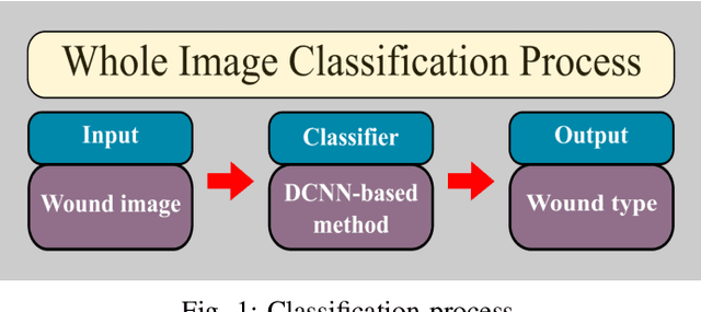
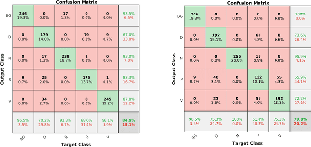
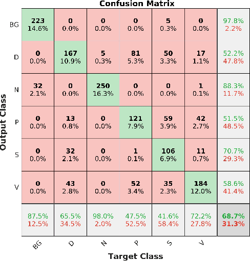
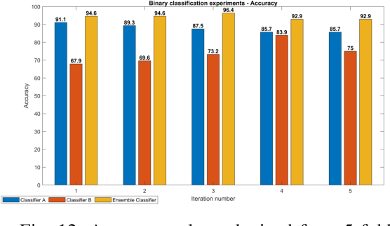
Abstract:Acute and chronic wounds are a challenge to healthcare systems around the world and affect many people's lives annually. Wound classification is a key step in wound diagnosis that would help clinicians to identify an optimal treatment procedure. Hence, having a high-performance classifier assists the specialists in the field to classify the wounds with less financial and time costs. Different machine learning and deep learning-based wound classification methods have been proposed in the literature. In this study, we have developed an ensemble Deep Convolutional Neural Network-based classifier to classify wound images including surgical, diabetic, and venous ulcers, into multi-classes. The output classification scores of two classifiers (patch-wise and image-wise) are fed into a Multi-Layer Perceptron to provide a superior classification performance. A 5-fold cross-validation approach is used to evaluate the proposed method. We obtained maximum and average classification accuracy values of 96.4% and 94.28% for binary and 91.9\% and 87.7\% for 3-class classification problems. The results show that our proposed method can be used effectively as a decision support system in classification of wound images or other related clinical applications.
Fully Automatic Wound Segmentation with Deep Convolutional Neural Networks
Oct 12, 2020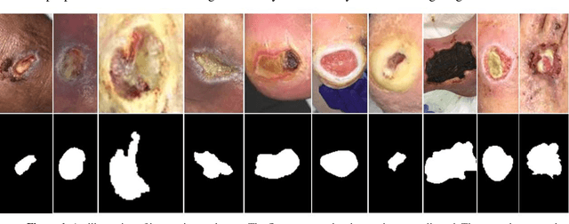

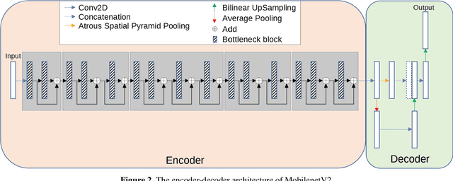

Abstract:Acute and chronic wounds have varying etiologies and are an economic burden to healthcare systems around the world. The advanced wound care market is expected to exceed $22 billion by 2024. Wound care professionals rely heavily on images and image documentation for proper diagnosis and treatment. Unfortunately lack of expertise can lead to improper diagnosis of wound etiology and inaccurate wound management and documentation. Fully automatic segmentation of wound areas in natural images is an important part of the diagnosis and care protocol since it is crucial to measure the area of the wound and provide quantitative parameters in the treatment. Various deep learning models have gained success in image analysis including semantic segmentation. Particularly, MobileNetV2 stands out among others due to its lightweight architecture and uncompromised performance. This manuscript proposes a novel convolutional framework based on MobileNetV2 and connected component labelling to segment wound regions from natural images. We build an annotated wound image dataset consisting of 1,109 foot ulcer images from 889 patients to train and test the deep learning models. We demonstrate the effectiveness and mobility of our method by conducting comprehensive experiments and analyses on various segmentation neural networks.
Fully Automatic Intervertebral Disc Segmentation Using Multimodal 3D U-Net
Sep 28, 2020


Abstract:Intervertebral discs (IVDs), as small joints lying between adjacent vertebrae, have played an important role in pressure buffering and tissue protection. The fully-automatic localization and segmentation of IVDs have been discussed in the literature for many years since they are crucial to spine disease diagnosis and provide quantitative parameters in the treatment. Traditionally hand-crafted features are derived based on image intensities and shape priors to localize and segment IVDs. With the advance of deep learning, various neural network models have gained great success in image analysis including the recognition of intervertebral discs. Particularly, U-Net stands out among other approaches due to its outstanding performance on biomedical images with a relatively small set of training data. This paper proposes a novel convolutional framework based on 3D U-Net to segment IVDs from multi-modality MRI images. We first localize the centers of intervertebral discs in each spine sample and then train the network based on the cropped small volumes centered at the localized intervertebral discs. A detailed comprehensive analysis of the results using various combinations of multi-modalities is presented. Furthermore, experiments conducted on 2D and 3D U-Nets with augmented and non-augmented datasets are demonstrated and compared in terms of Dice coefficient and Hausdorff distance. Our method has proved to be effective with a mean segmentation Dice coefficient of 89.0% and a standard deviation of 1.4%.
Image Based Artificial Intelligence in Wound Assessment: A Systematic Review
Sep 15, 2020


Abstract:Efficient and effective assessment of acute and chronic wounds can help wound care teams in clinical practice to greatly improve wound diagnosis, optimize treatment plans, ease the workload and achieve health related quality of life to the patient population. While artificial intelligence (AI) has found wide applications in health-related sciences and technology, AI-based systems remain to be developed clinically and computationally for high-quality wound care. To this end, we have carried out a systematic review of intelligent image-based data analysis and system developments for wound assessment. Specifically, we provide an extensive review of research methods on wound measurement (segmentation) and wound diagnosis (classification). We also reviewed recent work on wound assessment systems (including hardware, software, and mobile apps). More than 250 articles were retrieved from various publication databases and online resources, and 115 of them were carefully selected to cover the breadth and depth of most recent and relevant work to convey the current review to its fulfillment.
 Add to Chrome
 Add to Firefox
 Add to Edge