Thierry Judge
Reinforcement Learning for Unsupervised Domain Adaptation in Spatio-Temporal Echocardiography Segmentation
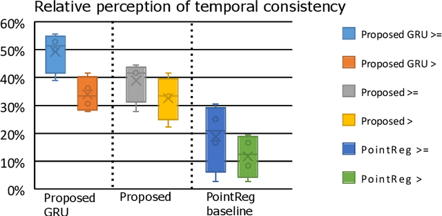
Oct 16, 2025Abstract:Domain adaptation methods aim to bridge the gap between datasets by enabling knowledge transfer across domains, reducing the need for additional expert annotations. However, many approaches struggle with reliability in the target domain, an issue particularly critical in medical image segmentation, where accuracy and anatomical validity are essential. This challenge is further exacerbated in spatio-temporal data, where the lack of temporal consistency can significantly degrade segmentation quality, and particularly in echocardiography, where the presence of artifacts and noise can further hinder segmentation performance. To address these issues, we present RL4Seg3D, an unsupervised domain adaptation framework for 2D + time echocardiography segmentation. RL4Seg3D integrates novel reward functions and a fusion scheme to enhance key landmark precision in its segmentations while processing full-sized input videos. By leveraging reinforcement learning for image segmentation, our approach improves accuracy, anatomical validity, and temporal consistency while also providing, as a beneficial side effect, a robust uncertainty estimator, which can be used at test time to further enhance segmentation performance. We demonstrate the effectiveness of our framework on over 30,000 echocardiographic videos, showing that it outperforms standard domain adaptation techniques without the need for any labels on the target domain. Code is available at https://github.com/arnaudjudge/RL4Seg3D.
Generation of realistic cardiac ultrasound sequences with ground truth motion and speckle decorrelation
Sep 05, 2025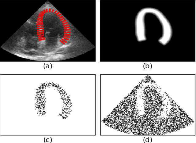

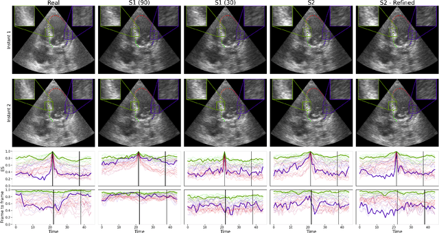
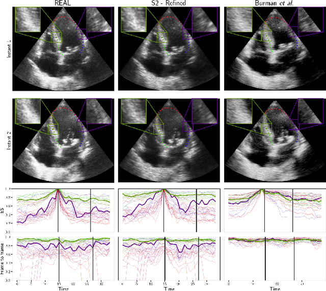
Abstract:Simulated ultrasound image sequences are key for training and validating machine learning algorithms for left ventricular strain estimation. Several simulation pipelines have been proposed to generate sequences with corresponding ground truth motion, but they suffer from limited realism as they do not consider speckle decorrelation. In this work, we address this limitation by proposing an improved simulation framework that explicitly accounts for speckle decorrelation. Our method builds on an existing ultrasound simulation pipeline by incorporating a dynamic model of speckle variation. Starting from real ultrasound sequences and myocardial segmentations, we generate meshes that guide image formation. Instead of applying a fixed ratio of myocardial and background scatterers, we introduce a coherence map that adapts locally over time. This map is derived from correlation values measured directly from the real ultrasound data, ensuring that simulated sequences capture the characteristic temporal changes observed in practice. We evaluated the realism of our approach using ultrasound data from 98 patients in the CAMUS database. Performance was assessed by comparing correlation curves from real and simulated images. The proposed method achieved lower mean absolute error compared to the baseline pipeline, indicating that it more faithfully reproduces the decorrelation behavior seen in clinical data.
Uncertainty Propagation for Echocardiography Clinical Metric Estimation via Contour Sampling
Feb 18, 2025Abstract:Echocardiography plays a fundamental role in the extraction of important clinical parameters (e.g. left ventricular volume and ejection fraction) required to determine the presence and severity of heart-related conditions. When deploying automated techniques for computing these parameters, uncertainty estimation is crucial for assessing their utility. Since clinical parameters are usually derived from segmentation maps, there is no clear path for converting pixel-wise uncertainty values into uncertainty estimates in the downstream clinical metric calculation. In this work, we propose a novel uncertainty estimation method based on contouring rather than segmentation. Our method explicitly predicts contour location uncertainty from which contour samples can be drawn. Finally, the sampled contours can be used to propagate uncertainty to clinical metrics. Our proposed method not only provides accurate uncertainty estimations for the task of contouring but also for the downstream clinical metrics on two cardiac ultrasound datasets. Code is available at: https://github.com/ThierryJudge/contouring-uncertainty.
Domain Adaptation of Echocardiography Segmentation Via Reinforcement Learning
Jun 25, 2024


Abstract:Performance of deep learning segmentation models is significantly challenged in its transferability across different medical imaging domains, particularly when aiming to adapt these models to a target domain with insufficient annotated data for effective fine-tuning. While existing domain adaptation (DA) methods propose strategies to alleviate this problem, these methods do not explicitly incorporate human-verified segmentation priors, compromising the potential of a model to produce anatomically plausible segmentations. We introduce RL4Seg, an innovative reinforcement learning framework that reduces the need to otherwise incorporate large expertly annotated datasets in the target domain, and eliminates the need for lengthy manual human review. Using a target dataset of 10,000 unannotated 2D echocardiographic images, RL4Seg not only outperforms existing state-of-the-art DA methods in accuracy but also achieves 99% anatomical validity on a subset of 220 expert-validated subjects from the target domain. Furthermore, our framework's reward network offers uncertainty estimates comparable with dedicated state-of-the-art uncertainty methods, demonstrating the utility and effectiveness of RL4Seg in overcoming domain adaptation challenges in medical image segmentation.
Left Ventricle Contouring of Apical Three-Chamber Views on 2D Echocardiography
Jul 13, 2022
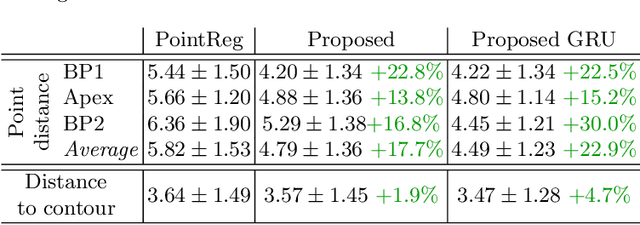
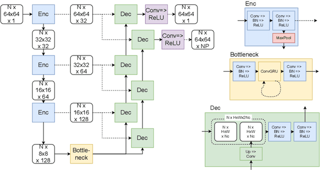
Abstract:We propose a new method to automatically contour the left ventricle on 2D echocardiographic images. Unlike most existing segmentation methods, which are based on predicting segmentation masks, we focus at predicting the endocardial contour and the key landmark points within this contour (basal points and apex). This provides a representation that is closer to how experts perform manual annotations and hence produce results that are physiologically more plausible. Our proposed method uses a two-headed network based on the U-Net architecture. One head predicts the 7 contour points, and the other head predicts a distance map to the contour. This approach was compared to the U-Net and to a point based approach, achieving performance gains of up to 30\% in terms of landmark localisation (<4.5mm) and distance to the ground truth contour (<3.5mm).
CRISP - Reliable Uncertainty Estimation for Medical Image Segmentation
Jun 15, 2022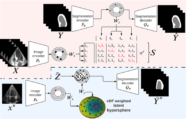
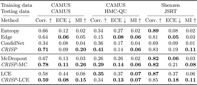
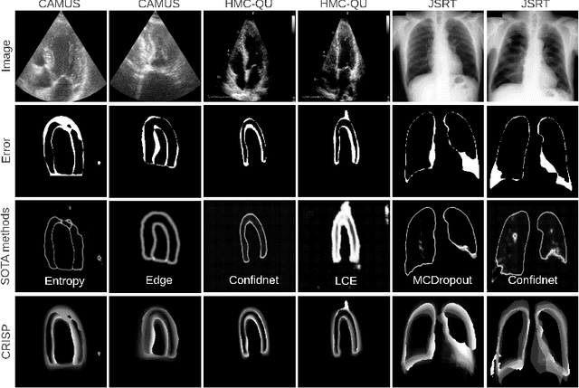
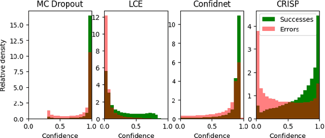
Abstract:Accurate uncertainty estimation is a critical need for the medical imaging community. A variety of methods have been proposed, all direct extensions of classification uncertainty estimations techniques. The independent pixel-wise uncertainty estimates, often based on the probabilistic interpretation of neural networks, do not take into account anatomical prior knowledge and consequently provide sub-optimal results to many segmentation tasks. For this reason, we propose CRISP a ContRastive Image Segmentation for uncertainty Prediction method. At its core, CRISP implements a contrastive method to learn a joint latent space which encodes a distribution of valid segmentations and their corresponding images. We use this joint latent space to compare predictions to thousands of latent vectors and provide anatomically consistent uncertainty maps. Comprehensive studies performed on four medical image databases involving different modalities and organs underlines the superiority of our method compared to state-of-the-art approaches.
Neural Teleportation
Dec 02, 2020
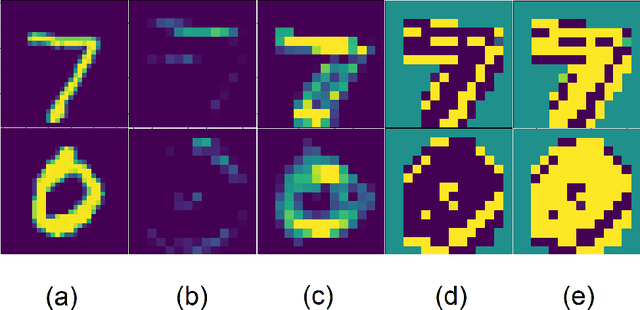
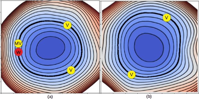
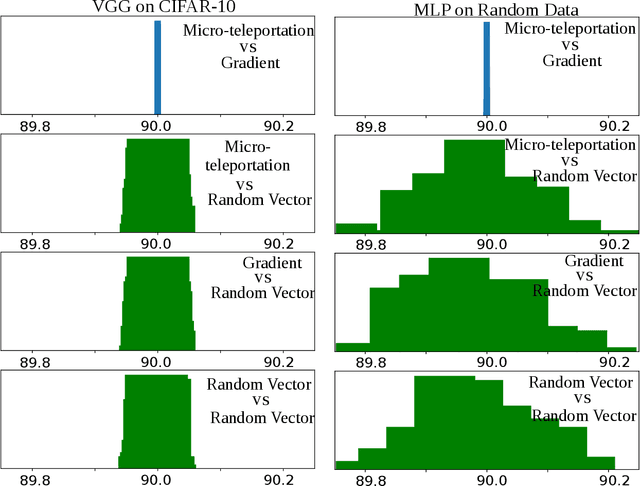
Abstract:In this paper, we explore a process called neural teleportation, a mathematical consequence of applying quiver representation theory to neural networks. Neural teleportation "teleports" a network to a new position in the weight space, while leaving its function unchanged. This concept generalizes the notion of positive scale invariance of ReLU networks to any network with any activation functions and any architecture. In this paper, we shed light on surprising and counter-intuitive consequences neural teleportation has on the loss landscape. In particular, we show that teleportation can be used to explore loss level curves, that it changes the loss landscape, sharpens global minima and boosts back-propagated gradients. From these observations, we demonstrate that teleportation accelerates training when used during initialization regardless of the model, its activation function, the loss function, and the training data. Our results can be reproduced with the code available here: https://github.com/vitalab/neuralteleportation.
Cardiac Segmentation with Strong Anatomical Guarantees
Jun 15, 2020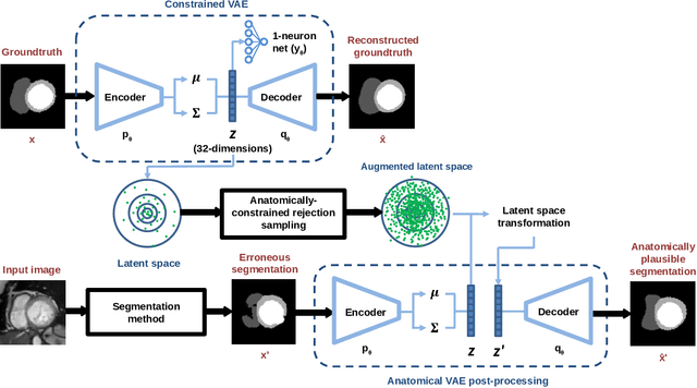
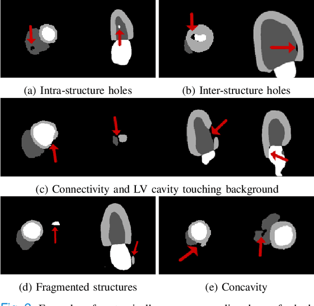
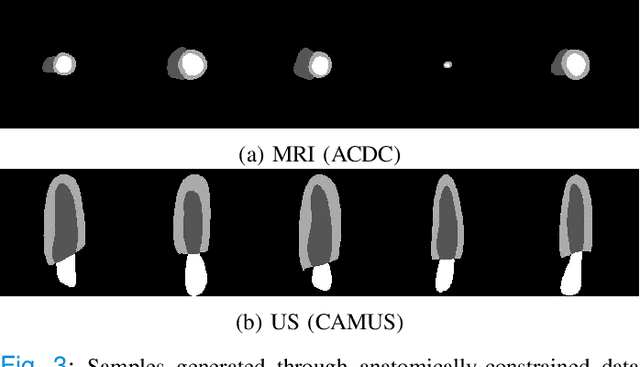
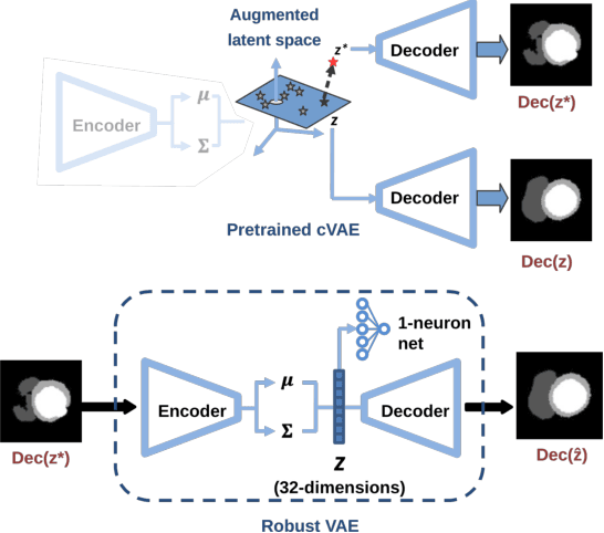
Abstract:Convolutional neural networks (CNN) have had unprecedented success in medical imaging and, in particular, in medical image segmentation. However, despite the fact that segmentation results are closer than ever to the inter-expert variability, CNNs are not immune to producing anatomically inaccurate segmentations, even when built upon a shape prior. In this paper, we present a framework for producing cardiac image segmentation maps that are guaranteed to respect pre-defined anatomical criteria, while remaining within the inter-expert variability. The idea behind our method is to use a well-trained CNN, have it process cardiac images, identify the anatomically implausible results and warp these results toward the closest anatomically valid cardiac shape. This warping procedure is carried out with a constrained variational autoencoder (cVAE) trained to learn a representation of valid cardiac shapes through a smooth, yet constrained, latent space. With this cVAE, we can project any implausible shape into the cardiac latent space and steer it toward the closest correct shape. We tested our framework on short-axis MRI as well as apical two and four-chamber view ultrasound images, two modalities for which cardiac shapes are drastically different. With our method, CNNs can now produce results that are both within the inter-expert variability and always anatomically plausible without having to rely on a shape prior.
Cardiac MRI Segmentation with Strong Anatomical Guarantees
Jul 05, 2019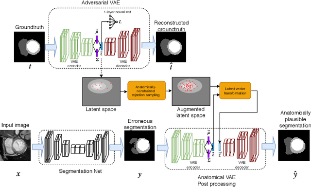
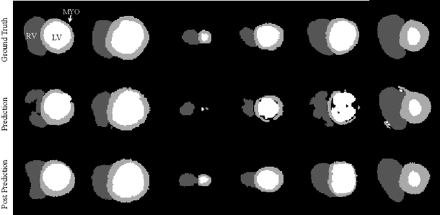
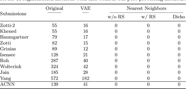
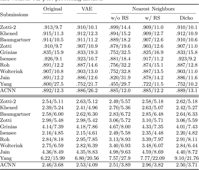
Abstract:Recent publications have shown that the segmentation accuracy of modern-day convolutional neural networks (CNN) applied on cardiac MRI can reach the inter-expert variability, a great achievement in this area of research. However, despite these successes, CNNs still produce anatomically inaccurate segmentations as they provide no guarantee on the anatomical plausibility of their outcome, even when using a shape prior. In this paper, we propose a cardiac MRI segmentation method which always produces anatomically plausible results. At the core of the method is an adversarial variational autoencoder (aVAE) whose latent space encodes a smooth manifold on which lies a large spectrum of valid cardiac shapes. This aVAE is used to automatically warp anatomically inaccurate cardiac shapes towards a close but correct shape. Our method can accommodate any cardiac segmentation method and convert its anatomically implausible results to plausible ones without affecting its overall geometric and clinical metrics. With our method, CNNs can now produce results that are both within the inter-expert variability and always anatomically plausible.
 Add to Chrome
 Add to Firefox
 Add to Edge