 Add to Chrome
 Add to Firefox
 Add to Edge
Mitosis Detection in the Wild: Multi-Tumor and Context-Aware Generalization in the MIDOG 2025 Challenge
Jun 05, 2026Automated mitosis detection is a well-established task in computational pathology. While previous benchmarks focused on scanner-induced domain shift, clinical "real-world" application requires models to be robust across the vast variance to be expected in the histological landscape. The MItosis DOmain Generalization (MIDOG) 2025 challenge was designed to evaluate algorithmic performance across unprecedented biological and contextual diversity. We curated a test dataset of 365 cases, encompassing 12 distinct human, canine and feline tumor types, digitized across multiple scanning platforms. Moving beyond hand-selected hotspots, the challenge required detection also in random tissue areas (representative of the whole slide detection situation) and challenging areas (areas rich in hard negatives). In the second track, we introduced the classification of atypical mitotic figures (AMFs). There were 18 teams submitting to the detection track, with F1 scores ranging up to 0.740. In the AMF detection track, we had 21 submissions with balanced accuracy values up to 0.908. Our analysis reveals that while most models perform reliably in traditional hotspots, significant performance degradation occurs in challenging ROIs, where false positive rates tripled. Furthermore, performance varied significantly across the 12 tumor types, highlighting "blind spots" in current state-of-the-art architectures when encountering rare or highly pleomorphic malignancies. Moreover, we evaluated the effectiveness of ensembling and found a mean increases of 1.5 and 1.3 percentage points in F1 score and balanced accuracy, respectively. In contrast, TTA showed no relevant improvement. MIDOG 2025 demonstrates that "in the wild" mitosis detection remains a significant hurdle. The transition from hotspot-only evaluation to a multi-contextual framework provides a more realistic proxy for clinical reliability.
A deep learning framework for glomeruli segmentation with boundary attention
Apr 15, 2026Accurate detection and segmentation of glomeruli in kidney tissue are essential for diagnostic applications. Traditional deep learning methods primarily rely on semantic segmentation, which often fails to precisely delineate adjacent glomeruli. To address this challenge, we propose a novel glomerulus detection and segmentation model that emphasises boundary separation. Leveraging pathology foundation models, the proposed U-Net-based architecture incorporates a specialised attention decoder designed to highlight critical regions and improve instancelevel segmentation. Experimental evaluations demonstrate that our approach surpasses state-of-the-art methods in both Dice score and Intersection over Union, indicating superior performance in glomerular delineation.
CORE - A Cell-Level Coarse-to-Fine Image Registration Engine for Multi-stain Image Alignment
Nov 12, 2025Accurate and efficient registration of whole slide images (WSIs) is essential for high-resolution, nuclei-level analysis in multi-stained tissue slides. We propose a novel coarse-to-fine framework CORE for accurate nuclei-level registration across diverse multimodal whole-slide image (WSI) datasets. The coarse registration stage leverages prompt-based tissue mask extraction to effectively filter out artefacts and non-tissue regions, followed by global alignment using tissue morphology and ac- celerated dense feature matching with a pre-trained feature extractor. From the coarsely aligned slides, nuclei centroids are detected and subjected to fine-grained rigid registration using a custom, shape-aware point-set registration model. Finally, non-rigid alignment at the cellular level is achieved by estimating a non-linear dis- placement field using Coherent Point Drift (CPD). Our approach benefits from automatically generated nuclei that enhance the accuracy of deformable registra- tion and ensure precise nuclei-level correspondence across modalities. The pro- posed model is evaluated on three publicly available WSI registration datasets, and two private datasets. We show that CORE outperforms current state-of-the-art methods in terms of generalisability, precision, and robustness in bright-field and immunofluorescence microscopy WSIs
MultiSurv: A Multimodal Deep Survival Framework for Prostrate and Bladder Cancer
Sep 05, 2025Accurate prediction of time-to-event outcomes is a central challenge in oncology, with significant implications for treatment planning and patient management. In this work, we present MultiSurv, a multimodal deep survival model utilising DeepHit with a projection layer and inter-modality cross-attention, which integrates heterogeneous patient data, including clinical, MRI, RNA-seq and whole-slide pathology features. The model is designed to capture complementary prognostic signals across modalities and estimate individualised time-to-biochemical recurrence in prostate cancer and time-to-cancer recurrence in bladder cancer. Our approach was evaluated in the context of the CHIMERA Grand Challenge, across two of the three provided tasks. For Task 1 (prostate cancer bio-chemical recurrence prediction), the proposed framework achieved a concordance index (C-index) of 0.843 on 5-folds cross-validation and 0.818 on CHIMERA development set, demonstrating robust discriminatory ability. For Task 3 (bladder cancer recurrence prediction), the model obtained a C-index of 0.662 on 5-folds cross-validation and 0.457 on development set, highlighting its adaptability and potential for clinical translation. These results suggest that leveraging multimodal integration with deep survival learning provides a promising pathway toward personalised risk stratification in prostate and bladder cancer. Beyond the challenge setting, our framework is broadly applicable to survival prediction tasks involving heterogeneous biomedical data.
From Traditional to Deep Learning Approaches in Whole Slide Image Registration: A Methodological Review
Feb 26, 2025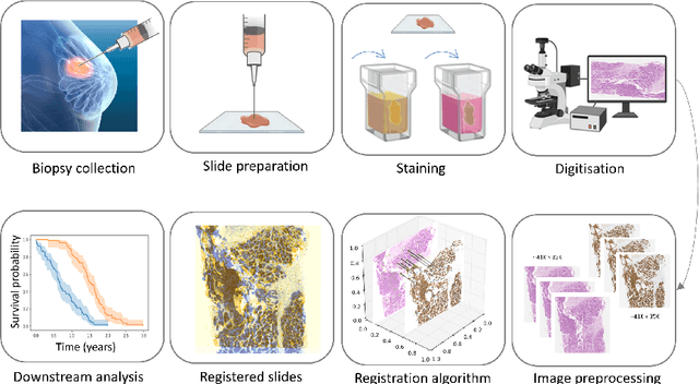
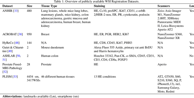
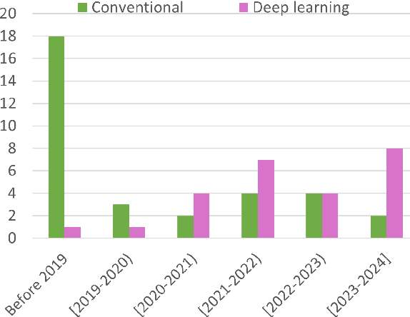
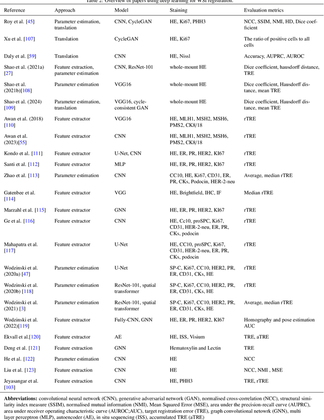
Whole slide image (WSI) registration is an essential task for analysing the tumour microenvironment (TME) in histopathology. It involves the alignment of spatial information between WSIs of the same section or serial sections of a tissue sample. The tissue sections are usually stained with single or multiple biomarkers before imaging, and the goal is to identify neighbouring nuclei along the Z-axis for creating a 3D image or identifying subclasses of cells in the TME. This task is considerably more challenging compared to radiology image registration, such as magnetic resonance imaging or computed tomography, due to various factors. These include gigapixel size of images, variations in appearance between differently stained tissues, changes in structure and morphology between non-consecutive sections, and the presence of artefacts, tears, and deformations. Currently, there is a noticeable gap in the literature regarding a review of the current approaches and their limitations, as well as the challenges and opportunities they present. We aim to provide a comprehensive understanding of the available approaches and their application for various purposes. Furthermore, we investigate current deep learning methods used for WSI registration, emphasising their diverse methodologies. We examine the available datasets and explore tools and software employed in the field. Finally, we identify open challenges and potential future trends in this area of research.
Gland Segmentation Using SAM With Cancer Grade as a Prompt
Jan 27, 2025Cancer grade is a critical clinical criterion that can be used to determine the degree of cancer malignancy. Revealing the condition of the glands, a precise gland segmentation can assist in a more effective cancer grade classification. In machine learning, binary classification information about glands (i.e., benign and malignant) can be utilized as a prompt for gland segmentation and cancer grade classification. By incorporating prior knowledge of the benign or malignant classification of the gland, the model can anticipate the likely appearance of the target, leading to better segmentation performance. We utilize Segment Anything Model to solve the segmentation task, by taking advantage of its prompt function and applying appropriate modifications to the model structure and training strategies. We improve the results from fine-tuned Segment Anything Model and produce SOTA results using this approach.
Deep Learning Based Segmentation of Blood Vessels from H&E Stained Oesophageal Adenocarcinoma Whole-Slide Images
Jan 21, 2025Blood vessels (BVs) play a critical role in the Tumor Micro-Environment (TME), potentially influencing cancer progression and treatment response. However, manually quantifying BVs in Hematoxylin and Eosin (H&E) stained images is challenging and labor-intensive due to their heterogeneous appearances. We propose a novel approach of constructing guiding maps to improve the performance of state-of-the-art segmentation models for BV segmentation, the guiding maps encourage the models to learn representative features of BVs. This is particularly beneficial for computational pathology, where labeled training data is often limited and large models are prone to overfitting. We have quantitative and qualitative results to demonstrate the efficacy of our approach in improving segmentation accuracy. In future, we plan to validate this method to segment BVs across various tissue types and investigate the role of cellular structures in relation to BVs in the TME.
CellOMaps: A Compact Representation for Robust Classification of Lung Adenocarcinoma Growth Patterns
Jan 14, 2025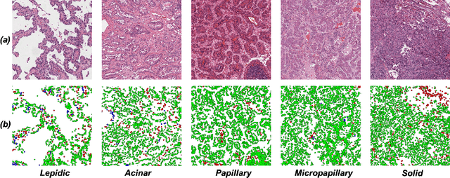


Lung adenocarcinoma (LUAD) is a morphologically heterogeneous disease, characterized by five primary histological growth patterns. The classification of such patterns is crucial due to their direct relation to prognosis but the high subjectivity and observer variability pose a major challenge. Although several studies have developed machine learning methods for growth pattern classification, they either only report the predominant pattern per slide or lack proper evaluation. We propose a generalizable machine learning pipeline capable of classifying lung tissue into one of the five patterns or as non-tumor. The proposed pipeline's strength lies in a novel compact Cell Organization Maps (cellOMaps) representation that captures the cellular spatial patterns from Hematoxylin and Eosin whole slide images (WSIs). The proposed pipeline provides state-of-the-art performance on LUAD growth pattern classification when evaluated on both internal unseen slides and external datasets, significantly outperforming the current approaches. In addition, our preliminary results show that the model's outputs can be used to predict patients Tumor Mutational Burden (TMB) levels.
An Attention Based Pipeline for Identifying Pre-Cancer Lesions in Head and Neck Clinical Images
May 07, 2024


Early detection of cancer can help improve patient prognosis by early intervention. Head and neck cancer is diagnosed in specialist centres after a surgical biopsy, however, there is a potential for these to be missed leading to delayed diagnosis. To overcome these challenges, we present an attention based pipeline that identifies suspected lesions, segments, and classifies them as non-dysplastic, dysplastic and cancerous lesions. We propose (a) a vision transformer based Mask R-CNN network for lesion detection and segmentation of clinical images, and (b) Multiple Instance Learning (MIL) based scheme for classification. Current results show that the segmentation model produces segmentation masks and bounding boxes with up to 82% overlap accuracy score on unseen external test data and surpassing reviewed segmentation benchmarks. Next, a classification F1-score of 85% on the internal cohort test set. An app has been developed to perform lesion segmentation taken via a smart device. Future work involves employing endoscopic video data for precise early detection and prognosis.
Nuclei-Location Based Point Set Registration of Multi-Stained Whole Slide Images
Apr 25, 2024


Whole Slide Images (WSIs) provide exceptional detail for studying tissue architecture at the cell level. To study tumour microenvironment (TME) with the context of various protein biomarkers and cell sub-types, analysis and registration of features using multi-stained WSIs is often required. Multi-stained WSI pairs normally suffer from rigid and non-rigid deformities in addition to slide artefacts and control tissue which present challenges at precise registration. Traditional registration methods mainly focus on global rigid/non-rigid registration but struggle with aligning slides with complex tissue deformations at the nuclei level. However, nuclei level non-rigid registration is essential for downstream tasks such as cell sub-type analysis in the context of protein biomarker signatures. This paper focuses on local level non-rigid registration using a nuclei-location based point set registration approach for aligning multi-stained WSIs. We exploit the spatial distribution of nuclei that is prominent and consistent (to a large level) across different stains to establish a spatial correspondence. We evaluate our approach using the HYRECO dataset consisting of 54 re-stained images of H\&E and PHH3 image pairs. The approach can be extended to other IHC and IF stained WSIs considering a good nuclei detection algorithm is accessible. The performance of the model is tested against established registration algorithms and is shown to outperform the model for nuclei level registration.