Lingling Sun
Anatomy-Guided Parallel Bottleneck Transformer Network for Automated Evaluation of Root Canal Therapy
May 02, 2021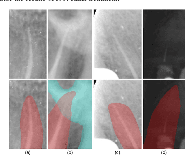

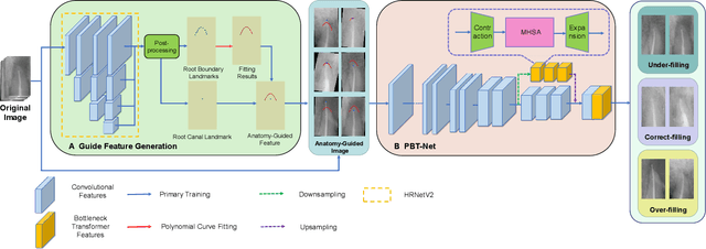

Abstract:Objective: Accurate evaluation of the root canal filling result in X-ray image is a significant step for the root canal therapy, which is based on the relative position between the apical area boundary of tooth root and the top of filled gutta-percha in root canal as well as the shape of the tooth root and so on to classify the result as correct-filling, under-filling or over-filling. Methods: We propose a novel anatomy-guided Transformer diagnosis network. For obtaining accurate anatomy-guided features, a polynomial curve fitting segmentation is proposed to segment the fuzzy boundary. And a Parallel Bottleneck Transformer network (PBT-Net) is introduced as the classification network for the final evaluation. Results, and conclusion: Our numerical experiments show that our anatomy-guided PBT-Net improves the accuracy from 40\% to 85\% relative to the baseline classification network. Comparing with the SOTA segmentation network indicates that the ASD is significantly reduced by 30.3\% through our fitting segmentation. Significance: Polynomial curve fitting segmentation has a great segmentation effect for extremely fuzzy boundaries. The prior knowledge guided classification network is suitable for the evaluation of root canal therapy greatly. And the new proposed Parallel Bottleneck Transformer for realizing self-attention is general in design, facilitating a broad use in most backbone networks.
High-Resolution Segmentation of Tooth Root Fuzzy Edge Based on Polynomial Curve Fitting with Landmark Detection
Mar 07, 2021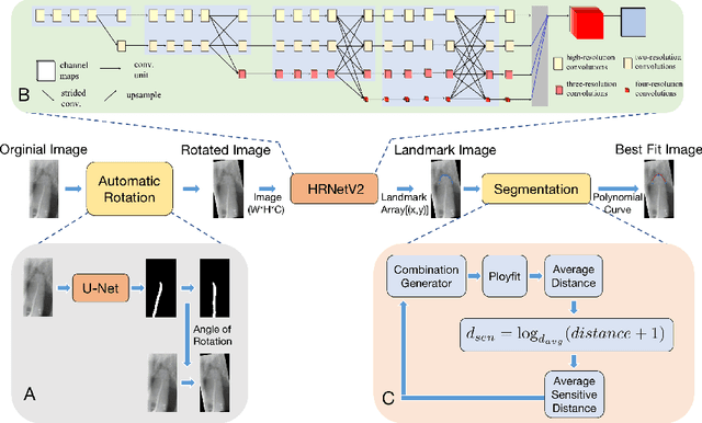

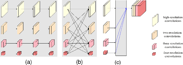
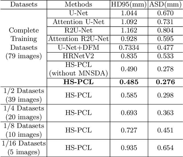
Abstract:As the most economical and routine auxiliary examination in the diagnosis of root canal treatment, oral X-ray has been widely used by stomatologists. It is still challenging to segment the tooth root with a blurry boundary for the traditional image segmentation method. To this end, we propose a model for high-resolution segmentation based on polynomial curve fitting with landmark detection (HS-PCL). It is based on detecting multiple landmarks evenly distributed on the edge of the tooth root to fit a smooth polynomial curve as the segmentation of the tooth root, thereby solving the problem of fuzzy edge. In our model, a maximum number of the shortest distances algorithm (MNSDA) is proposed to automatically reduce the negative influence of the wrong landmarks which are detected incorrectly and deviate from the tooth root on the fitting result. Our numerical experiments demonstrate that the proposed approach not only reduces Hausdorff95 (HD95) by 33.9% and Average Surface Distance (ASD) by 42.1% compared with the state-of-the-art method, but it also achieves excellent results on the minute quantity of datasets, which greatly improves the feasibility of automatic root canal therapy evaluation by medical image computing.
Multiscale Attention Guided Network for COVID-19 Detection Using Chest X-ray Images
Nov 11, 2020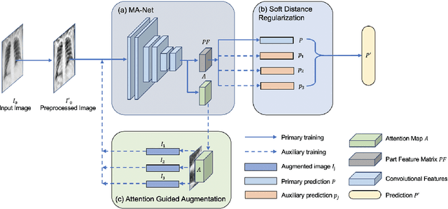
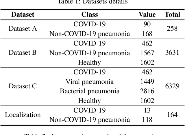
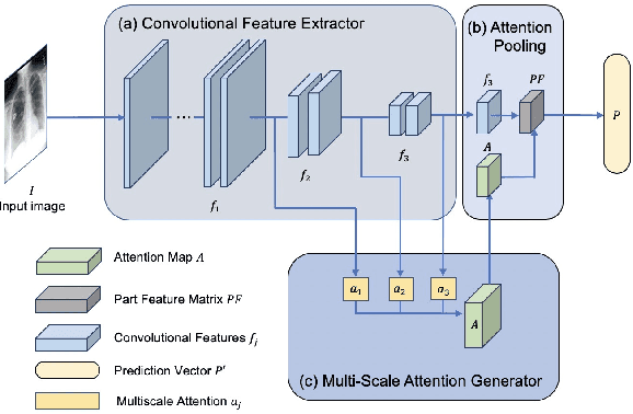
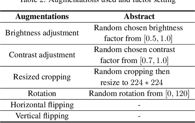
Abstract:Coronavirus disease 2019 (COVID-19) is one of the most destructive pandemic after millennium, forcing the world to tackle a health crisis. Automated classification of lung infections from chest X-ray (CXR) images strengthened traditional healthcare strategy to handle COVID-19. However, classifying COVID-19 from pneumonia cases using CXR image is challenging because of shared spatial characteristics, high feature variation in infections and contrast diversity between cases. Moreover, massive data collection is impractical for a newly emerged disease, which limited the performance of common deep learning models. To address this challenging topic, Multiscale Attention Guided deep network with Soft Distance regularization (MAG-SD) is proposed to automatically classify COVID-19 from pneumonia CXR images. In MAG-SD, MA-Net is used to produce prediction vector and attention map from multiscale feature maps. To relieve the shortage of training data, attention guided augmentations along with a soft distance regularization are posed, which requires a few labeled data to generate meaningful augmentations and reduce noise. Our multiscale attention model achieves better classification performance on our pneumonia CXR image dataset. Plentiful experiments are proposed for MAG-SD which demonstrates that it has its unique advantage in pneumonia classification over cuttingedge models. The code is available at https://github.com/ JasonLeeGHub/MAG-SD.
An Adaptive Enhancement Based Hybrid CNN Model for Digital Dental X-ray Positions Classification
May 01, 2020

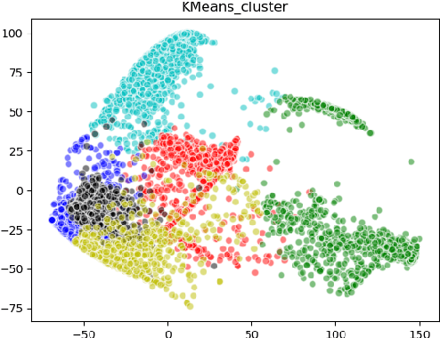
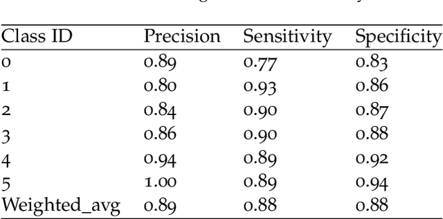
Abstract:Analysis of dental radiographs is an important part of the diagnostic process in daily clinical practice. Interpretation by an expert includes teeth detection and numbering. In this project, a novel solution based on adaptive histogram equalization and convolution neural network (CNN) is proposed, which automatically performs the task for dental x-rays. In order to improve the detection accuracy, we propose three pre-processing techniques to supplement the baseline CNN based on some prior domain knowledge. Firstly, image sharpening and median filtering are used to remove impulse noise, and the edge is enhanced to some extent. Next, adaptive histogram equalization is used to overcome the problem of excessive amplification noise of HE. Finally, a multi-CNN hybrid model is proposed to classify six different locations of dental slices. The results showed that the accuracy and specificity of the test set exceeded 90\%, and the AUC reached 0.97. In addition, four dentists were invited to manually annotate the test data set (independently) and then compare it with the labels obtained by our proposed algorithm. The results show that our method can effectively identify the X-ray location of teeth.
A cascade network for Detecting COVID-19 using chest x-rays
May 01, 2020


Abstract:The worldwide spread of pneumonia caused by a novel coronavirus poses an unprecedented challenge to the world's medical resources and prevention and control measures. Covid-19 attacks not only the lungs, making it difficult to breathe and life-threatening, but also the heart, kidneys, brain and other vital organs of the body, with possible sequela. At present, the detection of COVID-19 needs to be realized by the reverse transcription-polymerase Chain Reaction (RT-PCR). However, many countries are in the outbreak period of the epidemic, and the medical resources are very limited. They cannot provide sufficient numbers of gene sequence detection, and many patients may not be isolated and treated in time. Given this situation, we researched the analytical and diagnostic capabilities of deep learning on chest radiographs and proposed Cascade-SEMEnet which is cascaded with SEME-ResNet50 and SEME-DenseNet169. The two cascade networks of Cascade - SEMEnet both adopt large input sizes and SE-Structure and use MoEx and histogram equalization to enhance the data. We first used SEME-ResNet50 to screen chest X-ray and diagnosed three classes: normal, bacterial, and viral pneumonia. Then we used SEME-DenseNet169 for fine-grained classification of viral pneumonia and determined if it is caused by COVID-19. To exclude the influence of non-pathological features on the network, we preprocessed the data with U-Net during the training of SEME-DenseNet169. The results showed that our network achieved an accuracy of 85.6\% in determining the type of pneumonia infection and 97.1\% in the fine-grained classification of COVID-19. We used Grad-CAM to visualize the judgment based on the model and help doctors understand the chest radiograph while verifying the effectivene.
BACH: Grand Challenge on Breast Cancer Histology Images
Aug 13, 2018
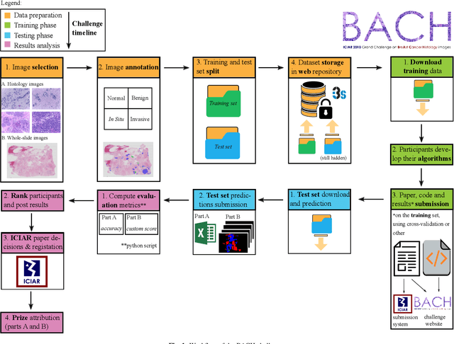

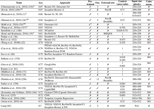
Abstract:Breast cancer is the most common invasive cancer in women, affecting more than 10% of women worldwide. Microscopic analysis of a biopsy remains one of the most important methods to diagnose the type of breast cancer. This requires specialized analysis by pathologists, in a task that i) is highly time- and cost-consuming and ii) often leads to nonconsensual results. The relevance and potential of automatic classification algorithms using hematoxylin-eosin stained histopathological images has already been demonstrated, but the reported results are still sub-optimal for clinical use. With the goal of advancing the state-of-the-art in automatic classification, the Grand Challenge on BreAst Cancer Histology images (BACH) was organized in conjunction with the 15th International Conference on Image Analysis and Recognition (ICIAR 2018). A large annotated dataset, composed of both microscopy and whole-slide images, was specifically compiled and made publicly available for the BACH challenge. Following a positive response from the scientific community, a total of 64 submissions, out of 677 registrations, effectively entered the competition. From the submitted algorithms it was possible to push forward the state-of-the-art in terms of accuracy (87%) in automatic classification of breast cancer with histopathological images. Convolutional neuronal networks were the most successful methodology in the BACH challenge. Detailed analysis of the collective results allowed the identification of remaining challenges in the field and recommendations for future developments. The BACH dataset remains publically available as to promote further improvements to the field of automatic classification in digital pathology.
 Add to Chrome
 Add to Firefox
 Add to Edge