Ruizi Peng
Anatomy-Guided Parallel Bottleneck Transformer Network for Automated Evaluation of Root Canal Therapy
May 02, 2021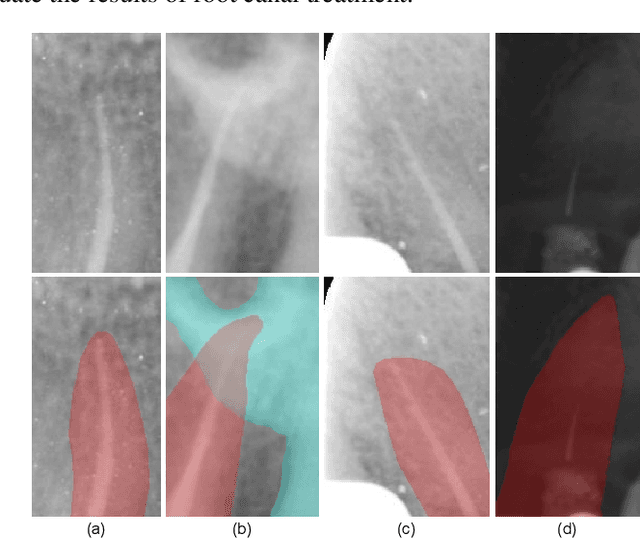

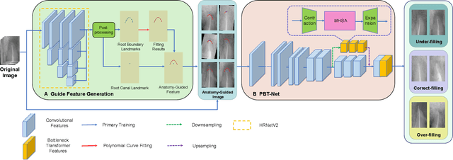

Abstract:Objective: Accurate evaluation of the root canal filling result in X-ray image is a significant step for the root canal therapy, which is based on the relative position between the apical area boundary of tooth root and the top of filled gutta-percha in root canal as well as the shape of the tooth root and so on to classify the result as correct-filling, under-filling or over-filling. Methods: We propose a novel anatomy-guided Transformer diagnosis network. For obtaining accurate anatomy-guided features, a polynomial curve fitting segmentation is proposed to segment the fuzzy boundary. And a Parallel Bottleneck Transformer network (PBT-Net) is introduced as the classification network for the final evaluation. Results, and conclusion: Our numerical experiments show that our anatomy-guided PBT-Net improves the accuracy from 40\% to 85\% relative to the baseline classification network. Comparing with the SOTA segmentation network indicates that the ASD is significantly reduced by 30.3\% through our fitting segmentation. Significance: Polynomial curve fitting segmentation has a great segmentation effect for extremely fuzzy boundaries. The prior knowledge guided classification network is suitable for the evaluation of root canal therapy greatly. And the new proposed Parallel Bottleneck Transformer for realizing self-attention is general in design, facilitating a broad use in most backbone networks.
High-Resolution Segmentation of Tooth Root Fuzzy Edge Based on Polynomial Curve Fitting with Landmark Detection
Mar 07, 2021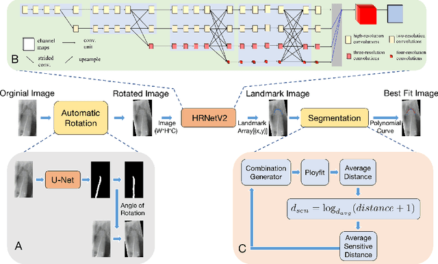

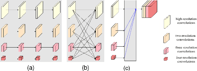
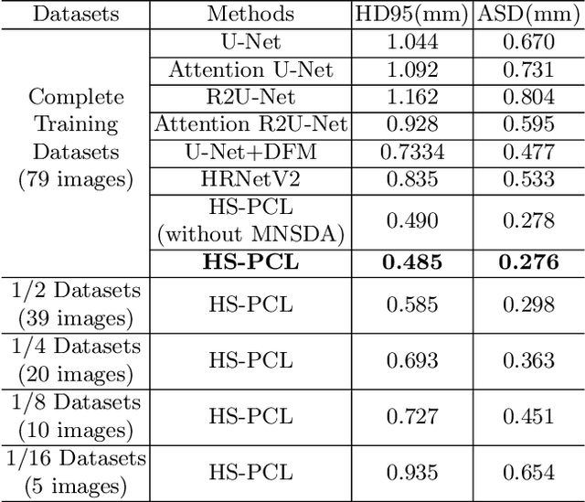
Abstract:As the most economical and routine auxiliary examination in the diagnosis of root canal treatment, oral X-ray has been widely used by stomatologists. It is still challenging to segment the tooth root with a blurry boundary for the traditional image segmentation method. To this end, we propose a model for high-resolution segmentation based on polynomial curve fitting with landmark detection (HS-PCL). It is based on detecting multiple landmarks evenly distributed on the edge of the tooth root to fit a smooth polynomial curve as the segmentation of the tooth root, thereby solving the problem of fuzzy edge. In our model, a maximum number of the shortest distances algorithm (MNSDA) is proposed to automatically reduce the negative influence of the wrong landmarks which are detected incorrectly and deviate from the tooth root on the fitting result. Our numerical experiments demonstrate that the proposed approach not only reduces Hausdorff95 (HD95) by 33.9% and Average Surface Distance (ASD) by 42.1% compared with the state-of-the-art method, but it also achieves excellent results on the minute quantity of datasets, which greatly improves the feasibility of automatic root canal therapy evaluation by medical image computing.
 Add to Chrome
 Add to Firefox
 Add to Edge