Kivanc Kose
On the Role of Calibration in Benchmarking Algorithmic Fairness for Skin Cancer Detection
Nov 10, 2025Abstract:Artificial Intelligence (AI) models have demonstrated expert-level performance in melanoma detection, yet their clinical adoption is hindered by performance disparities across demographic subgroups such as gender, race, and age. Previous efforts to benchmark the performance of AI models have primarily focused on assessing model performance using group fairness metrics that rely on the Area Under the Receiver Operating Characteristic curve (AUROC), which does not provide insights into a model's ability to provide accurate estimates. In line with clinical assessments, this paper addresses this gap by incorporating calibration as a complementary benchmarking metric to AUROC-based fairness metrics. Calibration evaluates the alignment between predicted probabilities and observed event rates, offering deeper insights into subgroup biases. We assess the performance of the leading skin cancer detection algorithm of the ISIC 2020 Challenge on the ISIC 2020 Challenge dataset and the PROVE-AI dataset, and compare it with the second and third place models, focusing on subgroups defined by sex, race (Fitzpatrick Skin Tone), and age. Our findings reveal that while existing models enhance discriminative accuracy, they often over-diagnose risk and exhibit calibration issues when applied to new datasets. This study underscores the necessity for comprehensive model auditing strategies and extensive metadata collection to achieve equitable AI-driven healthcare solutions. All code is publicly available at https://github.com/bdominique/testing_strong_calibration.
* 19 pages, 4 figures. Accepted for publication at the Journal of Machine Learning for Biomedical Imaging (MELBA) https://melba-journal.org/2025:027
SmoothHess: ReLU Network Feature Interactions via Stein's Lemma
Nov 01, 2023


Abstract:Several recent methods for interpretability model feature interactions by looking at the Hessian of a neural network. This poses a challenge for ReLU networks, which are piecewise-linear and thus have a zero Hessian almost everywhere. We propose SmoothHess, a method of estimating second-order interactions through Stein's Lemma. In particular, we estimate the Hessian of the network convolved with a Gaussian through an efficient sampling algorithm, requiring only network gradient calls. SmoothHess is applied post-hoc, requires no modifications to the ReLU network architecture, and the extent of smoothing can be controlled explicitly. We provide a non-asymptotic bound on the sample complexity of our estimation procedure. We validate the superior ability of SmoothHess to capture interactions on benchmark datasets and a real-world medical spirometry dataset.
Unsupervised Approaches for Out-Of-Distribution Dermoscopic Lesion Detection
Nov 08, 2021

Abstract:There are limited works showing the efficacy of unsupervised Out-of-Distribution (OOD) methods on complex medical data. Here, we present preliminary findings of our unsupervised OOD detection algorithm, SimCLR-LOF, as well as a recent state of the art approach (SSD), applied on medical images. SimCLR-LOF learns semantically meaningful features using SimCLR and uses LOF for scoring if a test sample is OOD. We evaluated on the multi-source International Skin Imaging Collaboration (ISIC) 2019 dataset, and show results that are competitive with SSD as well as with recent supervised approaches applied on the same data.
A Patient-Centric Dataset of Images and Metadata for Identifying Melanomas Using Clinical Context
Aug 07, 2020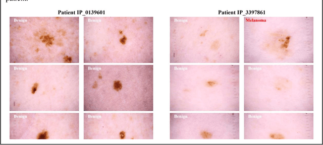
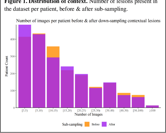
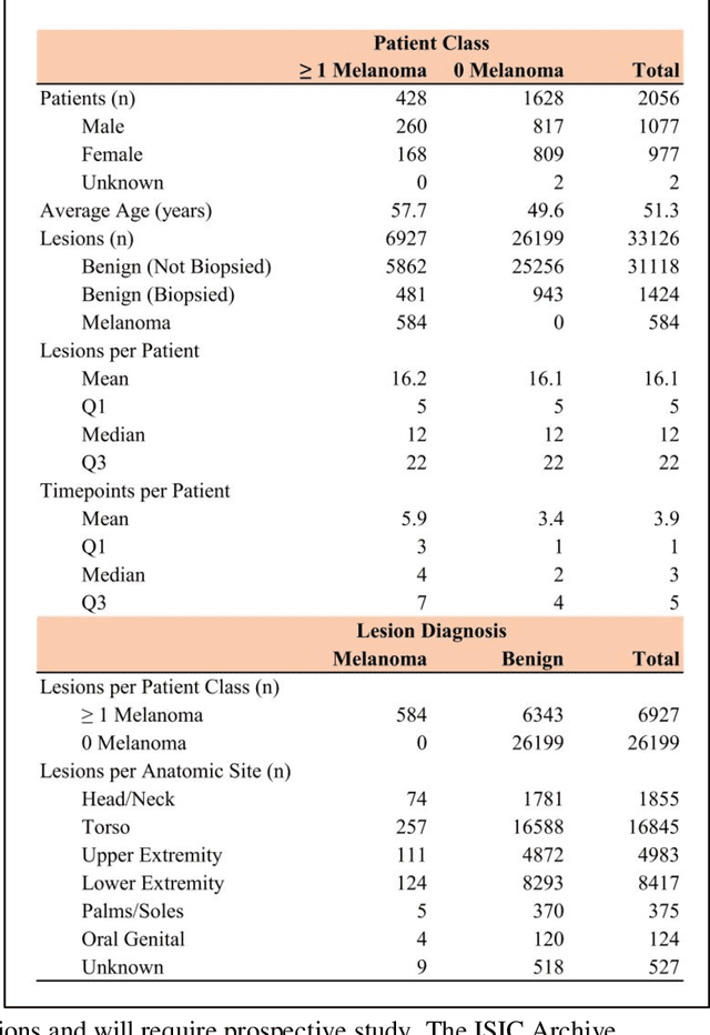
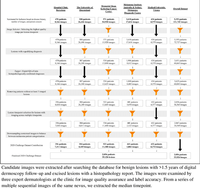
Abstract:Prior skin image datasets have not addressed patient-level information obtained from multiple skin lesions from the same patient. Though artificial intelligence classification algorithms have achieved expert-level performance in controlled studies examining single images, in practice dermatologists base their judgment holistically from multiple lesions on the same patient. The 2020 SIIM-ISIC Melanoma Classification challenge dataset described herein was constructed to address this discrepancy between prior challenges and clinical practice, providing for each image in the dataset an identifier allowing lesions from the same patient to be mapped to one another. This patient-level contextual information is frequently used by clinicians to diagnose melanoma and is especially useful in ruling out false positives in patients with many atypical nevi. The dataset represents 2,056 patients from three continents with an average of 16 lesions per patient, consisting of 33,126 dermoscopic images and 584 histopathologically confirmed melanomas compared with benign melanoma mimickers.
Segmentation of Cellular Patterns in Confocal Images of Melanocytic Lesions in vivo via a Multiscale Encoder-Decoder Network (MED-Net)
Jan 03, 2020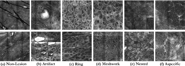
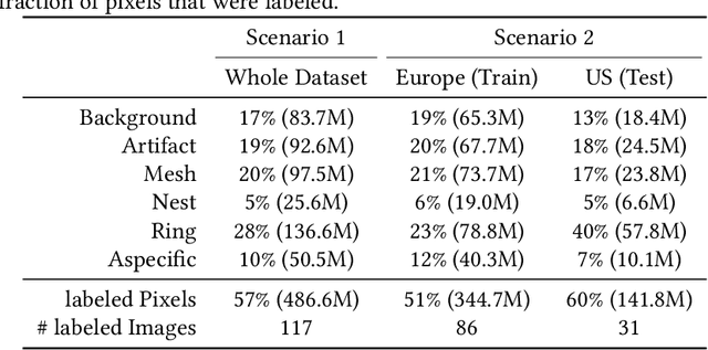
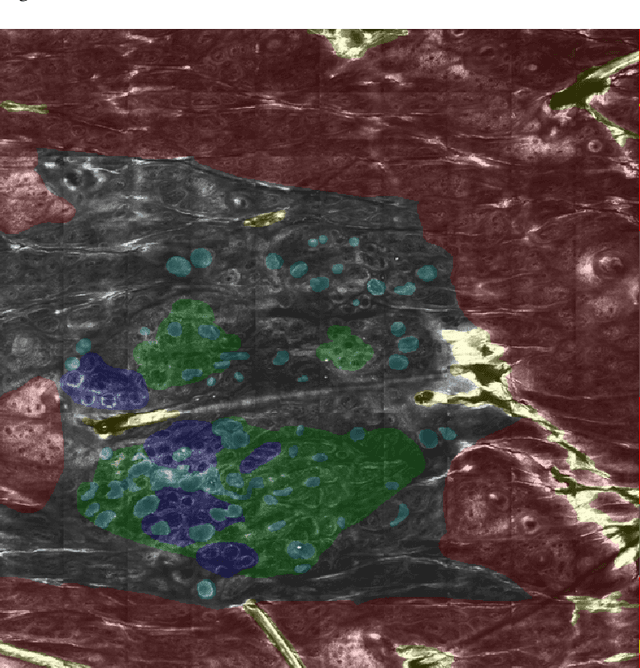
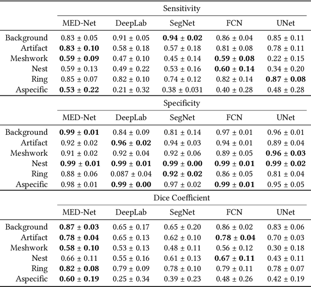
Abstract:In-vivo optical microscopy is advancing into routine clinical practice for non-invasively guiding diagnosis and treatment of cancer and other diseases, and thus beginning to reduce the need for traditional biopsy. However, reading and analysis of the optical microscopic images are generally still qualitative, relying mainly on visual examination. Here we present an automated semantic segmentation method called "Multiscale Encoder-Decoder Network (MED-Net)" that provides pixel-wise labeling into classes of patterns in a quantitative manner. The novelty in our approach is the modeling of textural patterns at multiple scales. This mimics the procedure for examining pathology images, which routinely starts with low magnification (low resolution, large field of view) followed by closer inspection of suspicious areas with higher magnification (higher resolution, smaller fields of view). We trained and tested our model on non-overlapping partitions of 117 reflectance confocal microscopy (RCM) mosaics of melanocytic lesions, an extensive dataset for this application, collected at four clinics in the US, and two in Italy. With patient-wise cross-validation, we achieved pixel-wise mean sensitivity and specificity of $70\pm11\%$ and $95\pm2\%$, respectively, with $0.71\pm0.09$ Dice coefficient over six classes. In the scenario, we partitioned the data clinic-wise and tested the generalizability of the model over multiple clinics. In this setting, we achieved pixel-wise mean sensitivity and specificity of $74\%$ and $95\%$, respectively, with $0.75$ Dice coefficient. We compared MED-Net against the state-of-the-art semantic segmentation models and achieved better quantitative segmentation performance. Our results also suggest that, due to its nested multiscale architecture, the MED-Net model annotated RCM mosaics more coherently, avoiding unrealistic-fragmented annotations.
A Multiresolution Convolutional Neural Network with Partial Label Training for Annotating Reflectance Confocal Microscopy Images of Skin
Aug 23, 2018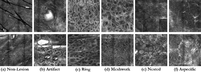
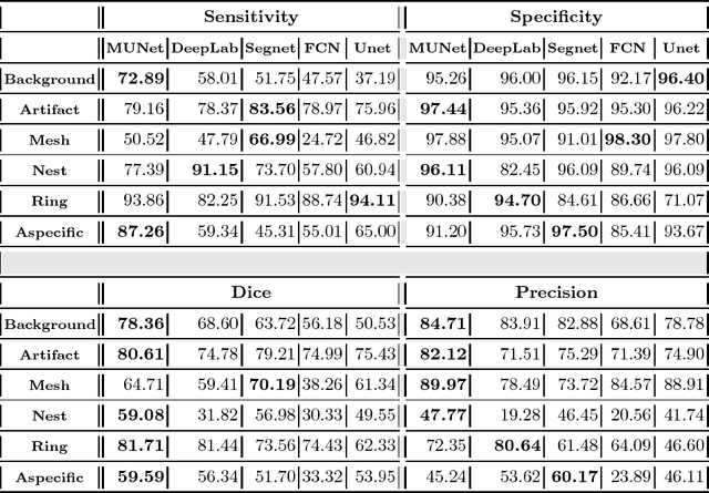


Abstract:We describe a new multiresolution "nested encoder-decoder" convolutional network architecture and use it to annotate morphological patterns in reflectance confocal microscopy (RCM) images of human skin for aiding cancer diagnosis. Skin cancers are the most common types of cancers, melanoma being the deadliest among them. RCM is an effective, non-invasive pre-screening tool for skin cancer diagnosis, with the required cellular resolution. However, images are complex, low-contrast, and highly variable, so that clinicians require months to years of expert-level training to be able to make accurate assessments. In this paper, we address classifying 4 key clinically important structural/textural patterns in RCM images. The occurrence and morphology of these patterns are used by clinicians for diagnosis of melanomas. The large size of RCM images, the large variance of pattern size, the large-scale range over which patterns appear, the class imbalance in collected images, and the lack of fully-labeled images all make this a challenging problem to address, even with automated machine learning tools. We designed a novel nested U-net architecture to cope with these challenges, and a selective loss function to handle partial labeling. Trained and tested on 56 melanoma-suspicious, partially labeled, 12k x 12k pixel images, our network automatically annotated diagnostic patterns with high sensitivity and specificity, providing consistent labels for unlabeled sections of the test images. Providing such annotation will aid clinicians in achieving diagnostic accuracy, and perhaps more important, dramatically facilitate clinical training, thus enabling much more rapid adoption of RCM into widespread clinical use process. In addition, our adaptation of U-net architecture provides an intrinsically multiresolution deep network that may be useful in other challenging biomedical image analysis applications.
Delineation of Skin Strata in Reflectance Confocal Microscopy Images using Recurrent Convolutional Networks with Toeplitz Attention
Dec 01, 2017

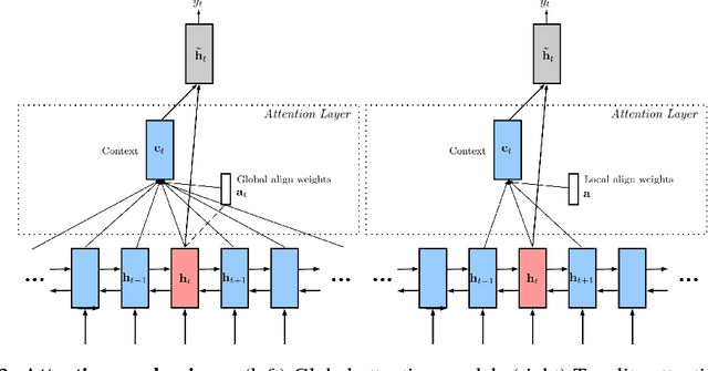
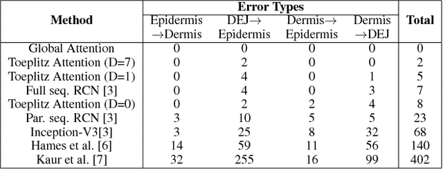
Abstract:Reflectance confocal microscopy (RCM) is an effective, non-invasive pre-screening tool for skin cancer diagnosis, but it requires extensive training and experience to assess accurately. There are few quantitative tools available to standardize image acquisition and analysis, and the ones that are available are not interpretable. In this study, we use a recurrent neural network with attention on convolutional network features. We apply it to delineate skin strata in vertically-oriented stacks of transverse RCM image slices in an interpretable manner. We introduce a new attention mechanism called Toeplitz attention, which constrains the attention map to have a Toeplitz structure. Testing our model on an expert labeled dataset of 504 RCM stacks, we achieve 88.17% image-wise classification accuracy, which is the current state-of-art.
Signal Reconstruction Framework Based On Projections Onto Epigraph Set Of A Convex Cost Function (PESC)
Feb 10, 2014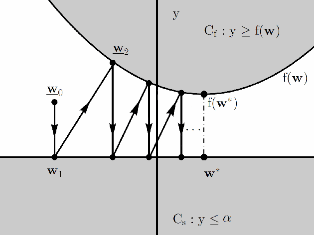
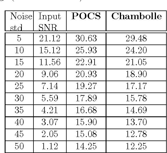
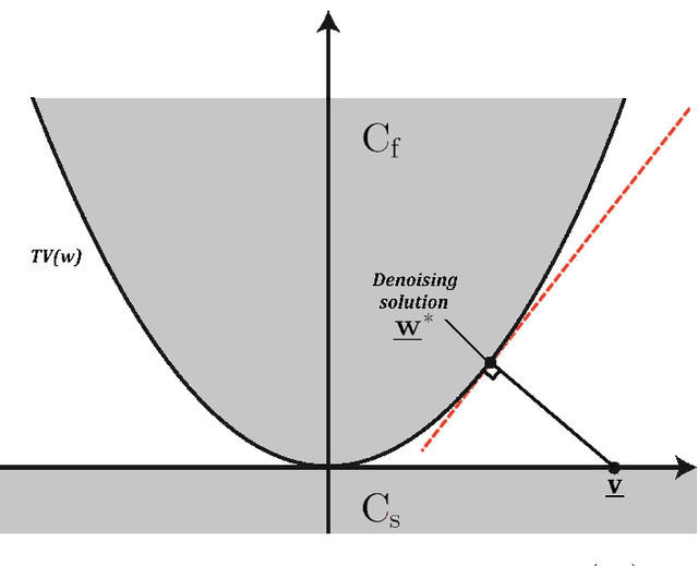
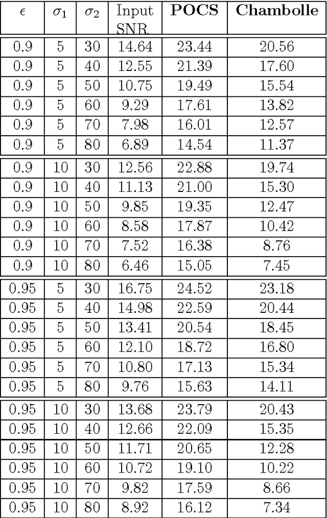
Abstract:A new signal processing framework based on making orthogonal Projections onto the Epigraph Set of a Convex cost function (PESC) is developed. In this way it is possible to solve convex optimization problems using the well-known Projections onto Convex Set (POCS) approach. In this algorithm, the dimension of the minimization problem is lifted by one and a convex set corresponding to the epigraph of the cost function is defined. If the cost function is a convex function in $R^N$, the corresponding epigraph set is also a convex set in R^{N+1}. The PESC method provides globally optimal solutions for total-variation (TV), filtered variation (FV), L_1, L_2, and entropic cost function based convex optimization problems. In this article, the PESC based denoising and compressive sensing algorithms are developed. Simulation examples are presented.
Online Adaptive Decision Fusion Framework Based on Entropic Projections onto Convex Sets with Application to Wildfire Detection in Video
Jan 25, 2011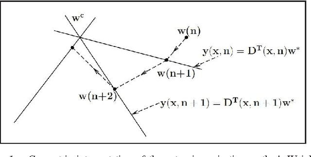
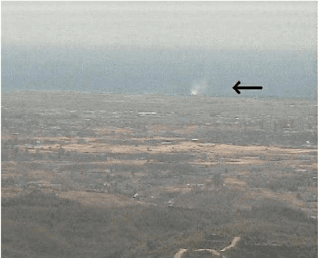
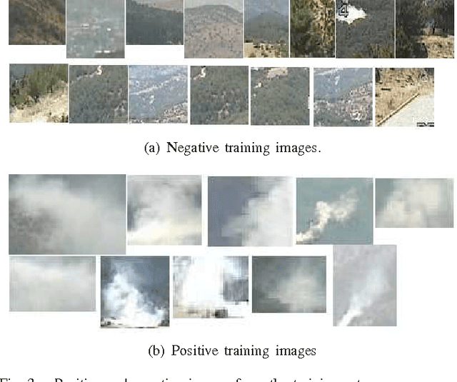
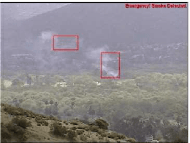
Abstract:In this paper, an Entropy functional based online Adaptive Decision Fusion (EADF) framework is developed for image analysis and computer vision applications. In this framework, it is assumed that the compound algorithm consists of several sub-algorithms each of which yielding its own decision as a real number centered around zero, representing the confidence level of that particular sub-algorithm. Decision values are linearly combined with weights which are updated online according to an active fusion method based on performing entropic projections onto convex sets describing sub-algorithms. It is assumed that there is an oracle, who is usually a human operator, providing feedback to the decision fusion method. A video based wildfire detection system is developed to evaluate the performance of the algorithm in handling the problems where data arrives sequentially. In this case, the oracle is the security guard of the forest lookout tower verifying the decision of the combined algorithm. Simulation results are presented. The EADF framework is also tested with a standard dataset.
 Add to Chrome
 Add to Firefox
 Add to Edge