Xiaolong Luo
The CRITICAL Records Integrated Standardization Pipeline (CRISP): End-to-End Processing of Large-scale Multi-institutional OMOP CDM Data
Sep 10, 2025Abstract:While existing critical care EHR datasets such as MIMIC and eICU have enabled significant advances in clinical AI research, the CRITICAL dataset opens new frontiers by providing extensive scale and diversity -- containing 1.95 billion records from 371,365 patients across four geographically diverse CTSA institutions. CRITICAL's unique strength lies in capturing full-spectrum patient journeys, including pre-ICU, ICU, and post-ICU encounters across both inpatient and outpatient settings. This multi-institutional, longitudinal perspective creates transformative opportunities for developing generalizable predictive models and advancing health equity research. However, the richness of this multi-site resource introduces substantial complexity in data harmonization, with heterogeneous collection practices and diverse vocabulary usage patterns requiring sophisticated preprocessing approaches. We present CRISP to unlock the full potential of this valuable resource. CRISP systematically transforms raw Observational Medical Outcomes Partnership Common Data Model data into ML-ready datasets through: (1) transparent data quality management with comprehensive audit trails, (2) cross-vocabulary mapping of heterogeneous medical terminologies to unified SNOMED-CT standards, with deduplication and unit standardization, (3) modular architecture with parallel optimization enabling complete dataset processing in $<$1 day even on standard computing hardware, and (4) comprehensive baseline model benchmarks spanning multiple clinical prediction tasks to establish reproducible performance standards. By providing processing pipeline, baseline implementations, and detailed transformation documentation, CRISP saves researchers months of preprocessing effort and democratizes access to large-scale multi-institutional critical care data, enabling them to focus on advancing clinical AI.
BFA-YOLO: Balanced multiscale object detection network for multi-view building facade attachments detection
Sep 06, 2024

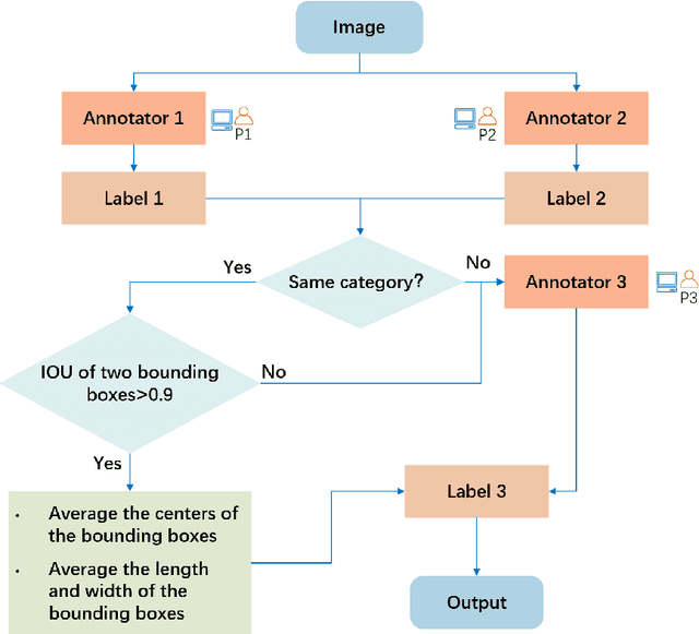

Abstract:Detection of building facade attachments such as doors, windows, balconies, air conditioner units, billboards, and glass curtain walls plays a pivotal role in numerous applications. Building facade attachments detection aids in vbuilding information modeling (BIM) construction and meeting Level of Detail 3 (LOD3) standards. Yet, it faces challenges like uneven object distribution, small object detection difficulty, and background interference. To counter these, we propose BFA-YOLO, a model for detecting facade attachments in multi-view images. BFA-YOLO incorporates three novel innovations: the Feature Balanced Spindle Module (FBSM) for addressing uneven distribution, the Target Dynamic Alignment Task Detection Head (TDATH) aimed at improving small object detection, and the Position Memory Enhanced Self-Attention Mechanism (PMESA) to combat background interference, with each component specifically designed to solve its corresponding challenge. Detection efficacy of deep network models deeply depends on the dataset's characteristics. Existing open source datasets related to building facades are limited by their single perspective, small image pool, and incomplete category coverage. We propose a novel method for building facade attachments detection dataset construction and construct the BFA-3D dataset for facade attachments detection. The BFA-3D dataset features multi-view, accurate labels, diverse categories, and detailed classification. BFA-YOLO surpasses YOLOv8 by 1.8% and 2.9% in mAP@0.5 on the multi-view BFA-3D and street-view Facade-WHU datasets, respectively. These results underscore BFA-YOLO's superior performance in detecting facade attachments.
Semantic decomposition Network with Contrastive and Structural Constraints for Dental Plaque Segmentation
Aug 12, 2022
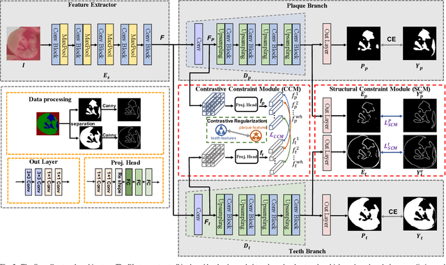
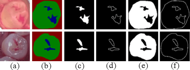
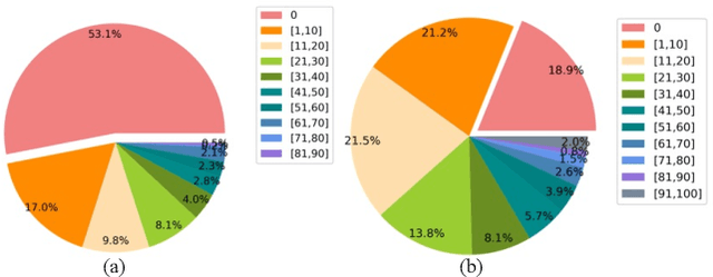
Abstract:Segmenting dental plaque from images of medical reagent staining provides valuable information for diagnosis and the determination of follow-up treatment plan. However, accurate dental plaque segmentation is a challenging task that requires identifying teeth and dental plaque subjected to semantic-blur regions (i.e., confused boundaries in border regions between teeth and dental plaque) and complex variations of instance shapes, which are not fully addressed by existing methods. Therefore, we propose a semantic decomposition network (SDNet) that introduces two single-task branches to separately address the segmentation of teeth and dental plaque and designs additional constraints to learn category-specific features for each branch, thus facilitating the semantic decomposition and improving the performance of dental plaque segmentation. Specifically, SDNet learns two separate segmentation branches for teeth and dental plaque in a divide-and-conquer manner to decouple the entangled relation between them. Each branch that specifies a category tends to yield accurate segmentation. To help these two branches better focus on category-specific features, two constraint modules are further proposed: 1) contrastive constraint module (CCM) to learn discriminative feature representations by maximizing the distance between different category representations, so as to reduce the negative impact of semantic-blur regions on feature extraction; 2) structural constraint module (SCM) to provide complete structural information for dental plaque of various shapes by the supervision of an boundary-aware geometric constraint. Besides, we construct a large-scale open-source Stained Dental Plaque Segmentation dataset (SDPSeg), which provides high-quality annotations for teeth and dental plaque. Experimental results on SDPSeg datasets show SDNet achieves state-of-the-art performance.
Deep Learning-enabled Spatial Phase Unwrapping for 3D Measurement
Aug 06, 2022


Abstract:In terms of 3D imaging speed and system cost, the single-camera system projecting single-frequency patterns is the ideal option among all proposed Fringe Projection Profilometry (FPP) systems. This system necessitates a robust spatial phase unwrapping (SPU) algorithm. However, robust SPU remains a challenge in complex scenes. Quality-guided SPU algorithms need more efficient ways to identify the unreliable points in phase maps before unwrapping. End-to-end deep learning SPU methods face generality and interpretability problems. This paper proposes a hybrid method combining deep learning and traditional path-following for robust SPU in FPP. This hybrid SPU scheme demonstrates better robustness than traditional quality-guided SPU methods, better interpretability than end-to-end deep learning scheme, and generality on unseen data. Experiments on the real dataset of multiple illumination conditions and multiple FPP systems differing in image resolution, the number of fringes, fringe direction, and optics wavelength verify the effectiveness of the proposed method.
Masked Co-attentional Transformer reconstructs 100x ultra-fast/low-dose whole-body PET from longitudinal images and anatomically guided MRI
May 09, 2022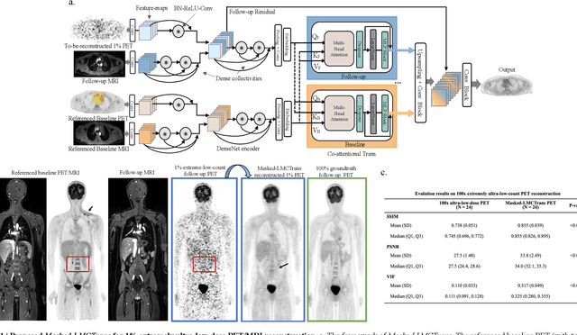
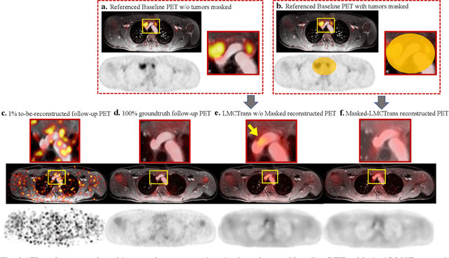
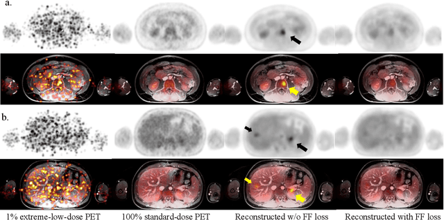
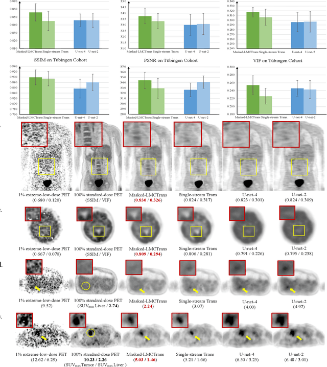
Abstract:Despite its tremendous value for the diagnosis, treatment monitoring and surveillance of children with cancer, whole body staging with positron emission tomography (PET) is time consuming and associated with considerable radiation exposure. 100x (1% of the standard clinical dosage) ultra-low-dose/ultra-fast whole-body PET reconstruction has the potential for cancer imaging with unprecedented speed and improved safety, but it cannot be achieved by the naive use of machine learning techniques. In this study, we utilize the global similarity between baseline and follow-up PET and magnetic resonance (MR) images to develop Masked-LMCTrans, a longitudinal multi-modality co-attentional CNN-Transformer that provides interaction and joint reasoning between serial PET/MRs of the same patient. We mask the tumor area in the referenced baseline PET and reconstruct the follow-up PET scans. In this manner, Masked-LMCTrans reconstructs 100x almost-zero radio-exposure whole-body PET that was not possible before. The technique also opens a new pathway for longitudinal radiology imaging reconstruction, a significantly under-explored area to date. Our model was trained and tested with Stanford PET/MRI scans of pediatric lymphoma patients and evaluated externally on PET/MRI images from T\"ubingen University. The high image quality of the reconstructed 100x whole-body PET images resulting from the application of Masked-LMCTrans will substantially advance the development of safer imaging approaches and shorter exam-durations for pediatric patients, as well as expand the possibilities for frequent longitudinal monitoring of these patients by PET.
 Add to Chrome
 Add to Firefox
 Add to Edge