Daoying Geng
MOSMOS: Multi-organ segmentation facilitated by medical report supervision
Sep 04, 2024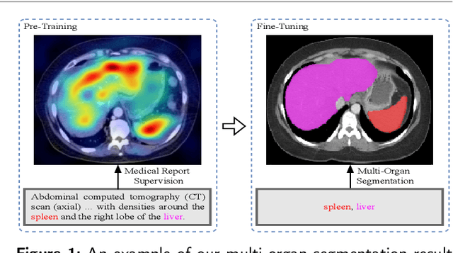
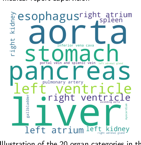
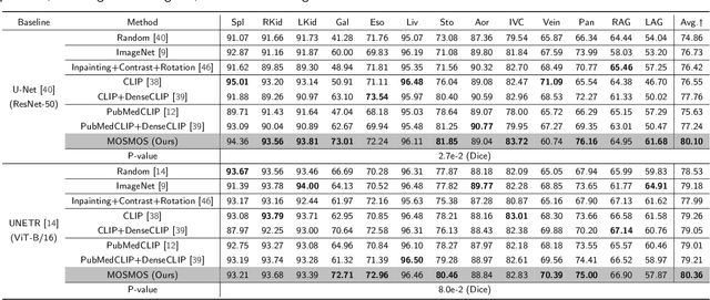
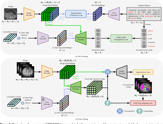
Abstract:Owing to a large amount of multi-modal data in modern medical systems, such as medical images and reports, Medical Vision-Language Pre-training (Med-VLP) has demonstrated incredible achievements in coarse-grained downstream tasks (i.e., medical classification, retrieval, and visual question answering). However, the problem of transferring knowledge learned from Med-VLP to fine-grained multi-organ segmentation tasks has barely been investigated. Multi-organ segmentation is challenging mainly due to the lack of large-scale fully annotated datasets and the wide variation in the shape and size of the same organ between individuals with different diseases. In this paper, we propose a novel pre-training & fine-tuning framework for Multi-Organ Segmentation by harnessing Medical repOrt Supervision (MOSMOS). Specifically, we first introduce global contrastive learning to maximally align the medical image-report pairs in the pre-training stage. To remedy the granularity discrepancy, we further leverage multi-label recognition to implicitly learn the semantic correspondence between image pixels and organ tags. More importantly, our pre-trained models can be transferred to any segmentation model by introducing the pixel-tag attention maps. Different network settings, i.e., 2D U-Net and 3D UNETR, are utilized to validate the generalization. We have extensively evaluated our approach using different diseases and modalities on BTCV, AMOS, MMWHS, and BRATS datasets. Experimental results in various settings demonstrate the effectiveness of our framework. This framework can serve as the foundation to facilitate future research on automatic annotation tasks under the supervision of medical reports.
A Medical Multimodal Large Language Model for Pediatric Pneumonia
Sep 04, 2024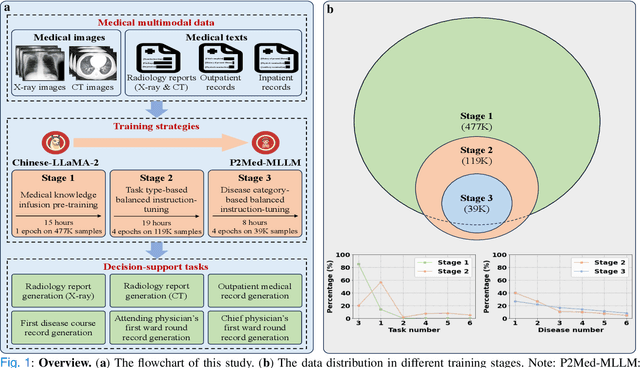
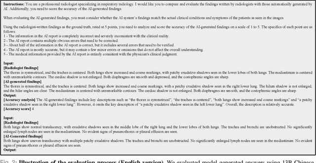
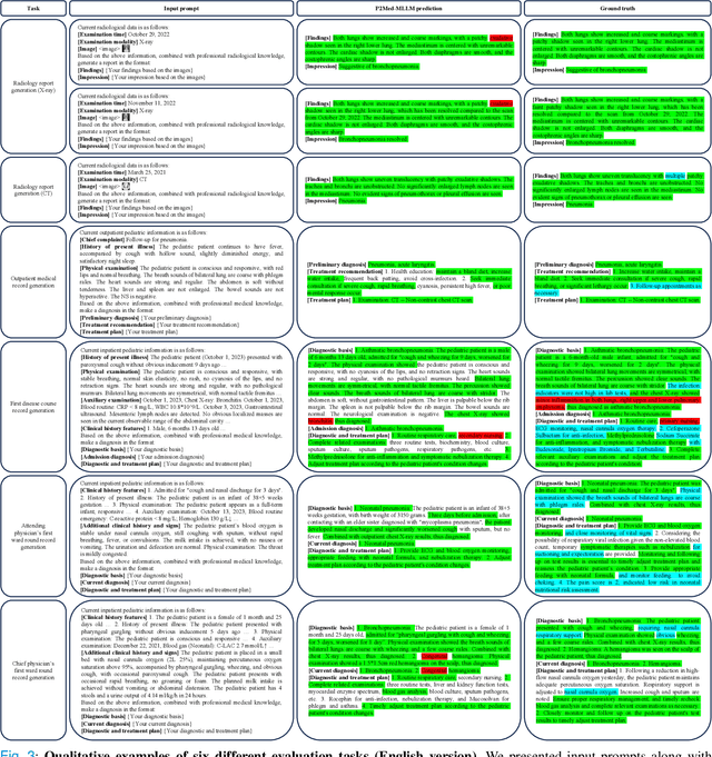
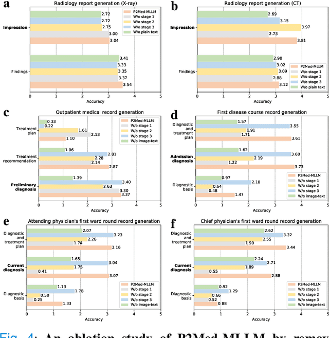
Abstract:Pediatric pneumonia is the leading cause of death among children under five years worldwide, imposing a substantial burden on affected families. Currently, there are three significant hurdles in diagnosing and treating pediatric pneumonia. Firstly, pediatric pneumonia shares similar symptoms with other respiratory diseases, making rapid and accurate differential diagnosis challenging. Secondly, primary hospitals often lack sufficient medical resources and experienced doctors. Lastly, providing personalized diagnostic reports and treatment recommendations is labor-intensive and time-consuming. To tackle these challenges, we proposed a Medical Multimodal Large Language Model for Pediatric Pneumonia (P2Med-MLLM). It was capable of handling diverse clinical tasks, such as generating free-text radiology reports and medical records within a unified framework. Specifically, P2Med-MLLM can process both pure text and image-text data, trained on an extensive and large-scale dataset (P2Med-MD), including real clinical information from 163,999 outpatient and 8,684 inpatient cases. This dataset comprised 2D chest X-ray images, 3D chest CT images, corresponding radiology reports, and outpatient and inpatient records. We designed a three-stage training strategy to enable P2Med-MLLM to comprehend medical knowledge and follow instructions for various clinical tasks. To rigorously evaluate P2Med-MLLM's performance, we developed P2Med-MBench, a benchmark consisting of 642 meticulously verified samples by pediatric pulmonology specialists, covering six clinical decision-support tasks and a balanced variety of diseases. The automated scoring results demonstrated the superiority of P2Med-MLLM. This work plays a crucial role in assisting primary care doctors with prompt disease diagnosis and treatment planning, reducing severe symptom mortality rates, and optimizing the allocation of medical resources.
An Automatic Detection Method Of Cerebral Aneurysms In Time-Of-Flight Magnetic Resonance Angiography Images Based On Attention 3D U-Net
Oct 26, 2021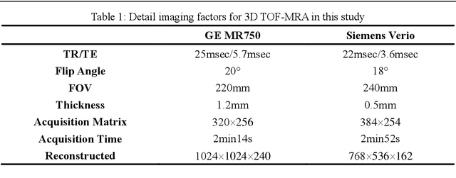
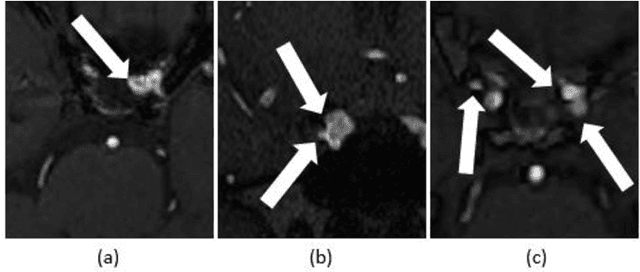
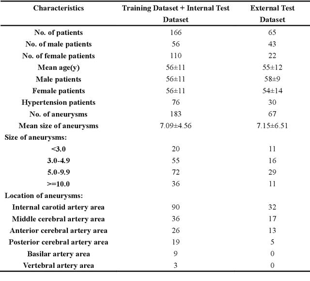
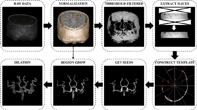
Abstract:Background:Subarachnoid hemorrhage caused by ruptured cerebral aneurysm often leads to fatal consequences.However,if the aneurysm can be found and treated during asymptomatic periods,the probability of rupture can be greatly reduced.At present,time-of-flight magnetic resonance angiography is one of the most commonly used non-invasive screening techniques for cerebral aneurysm,and the application of deep learning technology in aneurysm detection can effectively improve the screening effect of aneurysm.Existing studies have found that three-dimensional features play an important role in aneurysm detection,but they require a large amount of training data and have problems such as a high false positive rate. Methods:This paper proposed a novel method for aneurysm detection.First,a fully automatic cerebral artery segmentation algorithm without training data was used to extract the volume of interest,and then the 3D U-Net was improved by the 3D SENet module to establish an aneurysm detection model.Eventually a set of fully automated,end-to-end aneurysm detection methods have been formed. Results:A total of 231 magnetic resonance angiography image data were used in this study,among which 132 were training sets,34 were internal test sets and 65 were external test sets.The presented method obtained 97.89% sensitivity in the five-fold cross-validation and obtained 91.0% sensitivity with 2.48 false positives/case in the detection of the external test sets. Conclusions:Compared with the results of our previous studies and other studies,the method in this paper achieves a very competitive sensitivity with less training data and maintains a low false positive rate.As the only method currently using 3D U-Net for aneurysm detection,it proves the feasibility and superior performance of this network in aneurysm detection,and also explores the potential of the channel attention mechanism in this task.
 Add to Chrome
 Add to Firefox
 Add to Edge