 Add to Chrome
 Add to Firefox
 Add to Edge
Multi-scale gigapixel microscopy using a multi-camera array microscope
Nov 30, 2022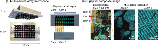
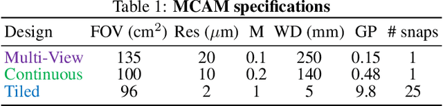
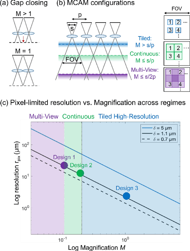
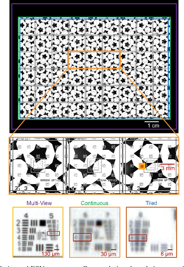
This article experimentally examines different configurations of a novel multi-camera array microscope (MCAM) imaging technology. The MCAM is based upon a densely packed array of "micro-cameras" to jointly image across a large field-of-view at high resolution. Each micro-camera within the array images a unique area of a sample of interest, and then all acquired data with 54 micro-cameras are digitally combined into composite frames, whose total pixel counts significantly exceed the pixel counts of standard microscope systems. We present results from three unique MCAM configurations for different use cases. First, we demonstrate a configuration that simultaneously images and estimates the 3D object depth across a 100 x 135 mm^2 field-of-view (FOV) at approximately 20 um resolution, which results in 0.15 gigapixels (GP) per snapshot. Second, we demonstrate an MCAM configuration that records video across a continuous 83 x 123 mm^2 FOV with two-fold increased resolution (0.48 GP per frame). Finally, we report a third high-resolution configuration (2 um resolution) that can rapidly produce 9.8 GP composites of large histopathology specimens.
Increasing a microscope's effective field of view via overlapped imaging and machine learning
Oct 10, 2021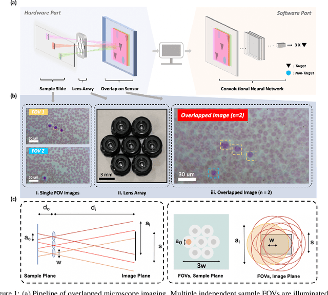
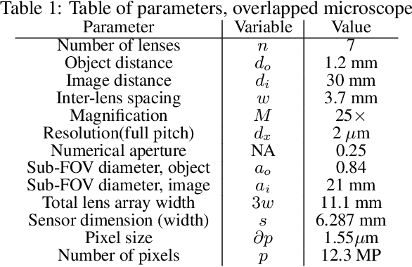
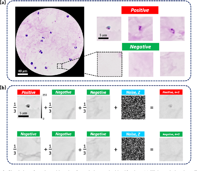
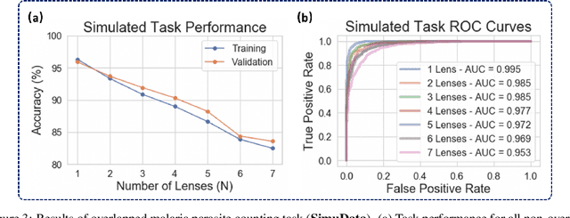
This work demonstrates a multi-lens microscopic imaging system that overlaps multiple independent fields of view on a single sensor for high-efficiency automated specimen analysis. Automatic detection, classification and counting of various morphological features of interest is now a crucial component of both biomedical research and disease diagnosis. While convolutional neural networks (CNNs) have dramatically improved the accuracy of counting cells and sub-cellular features from acquired digital image data, the overall throughput is still typically hindered by the limited space-bandwidth product (SBP) of conventional microscopes. Here, we show both in simulation and experiment that overlapped imaging and co-designed analysis software can achieve accurate detection of diagnostically-relevant features for several applications, including counting of white blood cells and the malaria parasite, leading to multi-fold increase in detection and processing throughput with minimal reduction in accuracy.
Mesoscopic photogrammetry with an unstabilized phone camera
Dec 11, 2020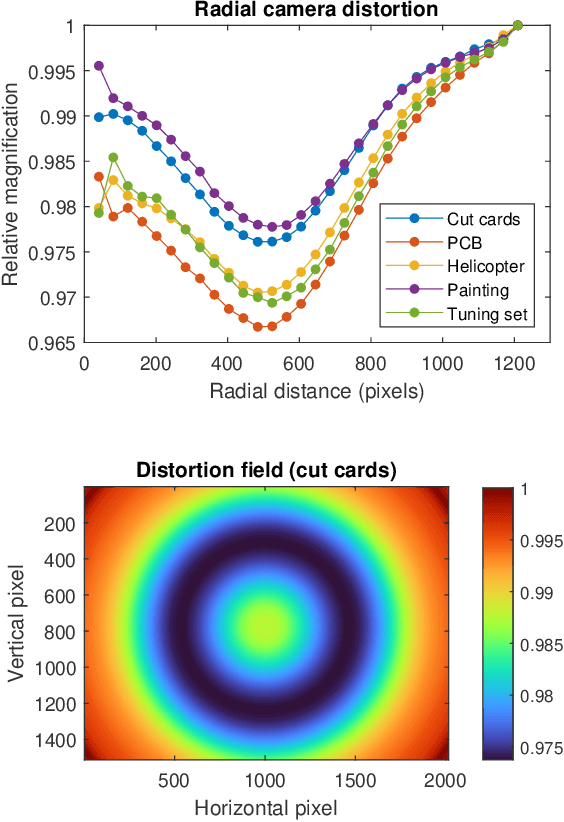
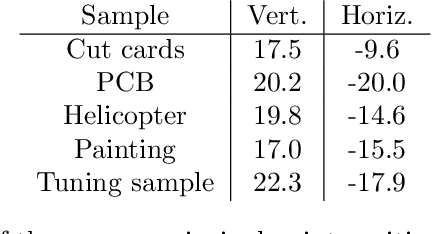
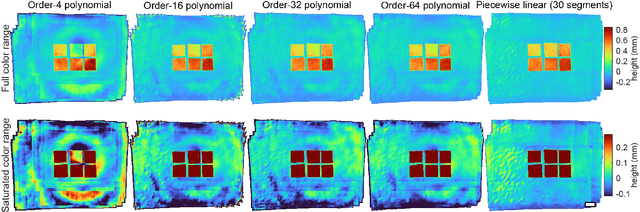
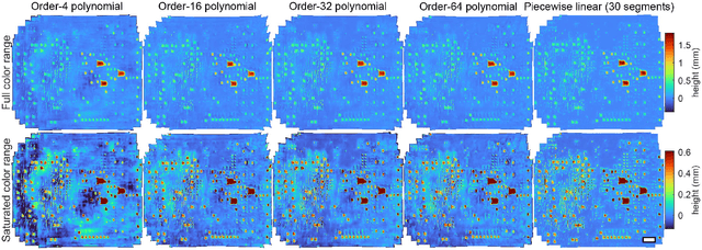
We present a feature-free photogrammetric technique that enables quantitative 3D mesoscopic (mm-scale height variation) imaging with tens-of-micron accuracy from sequences of images acquired by a smartphone at close range (several cm) under freehand motion without additional hardware. Our end-to-end, pixel-intensity-based approach jointly registers and stitches all the images by estimating a coaligned height map, which acts as a pixel-wise radial deformation field that orthorectifies each camera image to allow homographic registration. The height maps themselves are reparameterized as the output of an untrained encoder-decoder convolutional neural network (CNN) with the raw camera images as the input, which effectively removes many reconstruction artifacts. Our method also jointly estimates both the camera's dynamic 6D pose and its distortion using a nonparametric model, the latter of which is especially important in mesoscopic applications when using cameras not designed for imaging at short working distances, such as smartphone cameras. We also propose strategies for reducing computation time and memory, applicable to other multi-frame registration problems. Finally, we demonstrate our method using sequences of multi-megapixel images captured by an unstabilized smartphone on a variety of samples (e.g., painting brushstrokes, circuit board, seeds).