Chang Min Hyun
NeRF Solves Undersampled MRI Reconstruction
Feb 20, 2024


Abstract:This article presents a novel undersampled magnetic resonance imaging (MRI) technique that leverages the concept of Neural Radiance Field (NeRF). With radial undersampling, the corresponding imaging problem can be reformulated into an image modeling task from sparse-view rendered data; therefore, a high dimensional MR image is obtainable from undersampled $k$-space data by taking advantage of implicit neural representation. A multi-layer perceptron, which is designed to output an image intensity from a spatial coordinate, learns the MR physics-driven rendering relation between given measurement data and desired image. Effective undersampling strategies for high-quality neural representation are investigated. The proposed method serves two benefits: (i) The learning is based fully on single undersampled $k$-space data, not a bunch of measured data and target image sets. It can be used potentially for diagnostic MR imaging, such as fetal MRI, where data acquisition is relatively rare or limited against diversity of clinical images while undersampled reconstruction is highly demanded. (ii) A reconstructed MR image is a scan-specific representation highly adaptive to the given $k$-space measurement. Numerous experiments validate the feasibility and capability of the proposed approach.
Automatic 3D Registration of Dental CBCT and Face Scan Data using 2D Projection images
May 17, 2023


Abstract:This paper presents a fully automatic registration method of dental cone-beam computed tomography (CBCT) and face scan data. It can be used for a digital platform of 3D jaw-teeth-face models in a variety of applications, including 3D digital treatment planning and orthognathic surgery. Difficulties in accurately merging facial scans and CBCT images are due to the different image acquisition methods and limited area of correspondence between the two facial surfaces. In addition, it is difficult to use machine learning techniques because they use face-related 3D medical data with radiation exposure, which are difficult to obtain for training. The proposed method addresses these problems by reusing an existing machine-learning-based 2D landmark detection algorithm in an open-source library and developing a novel mathematical algorithm that identifies paired 3D landmarks from knowledge of the corresponding 2D landmarks. A main contribution of this study is that the proposed method does not require annotated training data of facial landmarks because it uses a pre-trained facial landmark detection algorithm that is known to be robust and generalized to various 2D face image models. Note that this reduces a 3D landmark detection problem to a 2D problem of identifying the corresponding landmarks on two 2D projection images generated from two different projection angles. Here, the 3D landmarks for registration were selected from the sub-surfaces with the least geometric change under the CBCT and face scan environments. For the final fine-tuning of the registration, the Iterative Closest Point method was applied, which utilizes geometrical information around the 3D landmarks. The experimental results show that the proposed method achieved an averaged surface distance error of 0.74 mm for three pairs of CBCT and face scan datasets.
Unsupervised sequence-to-sequence learning for automatic signal quality assessment in multi-channel electrical impedance-based hemodynamic monitoring
May 17, 2023Abstract:This study proposes an unsupervised sequence-to-sequence learning approach that automatically assesses the motion-induced reliability degradation of the cardiac volume signal (CVS) in multi-channel electrical impedance-based hemodynamic monitoring. The proposed method attempts to tackle shortcomings in existing learning-based assessment approaches, such as the requirement of manual annotation for motion influence and the lack of explicit mechanisms for realizing motion-induced abnormalities under contextual variations in CVS over time. By utilizing long-short term memory and variational auto-encoder structures, an encoder--decoder model is trained not only to self-reproduce an input sequence of the CVS but also to extrapolate the future in a parallel fashion. By doing so, the model can capture contextual knowledge lying in a temporal CVS sequence while being regularized to explore a general relationship over the entire time-series. A motion-influenced CVS of low-quality is detected, based on the residual between the input sequence and its neural representation with a cut--off value determined from the two-sigma rule of thumb over the training set. Our experimental observations validated two claims: (i) in the learning environment of label-absence, assessment performance is achievable at a competitive level to the supervised setting, and (ii) the contextual information across a time series of CVS is advantageous for effectively realizing motion-induced unrealistic distortions in signal amplitude and morphology. We also investigated the capability as a pseudo-labeling tool to minimize human-craft annotation by preemptively providing strong candidates for motion-induced anomalies. Empirical evidence has shown that machine-guided annotation can reduce inevitable human-errors during manual assessment while minimizing cumbersome and time-consuming processes.
Nonlinear ill-posed problem in low-dose dental cone-beam computed tomography
Mar 03, 2023


Abstract:This paper describes the mathematical structure of the ill-posed nonlinear inverse problem of low-dose dental cone-beam computed tomography (CBCT) and explains the advantages of a deep learning-based approach to the reconstruction of computed tomography images over conventional regularization methods. This paper explains the underlying reasons why dental CBCT is more ill-posed than standard computed tomography. Despite this severe ill-posedness, the demand for dental CBCT systems is rapidly growing because of their cost competitiveness and low radiation dose. We then describe the limitations of existing methods in the accurate restoration of the morphological structures of teeth using dental CBCT data severely damaged by metal implants. We further discuss the usefulness of panoramic images generated from CBCT data for accurate tooth segmentation. We also discuss the possibility of utilizing radiation-free intra-oral scan data as prior information in CBCT image reconstruction to compensate for the damage to data caused by metal implants.
Machine Learning-based Signal Quality Assessment for Cardiac Volume Monitoring in Electrical Impedance Tomography
Jan 04, 2023Abstract:Owing to recent advances in thoracic electrical impedance tomography, a patient's hemodynamic function can be noninvasively and continuously estimated in real-time by surveilling a cardiac volume signal associated with stroke volume and cardiac output. In clinical applications, however, a cardiac volume signal is often of low quality, mainly because of the patient's deliberate movements or inevitable motions during clinical interventions. This study aims to develop a signal quality indexing method that assesses the influence of motion artifacts on transient cardiac volume signals. The assessment is performed on each cardiac cycle to take advantage of the periodicity and regularity in cardiac volume changes. Time intervals are identified using the synchronized electrocardiography system. We apply divergent machine-learning methods, which can be sorted into discriminative-model and manifold-learning approaches. The use of machine-learning could be suitable for our real-time monitoring application that requires fast inference and automation as well as high accuracy. In the clinical environment, the proposed method can be utilized to provide immediate warnings so that clinicians can minimize confusion regarding patients' conditions, reduce clinical resource utilization, and improve the confidence level of the monitoring system. Numerous experiments using actual EIT data validate the capability of cardiac volume signals degraded by motion artifacts to be accurately and automatically assessed in real-time by machine learning. The best model achieved an accuracy of 0.95, positive and negative predictive values of 0.96 and 0.86, sensitivity of 0.98, specificity of 0.77, and AUC of 0.96.
Metal Artifact Reduction with Intra-Oral Scan Data for 3D Low Dose Maxillofacial CBCT Modeling
Feb 08, 2022


Abstract:Low-dose dental cone beam computed tomography (CBCT) has been increasingly used for maxillofacial modeling. However, the presence of metallic inserts, such as implants, crowns, and dental filling, causes severe streaking and shading artifacts in a CBCT image and loss of the morphological structures of the teeth, which consequently prevents accurate segmentation of bones. A two-stage metal artifact reduction method is proposed for accurate 3D low-dose maxillofacial CBCT modeling, where a key idea is to utilize explicit tooth shape prior information from intra-oral scan data whose acquisition does not require any extra radiation exposure. In the first stage, an image-to-image deep learning network is employed to mitigate metal-related artifacts. To improve the learning ability, the proposed network is designed to take advantage of the intra-oral scan data as side-inputs and perform multi-task learning of auxiliary tooth segmentation. In the second stage, a 3D maxillofacial model is constructed by segmenting the bones from the dental CBCT image corrected in the first stage. For accurate bone segmentation, weighted thresholding is applied, wherein the weighting region is determined depending on the geometry of the intra-oral scan data. Because acquiring a paired training dataset of metal-artifact-free and metal artifact-affected dental CBCT images is challenging in clinical practice, an automatic method of generating a realistic dataset according to the CBCT physics model is introduced. Numerical simulations and clinical experiments show the feasibility of the proposed method, which takes advantage of tooth surface information from intra-oral scan data in 3D low dose maxillofacial CBCT modeling.
Automated 3D cephalometric landmark identification using computerized tomography
Dec 16, 2020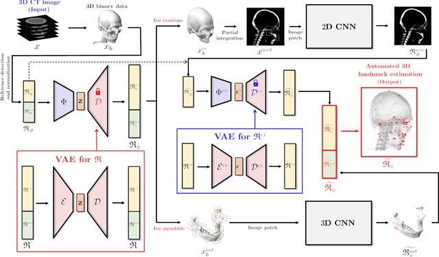

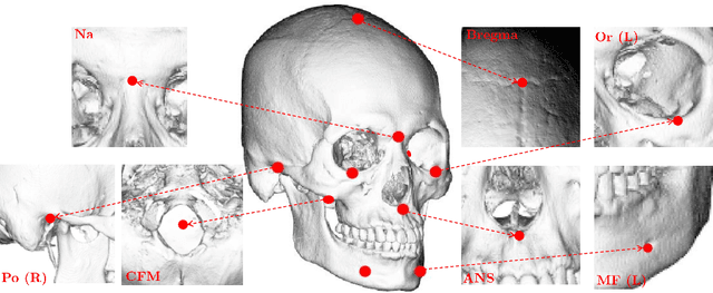

Abstract:Identification of 3D cephalometric landmarks that serve as proxy to the shape of human skull is the fundamental step in cephalometric analysis. Since manual landmarking from 3D computed tomography (CT) images is a cumbersome task even for the trained experts, automatic 3D landmark detection system is in a great need. Recently, automatic landmarking of 2D cephalograms using deep learning (DL) has achieved great success, but 3D landmarking for more than 80 landmarks has not yet reached a satisfactory level, because of the factors hindering machine learning such as the high dimensionality of the input data and limited amount of training data due to ethical restrictions on the use of medical data. This paper presents a semi-supervised DL method for 3D landmarking that takes advantage of anonymized landmark dataset with paired CT data being removed. The proposed method first detects a small number of easy-to-find reference landmarks, then uses them to provide a rough estimation of the entire landmarks by utilizing the low dimensional representation learned by variational autoencoder (VAE). Anonymized landmark dataset is used for training the VAE. Finally, coarse-to-fine detection is applied to the small bounding box provided by rough estimation, using separate strategies suitable for mandible and cranium. For mandibular landmarks, patch-based 3D CNN is applied to the segmented image of the mandible (separated from the maxilla), in order to capture 3D morphological features of mandible associated with the landmarks. We detect 6 landmarks around the condyle all at once, instead of one by one, because they are closely related to each other. For cranial landmarks, we again use VAE-based latent representation for more accurate annotation. In our experiment, the proposed method achieved an averaged 3D point-to-point error of 2.91 mm for 90 landmarks only with 15 paired training data.
Deep Learning-Based Solvability of Underdetermined Inverse Problems in Medical Imaging
Jan 07, 2020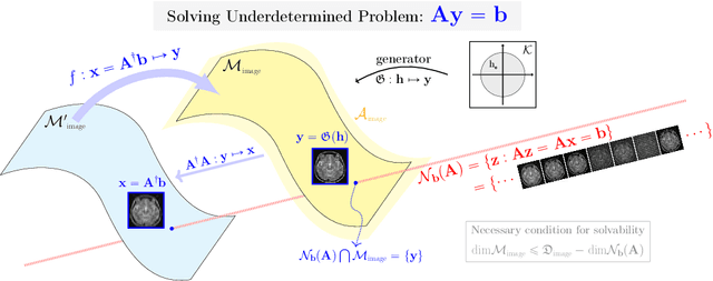
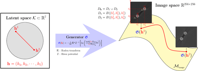
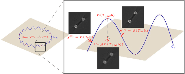
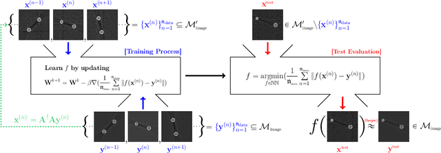
Abstract:Recently, with the significant developments in deep learning techniques, solving underdetermined inverse problems has become one of the major concerns in the medical imaging domain. Typical examples include undersampled magnetic resonance imaging, interior tomography, and sparse-view computed tomography, where deep learning techniques have achieved excellent performances. Although deep learning methods appear to overcome the limitations of existing mathematical methods when handling various underdetermined problems, there is a lack of rigorous mathematical foundations that would allow us to elucidate the reasons for the remarkable performance of deep learning methods. This study focuses on learning the causal relationship regarding the structure of the training data suitable for deep learning, to solve highly underdetermined inverse problems. We observe that a majority of the problems of solving underdetermined linear systems in medical imaging are highly non-linear. Furthermore, we analyze if a desired reconstruction map can be learnable from the training data and underdetermined system.
Framelet Pooling Aided Deep Learning Network : The Method to Process High Dimensional Medical Data
Jul 25, 2019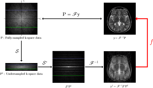

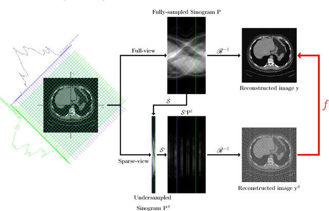
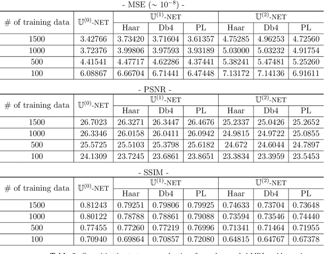
Abstract:Machine learning-based analysis of medical images often faces several hurdles, such as the lack of training data, the curse of dimensionality problem, and the generalization issues. One of the main difficulties is that there exists computational cost problem in dealing with input data of large size matrices which represent medical images. The purpose of this paper is to introduce a framelet-pooling aided deep learning method for mitigating computational bundle, caused by large dimensionality. By transforming high dimensional data into low dimensional components by filter banks with preserving detailed information, the proposed method aims to reduce the complexity of the neural network and computational costs significantly during the learning process. Various experiments show that our method is comparable to the standard unreduced learning method, while reducing computational burdens by decomposing large-sized learning tasks into several small-scale learning tasks.
Deep learning for undersampled MRI reconstruction
Sep 11, 2017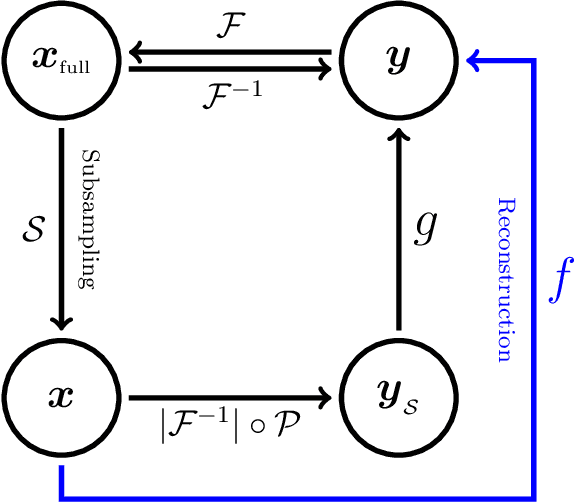


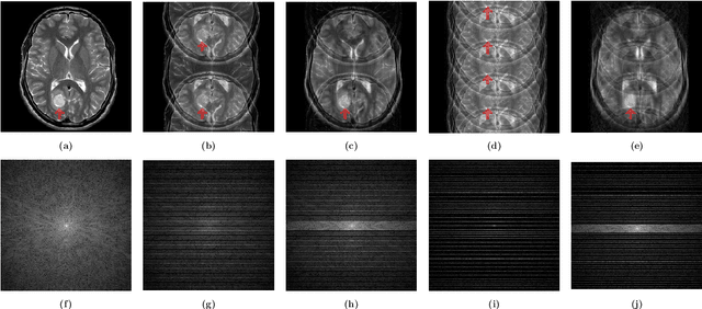
Abstract:This paper presents a deep learning method for faster magnetic resonance imaging (MRI) by reducing k-space data with sub-Nyquist sampling strategies and provides a rationale for why the proposed approach works well. Uniform subsampling is used in the time-consuming phase-encoding direction to capture high-resolution image information, while permitting the image-folding problem dictated by the Poisson summation formula. To deal with the localization uncertainty due to image folding, very few low-frequency k-space data are added. Training the deep learning net involves input and output images that are pairs of Fourier transforms of the subsampled and fully sampled k-space data. Numerous experiments show the remarkable performance of the proposed method; only 29% of k-space data can generate images of high quality as effectively as standard MRI reconstruction with fully sampled data.
 Add to Chrome
 Add to Firefox
 Add to Edge