Xingzhi Xie
A novel multiple instance learning framework for COVID-19 severity assessment via data augmentation and self-supervised learning
Feb 07, 2021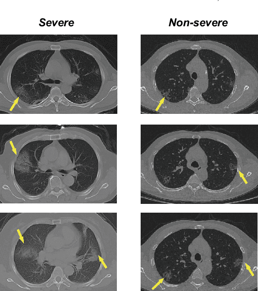
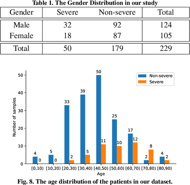
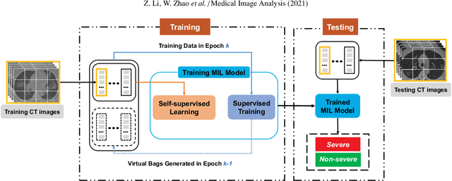
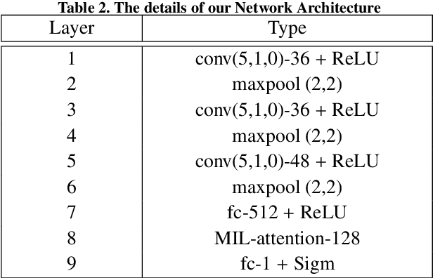
Abstract:How to fast and accurately assess the severity level of COVID-19 is an essential problem, when millions of people are suffering from the pandemic around the world. Currently, the chest CT is regarded as a popular and informative imaging tool for COVID-19 diagnosis. However, we observe that there are two issues -- weak annotation and insufficient data that may obstruct automatic COVID-19 severity assessment with CT images. To address these challenges, we propose a novel three-component method, i.e., 1) a deep multiple instance learning component with instance-level attention to jointly classify the bag and also weigh the instances, 2) a bag-level data augmentation component to generate virtual bags by reorganizing high confidential instances, and 3) a self-supervised pretext component to aid the learning process. We have systematically evaluated our method on the CT images of 229 COVID-19 cases, including 50 severe and 179 non-severe cases. Our method could obtain an average accuracy of 95.8%, with 93.6% sensitivity and 96.4% specificity, which outperformed previous works.
Synergistic Learning of Lung Lobe Segmentation and Hierarchical Multi-Instance Classification for Automated Severity Assessment of COVID-19 in CT Images
May 24, 2020
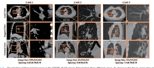
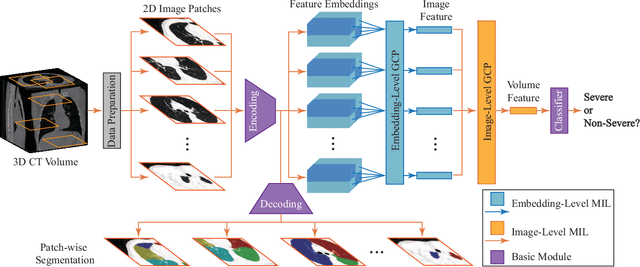
Abstract:Understanding chest CT imaging of the coronavirus disease 2019 (COVID-19) will help detect infections early and assess the disease progression. Especially, automated severity assessment of COVID-19 in CT images plays an essential role in identifying cases that are in great need of intensive clinical care. However, it is often challenging to accurately assess the severity of this disease in CT images, due to variable infection regions in the lungs, similar imaging biomarkers, and large inter-case variations. To this end, we propose a synergistic learning framework for automated severity assessment of COVID-19 in 3D CT images, by jointly performing lung lobe segmentation and multi-instance classification. Considering that only a few infection regions in a CT image are related to the severity assessment, we first represent each input image by a bag that contains a set of 2D image patches (with each cropped from a specific slice). A multi-task multi-instance deep network (called M$^2$UNet) is then developed to assess the severity of COVID-19 patients and also segment the lung lobe simultaneously. Our M$^2$UNet consists of a patch-level encoder, a segmentation sub-network for lung lobe segmentation, and a classification sub-network for severity assessment (with a unique hierarchical multi-instance learning strategy). Here, the context information provided by segmentation can be implicitly employed to improve the performance of severity assessment. Extensive experiments were performed on a real COVID-19 CT image dataset consisting of 666 chest CT images, with results suggesting the effectiveness of our proposed method compared to several state-of-the-art methods.
Severity Assessment of Coronavirus Disease 2019 (COVID-19) Using Quantitative Features from Chest CT Images
Mar 26, 2020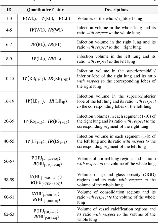
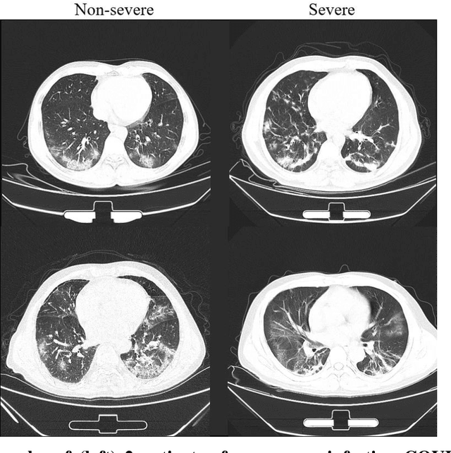
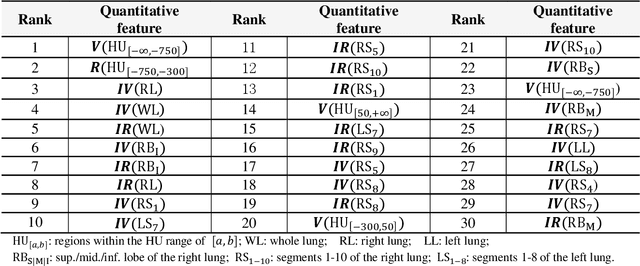
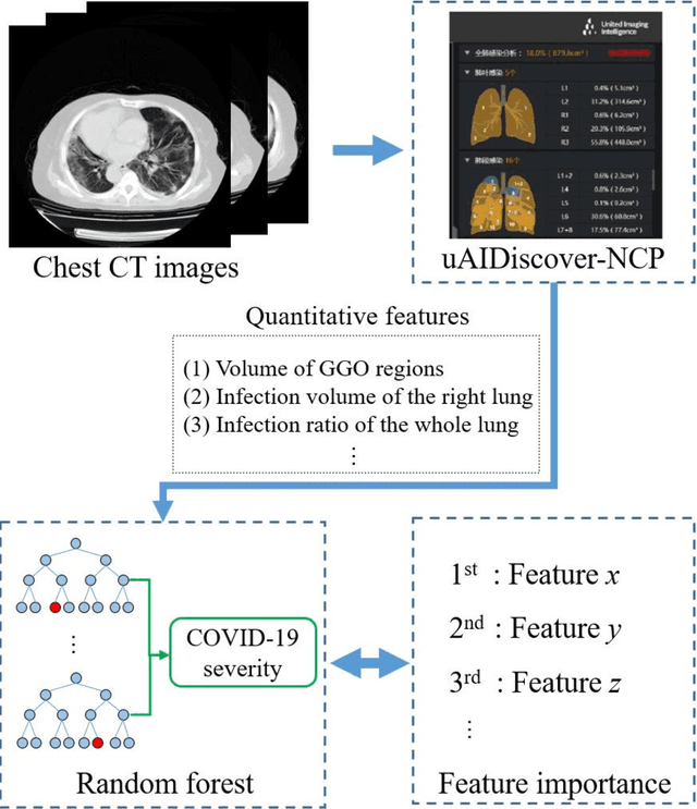
Abstract:Background: Chest computed tomography (CT) is recognized as an important tool for COVID-19 severity assessment. As the number of affected patients increase rapidly, manual severity assessment becomes a labor-intensive task, and may lead to delayed treatment. Purpose: Using machine learning method to realize automatic severity assessment (non-severe or severe) of COVID-19 based on chest CT images, and to explore the severity-related features from the resulting assessment model. Materials and Method: Chest CT images of 176 patients (age 45.3$\pm$16.5 years, 96 male and 80 female) with confirmed COVID-19 are used, from which 63 quantitative features, e.g., the infection volume/ratio of the whole lung and the volume of ground-glass opacity (GGO) regions, are calculated. A random forest (RF) model is trained to assess the severity (non-severe or severe) based on quantitative features. Importance of each quantitative feature, which reflects the correlation to the severity of COVID-19, is calculated from the RF model. Results: Using three-fold cross validation, the RF model shows promising results, i.e., 0.933 of true positive rate, 0.745 of true negative rate, 0.875 of accuracy, and 0.91 of area under receiver operating characteristic curve (AUC). The resulting importance of quantitative features shows that the volume and its ratio (with respect to the whole lung volume) of ground glass opacity (GGO) regions are highly related to the severity of COVID-19, and the quantitative features calculated from the right lung are more related to the severity assessment than those of the left lung. Conclusion: The RF based model can achieve automatic severity assessment (non-severe or severe) of COVID-19 infection, and the performance is promising. Several quantitative features, which have the potential to reflect the severity of COVID-19, were revealed.
 Add to Chrome
 Add to Firefox
 Add to Edge