 Add to Chrome
 Add to Firefox
 Add to Edge
++nnU-Net: Scaling nnU-Net with Prefix-Based Data Augmentation
Jun 09, 2026The nnU-Net has demonstrated continuous success in medical segmentation tasks, which heavily rely on the availability and diversity of annotated biomedical data. However, assembling medical imaging cohorts remains challenging due to numerous factors such as privacy regulations and annotation costs. As a result, data augmentation plays a crucial role in increasing data availability while maintaining anatomical feasibility. Hence, we propose the ++nnU-Net, a novel data augmentation module based on image registration that operates prior to preprocessing and training take place. Our framework was evaluated across five different 2D datasets. In this workflow, image data go through a two-stage registration process, generating new warped images. The transformations are then applied to the respective segmentation. In addition, the pipeline computes available disk space, generates supplementary binary synthetic masks and generates checkpoints. We demonstrate that the ++nnU-Net outperforms the nnU-Net baseline, yielding improvements in Dice Similarity Coefficient scores. In the most prominent cases, we observe performance gains of approximately 22\%. These findings highlight the effectiveness of registration-based data augmentation, particularly for 2D medical imaging datasets and suggest that the ++nnU-Net provides a practical and scalable approach for enhancing segmentation performance in data-limited settings. The source code for the ++nnU-Net is available at: https://github.com/sofia-adelie/plusplusnnunet.git
OSS: Open Suturing Skills Vision-Based Assessment Challenge 2024-2025
May 21, 2026Achieving high levels of surgical skill through effective training is essential for optimal patient outcomes. Automated, data-driven skill assessment holds significant potential to improve surgical training. While machine learning-based methods are increasingly popular for assessing skills in minimally invasive surgery, their application to open surgery remains limited. We present the results of a dedicated MICCAI challenge designed to benchmark and advance vision-based skill assessment in open surgery. The challenge dataset comprises videos of an open suturing training task recorded with a static GoPro camera in a dry-lab setting, with instrument trajectories available in addition to the primary video modality. The OSS Challenge was hosted over two consecutive years, comprising two and three independent tasks, respectively: (1) classifying skill level into four classes, (2) predicting the full Objective Structured Assessment of Technical Skills across eight categories, and (3) tracking hands and surgical tools. Participants submitted diverse solutions including deep learning-based video models, tracking-driven methods, and hybrid approaches. General-purpose spatiotemporal video models consistently achieved the strongest performance, though conceptually diverse approaches reached competitive levels when well-executed. Predicting fine-grained OSATS scores remains challenging but benefits substantially from increased training data. Keypoint tracking proves difficult given frequent occlusions and out-of-frame instances, limiting current applicability for motion-based skill analysis. This work benchmarks innovative and diverse solutions for surgical skill assessment, highlighting both the promise and current limitations of video-based evaluation in open surgery and identifying critical directions for advancing automated skill assessment toward clinical impact.
VS-DDPM: Efficient Low-Cost Diffusion Model for Medical Modality Translation
Apr 24, 2026Diffusion models produce high-quality synthetic data but suffer from slow inference. We propose 3D Variable-Step Denoising Diffusion Probabilistic Model (VS-DDPM) a framework engineered to maintain generative quality while accelerating inference by several factors. We tested our approach on four tasks (missing MRI, tumor removal, MRI-to-sCT, and CBCT-to-sCT) within the BraTS2025 and SynthRAD2025 challenges. Designed for high efficiency under hardware and time constrains imposed by both challenges. VS-DDPM achieved state-of-the-art (SOTA) performance in missing MRI synthesis, yielding Dice scores of 0.80, 0.83, and 0.88 for the enhancing tumor, tumor core, and whole tumor regions, respectively, alongside a structural similarity index (SSIM) of 0.95. For MRI tumor removal, the model attained a root mean squared error (RMSE) of 0.053, a peak signal-to-noise ratio (PSNR) of 26.77, and an SSIM of 0.918. While the framework demonstrated competitive performance in MRI-to-sCT and CBCT-to-sCT tasks, it did not reach SOTA benchmarks, potentially due to sensitivities in data pre and post-processing pipelines or specific loss function configurations. These results demonstrate that VS-DDPM provides a robust and tunable solution for high-fidelity 3D medical image synthesis. The code is available in https://github.com/andre-fs-ferreira/SynthRAD_by_Faking_it.
Curated endoscopic retrograde cholangiopancreatography images dataset
Jan 23, 2026Endoscopic Retrograde Cholangiopancreatography (ERCP) is a key procedure in the diagnosis and treatment of biliary and pancreatic diseases. Artificial intelligence has been pointed as one solution to automatize diagnosis. However, public ERCP datasets are scarce, which limits the use of such approach. Therefore, this study aims to help fill this gap by providing a large and curated dataset. The collection is composed of 19.018 raw images and 19.317 processed from 1.602 patients. 5.519 images are labeled, which provides a ready to use dataset. All images were manually inspected and annotated by two gastroenterologist with more than 5 years of experience and reviewed by another gastroenterologist with more than 20 years of experience, all with more than 400 ERCP procedures annually. The utility and validity of the dataset is proven by a classification experiment. This collection aims to provide or contribute for a benchmark in automatic ERCP analysis and diagnosis of biliary and pancreatic diseases.
Radiomics and Clinical Features in Predictive Modelling of Brain Metastases Recurrence
Dec 17, 2025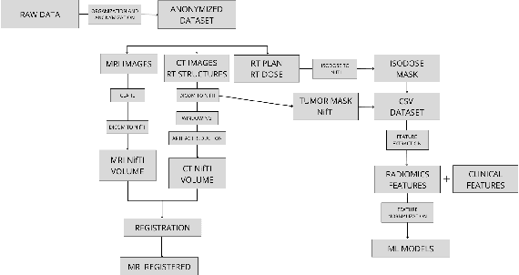


Brain metastases affect approximately between 20% and 40% of cancer patients and are commonly treated with radiotherapy or radiosurgery. Early prediction of recurrence following treatment could enable timely clinical intervention and improve patient outcomes. This study proposes an artificial intelligence based approach for predicting brain metastasis recurrence using multimodal imaging and clinical data. A retrospective cohort of 97 patients was collected, including Computed Tomography (CT) and Magnetic Resonance Imaging (MRI) acquired before treatment and at first follow-up, together with relevant clinical variables. Image preprocessing included CT windowing and artifact reduction, MRI enhancement, and multimodal CT MRI registration. After applying inclusion criteria, 53 patients were retained for analysis. Radiomics features were extracted from the imaging data, and delta radiomics was employed to characterize temporal changes between pre-treatment and follow-up scans. Multiple machine learning classifiers were trained and evaluated, including an analysis of discrepancies between treatment planning target volumes and delivered isodose volumes. Despite limitations related to sample size and class imbalance, the results demonstrate the feasibility of radiomics based models, namely ensemble models, for recurrence prediction and suggest a potential association between radiation dose discrepancies and recurrence risk. This work supports further investigation of AI-driven tools to assist clinical decision-making in brain metastasis management.
Enhancing Privacy: The Utility of Stand-Alone Synthetic CT and MRI for Tumor and Bone Segmentation
Jun 13, 2025AI requires extensive datasets, while medical data is subject to high data protection. Anonymization is essential, but poses a challenge for some regions, such as the head, as identifying structures overlap with regions of clinical interest. Synthetic data offers a potential solution, but studies often lack rigorous evaluation of realism and utility. Therefore, we investigate to what extent synthetic data can replace real data in segmentation tasks. We employed head and neck cancer CT scans and brain glioma MRI scans from two large datasets. Synthetic data were generated using generative adversarial networks and diffusion models. We evaluated the quality of the synthetic data using MAE, MS-SSIM, Radiomics and a Visual Turing Test (VTT) performed by 5 radiologists and their usefulness in segmentation tasks using DSC. Radiomics indicates high fidelity of synthetic MRIs, but fall short in producing highly realistic CT tissue, with correlation coefficient of 0.8784 and 0.5461 for MRI and CT tumors, respectively. DSC results indicate limited utility of synthetic data: tumor segmentation achieved DSC=0.064 on CT and 0.834 on MRI, while bone segmentation a mean DSC=0.841. Relation between DSC and correlation is observed, but is limited by the complexity of the task. VTT results show synthetic CTs' utility, but with limited educational applications. Synthetic data can be used independently for the segmentation task, although limited by the complexity of the structures to segment. Advancing generative models to better tolerate heterogeneous inputs and learn subtle details is essential for enhancing their realism and expanding their application potential.
A Simultaneous Approach for Training Neural Differential-Algebraic Systems of Equations
Apr 07, 2025


Scientific machine learning is an emerging field that broadly describes the combination of scientific computing and machine learning to address challenges in science and engineering. Within the context of differential equations, this has produced highly influential methods, such as neural ordinary differential equations (NODEs). Recent works extend this line of research to consider neural differential-algebraic systems of equations (DAEs), where some unknown relationships within the DAE are learned from data. Training neural DAEs, similarly to neural ODEs, is computationally expensive, as it requires the solution of a DAE for every parameter update. Further, the rigorous consideration of algebraic constraints is difficult within common deep learning training algorithms such as stochastic gradient descent. In this work, we apply the simultaneous approach to neural DAE problems, resulting in a fully discretized nonlinear optimization problem, which is solved to local optimality and simultaneously obtains the neural network parameters and the solution to the corresponding DAE. We extend recent work demonstrating the simultaneous approach for neural ODEs, by presenting a general framework to solve neural DAEs, with explicit consideration of hybrid models, where some components of the DAE are known, e.g. physics-informed constraints. Furthermore, we present a general strategy for improving the performance and convergence of the nonlinear programming solver, based on solving an auxiliary problem for initialization and approximating Hessian terms. We achieve promising results in terms of accuracy, model generalizability and computational cost, across different problem settings such as sparse data, unobserved states and multiple trajectories. Lastly, we provide several promising future directions to improve the scalability and robustness of our approach.
The Impact of Artificial Intelligence on Emergency Medicine: A Review of Recent Advances
Mar 17, 2025Artificial Intelligence (AI) is revolutionizing emergency medicine by enhancing diagnostic processes and improving patient outcomes. This article provides a review of the current applications of AI in emergency imaging studies, focusing on the last five years of advancements. AI technologies, particularly machine learning and deep learning, are pivotal in interpreting complex imaging data, offering rapid, accurate diagnoses and potentially surpassing traditional diagnostic methods. Studies highlighted within the article demonstrate AI's capabilities in accurately detecting conditions such as fractures, pneumothorax, and pulmonary diseases from various imaging modalities including X-rays, CT scans, and MRIs. Furthermore, AI's ability to predict clinical outcomes like mechanical ventilation needs illustrates its potential in crisis resource optimization. Despite these advancements, the integration of AI into clinical practice presents challenges such as data privacy, algorithmic bias, and the need for extensive validation across diverse settings. This review underscores the transformative potential of AI in emergency settings, advocating for a future where AI and clinical expertise synergize to elevate patient care standards.
RFUDS -- A Brain Metastases Imaging Dataset of Radiotherapy Follow-Up
Dec 21, 2024Brain metastases are a common diagnosis that affects between 20% and 40% of cancer patients. Subsequent to radiation therapy, patients with brain metastases undergo follow-up sessions during which the response to treatment is monitored. In this study, a dataset of medical images from 44 patients with at least one brain metastasis and different primary tumor locations was collected and processed. Each patient was treated with either a linear accelerator or a gamma knife. Computed Tomography (CT) and Magnetic Resonance Imaging (MRI) scans were collected at various time points, including before treatment and during follow-up sessions. The CT datasets were processed using windowing and artifact reduction techniques, while the MRI datasets were subjected to CLAHE. The NifTI files corresponding to the CT and MRI images were made public available. In order to align the datasets of each patient, a multimodal registration was performed between the CT and MRI datasets, with different software options being tested. The fusion matrices were provided together with the dataset. The aforementioned steps resulted in the creation of an optimized dataset, prepared for use in a range of studies related to brain metastases. RFUds is publicity available at zenodo under the DOI 10.5281/zenodo.14524784.
Comparative Analysis of nnUNet and MedNeXt for Head and Neck Tumor Segmentation in MRI-guided Radiotherapy
Nov 22, 2024Radiation therapy (RT) is essential in treating head and neck cancer (HNC), with magnetic resonance imaging(MRI)-guided RT offering superior soft tissue contrast and functional imaging. However, manual tumor segmentation is time-consuming and complex, and therfore remains a challenge. In this study, we present our solution as team TUMOR to the HNTS-MRG24 MICCAI Challenge which is focused on automated segmentation of primary gross tumor volumes (GTVp) and metastatic lymph node gross tumor volume (GTVn) in pre-RT and mid-RT MRI images. We utilized the HNTS-MRG2024 dataset, which consists of 150 MRI scans from patients diagnosed with HNC, including original and registered pre-RT and mid-RT T2-weighted images with corresponding segmentation masks for GTVp and GTVn. We employed two state-of-the-art models in deep learning, nnUNet and MedNeXt. For Task 1, we pretrained models on pre-RT registered and mid-RT images, followed by fine-tuning on original pre-RT images. For Task 2, we combined registered pre-RT images, registered pre-RT segmentation masks, and mid-RT data as a multi-channel input for training. Our solution for Task 1 achieved 1st place in the final test phase with an aggregated Dice Similarity Coefficient of 0.8254, and our solution for Task 2 ranked 8th with a score of 0.7005. The proposed solution is publicly available at Github Repository.