Luigi Cinque
LAGUNA: LAnguage Guided UNsupervised Adaptation with structured spaces
Nov 23, 2024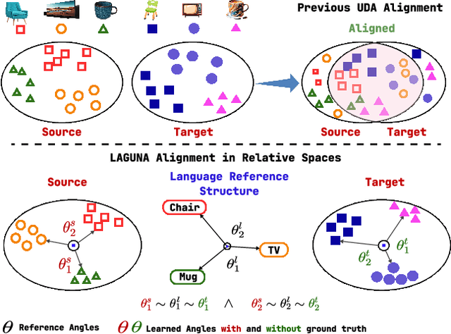
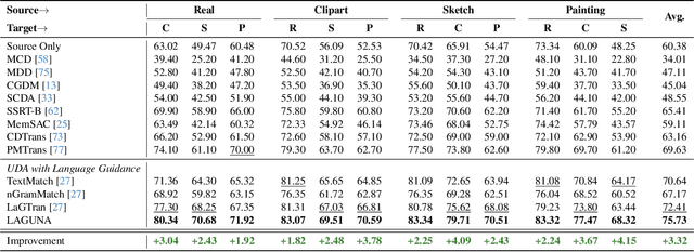
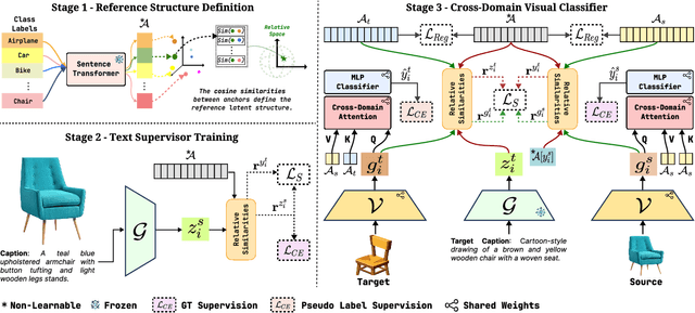
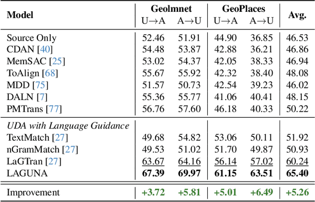
Abstract:Unsupervised domain adaptation remains a critical challenge in enabling the knowledge transfer of models across unseen domains. Existing methods struggle to balance the need for domain-invariant representations with preserving domain-specific features, which is often due to alignment approaches that impose the projection of samples with similar semantics close in the latent space despite their drastic domain differences. We introduce \mnamelong, a novel approach that shifts the focus from aligning representations in absolute coordinates to aligning the relative positioning of equivalent concepts in latent spaces. \mname defines a domain-agnostic structure upon the semantic/geometric relationships between class labels in language space and guides adaptation, ensuring that the organization of samples in visual space reflects reference inter-class relationships while preserving domain-specific characteristics. %We empirically demonstrate \mname's superiority in domain adaptation tasks across four diverse images and video datasets. Remarkably, \mname surpasses previous works in 18 different adaptation scenarios across four diverse image and video datasets with average accuracy improvements of +3.32% on DomainNet, +5.75% in GeoPlaces, +4.77% on GeoImnet, and +1.94% mean class accuracy improvement on EgoExo4D.
NT-ViT: Neural Transcoding Vision Transformers for EEG-to-fMRI Synthesis
Sep 18, 2024Abstract:This paper introduces the Neural Transcoding Vision Transformer (\modelname), a generative model designed to estimate high-resolution functional Magnetic Resonance Imaging (fMRI) samples from simultaneous Electroencephalography (EEG) data. A key feature of \modelname is its Domain Matching (DM) sub-module which effectively aligns the latent EEG representations with those of fMRI volumes, enhancing the model's accuracy and reliability. Unlike previous methods that tend to struggle with fidelity and reproducibility of images, \modelname addresses these challenges by ensuring methodological integrity and higher-quality reconstructions which we showcase through extensive evaluation on two benchmark datasets; \modelname outperforms the current state-of-the-art by a significant margin in both cases, e.g. achieving a $10\times$ reduction in RMSE and a $3.14\times$ increase in SSIM on the Oddball dataset. An ablation study also provides insights into the contribution of each component to the model's overall effectiveness. This development is critical in offering a new approach to lessen the time and financial constraints typically linked with high-resolution brain imaging, thereby aiding in the swift and precise diagnosis of neurological disorders. Although it is not a replacement for actual fMRI but rather a step towards making such imaging more accessible, we believe that it represents a pivotal advancement in clinical practice and neuroscience research. Code is available at \url{https://github.com/rom42pla/ntvit}.
ReViT: Enhancing Vision Transformers with Attention Residual Connections for Visual Recognition
Feb 17, 2024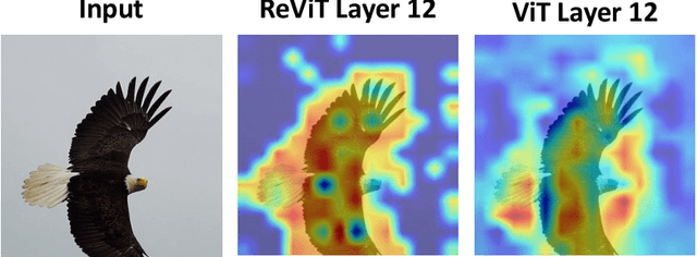

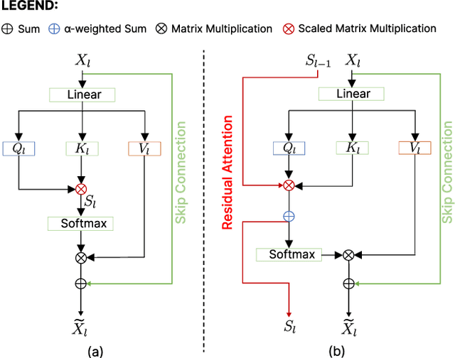
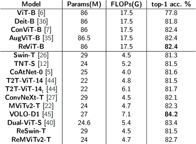
Abstract:Vision Transformer (ViT) self-attention mechanism is characterized by feature collapse in deeper layers, resulting in the vanishing of low-level visual features. However, such features can be helpful to accurately represent and identify elements within an image and increase the accuracy and robustness of vision-based recognition systems. Following this rationale, we propose a novel residual attention learning method for improving ViT-based architectures, increasing their visual feature diversity and model robustness. In this way, the proposed network can capture and preserve significant low-level features, providing more details about the elements within the scene being analyzed. The effectiveness and robustness of the presented method are evaluated on five image classification benchmarks, including ImageNet1k, CIFAR10, CIFAR100, Oxford Flowers-102, and Oxford-IIIT Pet, achieving improved performances. Additionally, experiments on the COCO2017 dataset show that the devised approach discovers and incorporates semantic and spatial relationships for object detection and instance segmentation when implemented into spatial-aware transformer models.
Medicinal Boxes Recognition on a Deep Transfer Learning Augmented Reality Mobile Application
Mar 26, 2022

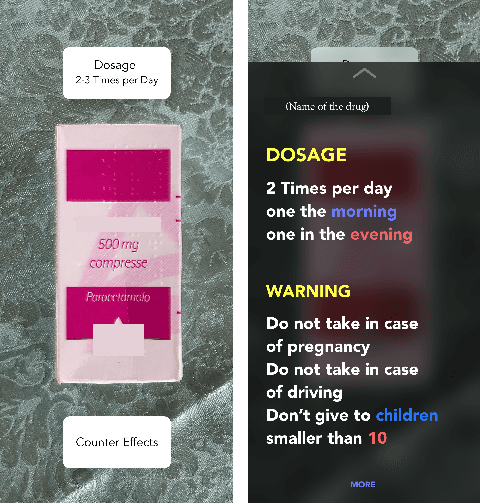

Abstract:Taking medicines is a fundamental aspect to cure illnesses. However, studies have shown that it can be hard for patients to remember the correct posology. More aggravating, a wrong dosage generally causes the disease to worsen. Although, all relevant instructions for a medicine are summarized in the corresponding patient information leaflet, the latter is generally difficult to navigate and understand. To address this problem and help patients with their medication, in this paper we introduce an augmented reality mobile application that can present to the user important details on the framed medicine. In particular, the app implements an inference engine based on a deep neural network, i.e., a densenet, fine-tuned to recognize a medicinal from its package. Subsequently, relevant information, such as posology or a simplified leaflet, is overlaid on the camera feed to help a patient when taking a medicine. Extensive experiments to select the best hyperparameters were performed on a dataset specifically collected to address this task; ultimately obtaining up to 91.30\% accuracy as well as real-time capabilities.
Analyzing EEG Data with Machine and Deep Learning: A Benchmark
Mar 18, 2022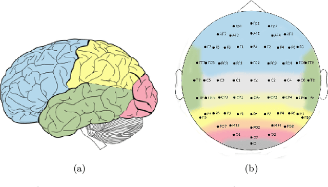
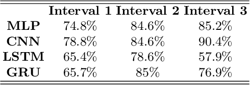
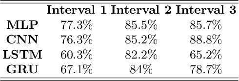
Abstract:Nowadays, machine and deep learning techniques are widely used in different areas, ranging from economics to biology. In general, these techniques can be used in two ways: trying to adapt well-known models and architectures to the available data, or designing custom architectures. In both cases, to speed up the research process, it is useful to know which type of models work best for a specific problem and/or data type. By focusing on EEG signal analysis, and for the first time in literature, in this paper a benchmark of machine and deep learning for EEG signal classification is proposed. For our experiments we used the four most widespread models, i.e., multilayer perceptron, convolutional neural network, long short-term memory, and gated recurrent unit, highlighting which one can be a good starting point for developing EEG classification models.
Human Silhouette and Skeleton Video Synthesis through Wi-Fi signals
Mar 11, 2022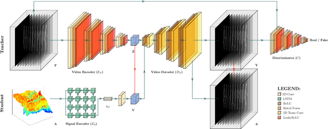
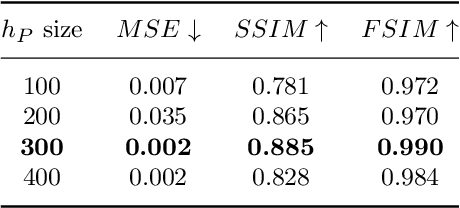
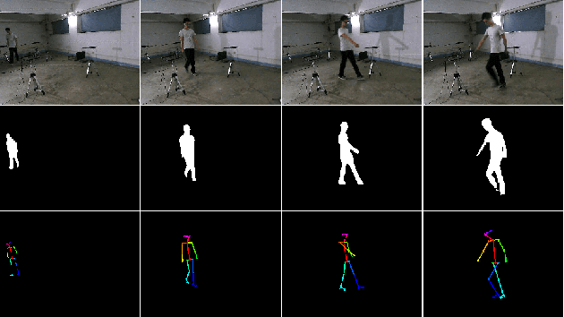
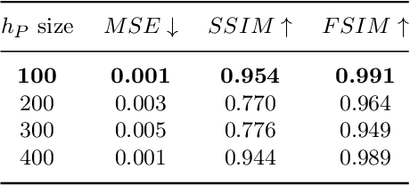
Abstract:The increasing availability of wireless access points (APs) is leading towards human sensing applications based on Wi-Fi signals as support or alternative tools to the widespread visual sensors, where the signals enable to address well-known vision-related problems such as illumination changes or occlusions. Indeed, using image synthesis techniques to translate radio frequencies to the visible spectrum can become essential to obtain otherwise unavailable visual data. This domain-to-domain translation is feasible because both objects and people affect electromagnetic waves, causing radio and optical frequencies variations. In literature, models capable of inferring radio-to-visual features mappings have gained momentum in the last few years since frequency changes can be observed in the radio domain through the channel state information (CSI) of Wi-Fi APs, enabling signal-based feature extraction, e.g., amplitude. On this account, this paper presents a novel two-branch generative neural network that effectively maps radio data into visual features, following a teacher-student design that exploits a cross-modality supervision strategy. The latter conditions signal-based features in the visual domain to completely replace visual data. Once trained, the proposed method synthesizes human silhouette and skeleton videos using exclusively Wi-Fi signals. The approach is evaluated on publicly available data, where it obtains remarkable results for both silhouette and skeleton videos generation, demonstrating the effectiveness of the proposed cross-modality supervision strategy.
Study on Transfer Learning Capabilities for Pneumonia Classification in Chest-X-Rays Image
Oct 06, 2021
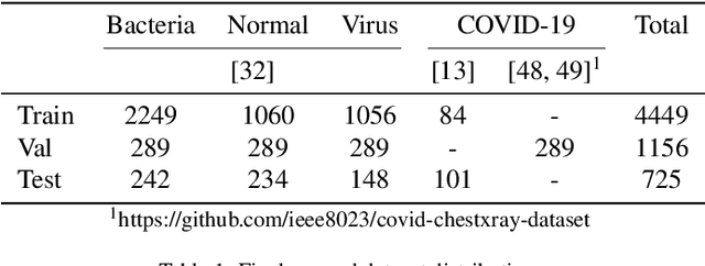
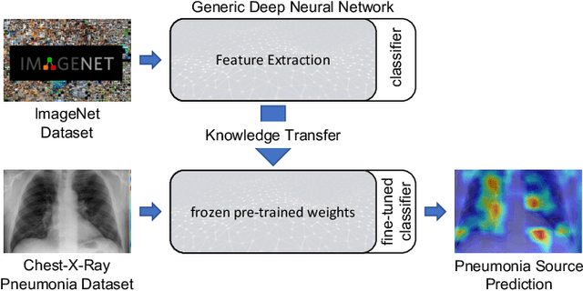
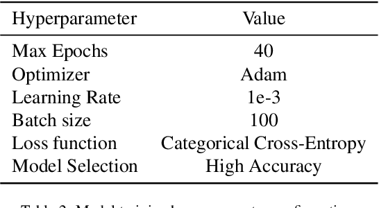
Abstract:Over the last year, the severe acute respiratory syndrome coronavirus-2 (SARS-CoV-2) and its variants have highlighted the importance of screening tools with high diagnostic accuracy for new illnesses such as COVID-19. To that regard, deep learning approaches have proven as effective solutions for pneumonia classification, especially when considering chest-x-rays images. However, this lung infection can also be caused by other viral, bacterial or fungi pathogens. Consequently, efforts are being poured toward distinguishing the infection source to help clinicians to diagnose the correct disease origin. Following this tendency, this study further explores the effectiveness of established neural network architectures on the pneumonia classification task through the transfer learning paradigm. To present a comprehensive comparison, 12 well-known ImageNet pre-trained models were fine-tuned and used to discriminate among chest-x-rays of healthy people, and those showing pneumonia symptoms derived from either a viral (i.e., generic or SARS-CoV-2) or bacterial source. Furthermore, since a common public collection distinguishing between such categories is currently not available, two distinct datasets of chest-x-rays images, describing the aforementioned sources, were combined and employed to evaluate the various architectures. The experiments were performed using a total of 6330 images split between train, validation and test sets. For all models, common classification metrics were computed (e.g., precision, f1-score) and most architectures obtained significant performances, reaching, among the others, up to 84.46% average f1-score when discriminating the 4 identified classes. Moreover, confusion matrices and activation maps computed via the Grad-CAM algorithm were also reported to present an informed discussion on the networks classifications.
SIRe-Networks: Skip Connections over Interlaced Multi-Task Learning and Residual Connections for Structure Preserving Object Classification
Oct 06, 2021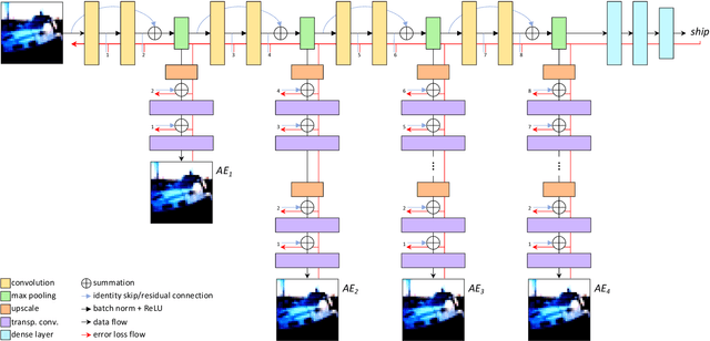
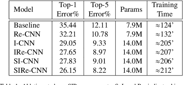
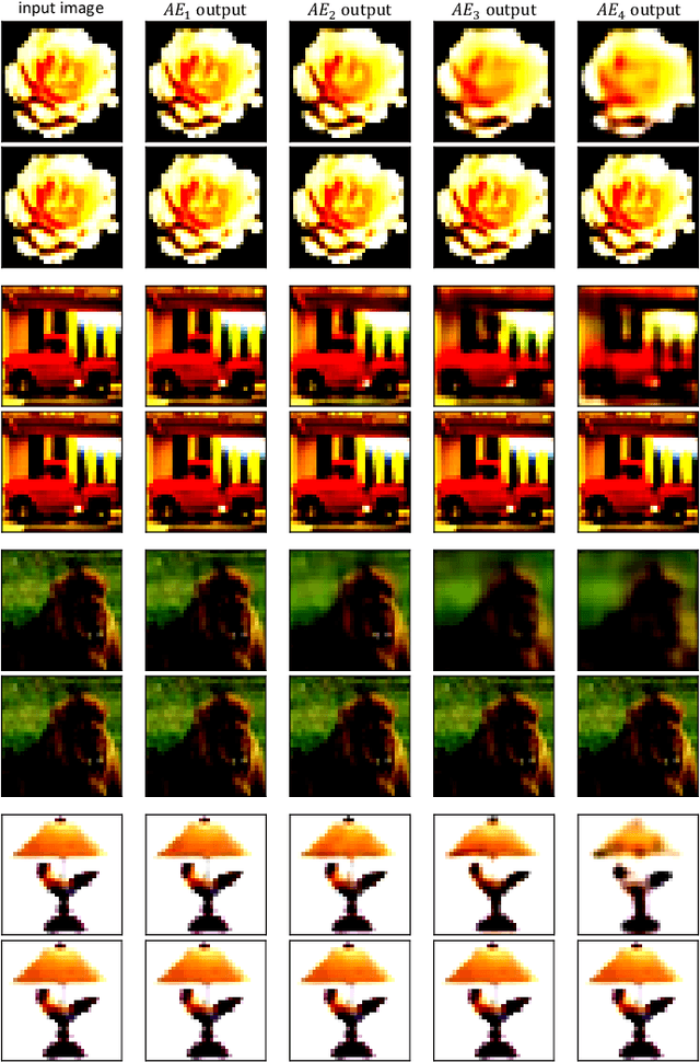
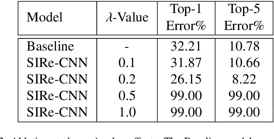
Abstract:Improving existing neural network architectures can involve several design choices such as manipulating the loss functions, employing a diverse learning strategy, exploiting gradient evolution at training time, optimizing the network hyper-parameters, or increasing the architecture depth. The latter approach is a straightforward solution, since it directly enhances the representation capabilities of a network; however, the increased depth generally incurs in the well-known vanishing gradient problem. In this paper, borrowing from different methods addressing this issue, we introduce an interlaced multi-task learning strategy, defined SIRe, to reduce the vanishing gradient in relation to the object classification task. The presented methodology directly improves a convolutional neural network (CNN) by enforcing the input image structure preservation through interlaced auto-encoders, and further refines the base network architecture by means of skip and residual connections. To validate the presented methodology, a simple CNN and various implementations of famous networks are extended via the SIRe strategy and extensively tested on the CIFAR100 dataset; where the SIRe-extended architectures achieve significantly increased performances across all models, thus confirming the presented approach effectiveness.
3D Hand Pose and Shape Estimation from RGB Images for Improved Keypoint-Based Hand-Gesture Recognition
Sep 28, 2021


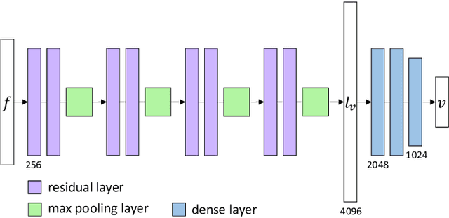
Abstract:Estimating the 3D hand pose from a 2D image is a well-studied problem and a requirement for several real-life applications such as virtual reality, augmented reality, and hand-gesture recognition. Currently, good estimations can be computed starting from single RGB images, especially when forcing the system to also consider, through a multi-task learning approach, the hand shape when the pose is determined. However, when addressing the aforementioned real-life tasks, performances can drop considerably depending on the hand representation, thus suggesting that stable descriptions are required to achieve satisfactory results. As a consequence, in this paper we present a keypoint-based end-to-end framework for the 3D hand and pose estimation, and successfully apply it to the hand-gesture recognition task as a study case. Specifically, after a pre-processing step where the images are normalized, the proposed pipeline comprises a multi-task semantic feature extractor generating 2D heatmaps and hand silhouettes from RGB images; a viewpoint encoder predicting hand and camera view parameters; a stable hand estimator producing the 3D hand pose and shape; and a loss function designed to jointly guide all of the components during the learning phase. To assess the proposed framework, tests were performed on a 3D pose and shape estimation benchmark dataset, obtaining state-of-the-art performances. What is more, the devised system was also evaluated on 2 hand-gesture recognition benchmark datasets, where the framework significantly outperforms other keypoint-based approaches; indicating that the presented method is an effective solution able to generate stable 3D estimates for the hand pose and shape.
Multiple Sclerosis Lesions Identification/Segmentation in Magnetic Resonance Imaging using Ensemble CNN and Uncertainty Classification
Aug 26, 2021
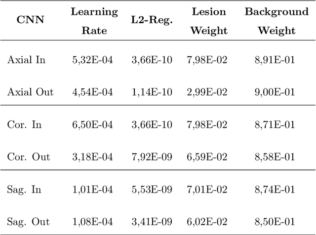


Abstract:To date, several automated strategies for identification/segmentation of Multiple Sclerosis (MS) lesions by Magnetic Resonance Imaging (MRI) have been presented which are either outperformed by human experts or, at least, whose results are well distinguishable from humans. This is due to the ambiguity originated by MRI instabilities, peculiar MS Heterogeneity and MRI unspecific nature with respect to MS. Physicians partially treat the uncertainty generated by ambiguity relying on personal radiological/clinical/anatomical background and experience. We present an automated framework for MS lesions identification/segmentation based on three pivotal concepts to better emulate human reasoning: the modeling of uncertainty; the proposal of two, separately trained, CNN, one optimized with respect to lesions themselves and the other to the environment surrounding lesions, respectively repeated for axial, coronal and sagittal directions; the ensemble of the CNN output. The proposed framework is trained, validated and tested on the 2016 MSSEG benchmark public data set from a single imaging modality, FLuid-Attenuated Inversion Recovery (FLAIR). The comparison, performed on the segmented lesions by means of most of the metrics normally used with respect to the ground-truth and the 7 human raters in MSSEG, prove that there is no significant difference between the proposed framework and the other raters. Results are also shown for the uncertainty, though a comparison with the other raters is impossible.
 Add to Chrome
 Add to Firefox
 Add to Edge