Lihong V. Wang
4KAgent: Agentic Any Image to 4K Super-Resolution
Jul 09, 2025Abstract:We present 4KAgent, a unified agentic super-resolution generalist system designed to universally upscale any image to 4K resolution (and even higher, if applied iteratively). Our system can transform images from extremely low resolutions with severe degradations, for example, highly distorted inputs at 256x256, into crystal-clear, photorealistic 4K outputs. 4KAgent comprises three core components: (1) Profiling, a module that customizes the 4KAgent pipeline based on bespoke use cases; (2) A Perception Agent, which leverages vision-language models alongside image quality assessment experts to analyze the input image and make a tailored restoration plan; and (3) A Restoration Agent, which executes the plan, following a recursive execution-reflection paradigm, guided by a quality-driven mixture-of-expert policy to select the optimal output for each step. Additionally, 4KAgent embeds a specialized face restoration pipeline, significantly enhancing facial details in portrait and selfie photos. We rigorously evaluate our 4KAgent across 11 distinct task categories encompassing a total of 26 diverse benchmarks, setting new state-of-the-art on a broad spectrum of imaging domains. Our evaluations cover natural images, portrait photos, AI-generated content, satellite imagery, fluorescence microscopy, and medical imaging like fundoscopy, ultrasound, and X-ray, demonstrating superior performance in terms of both perceptual (e.g., NIQE, MUSIQ) and fidelity (e.g., PSNR) metrics. By establishing a novel agentic paradigm for low-level vision tasks, we aim to catalyze broader interest and innovation within vision-centric autonomous agents across diverse research communities. We will release all the code, models, and results at: https://4kagent.github.io.
Rotational ultrasound and photoacoustic tomography of the human body
Apr 22, 2025Abstract:Imaging the human body's morphological and angiographic information is essential for diagnosing, monitoring, and treating medical conditions. Ultrasonography performs the morphological assessment of the soft tissue based on acoustic impedance variations, whereas photoacoustic tomography (PAT) can visualize blood vessels based on intrinsic hemoglobin absorption. Three-dimensional (3D) panoramic imaging of the vasculature is generally not practical in conventional ultrasonography with limited field-of-view (FOV) probes, and PAT does not provide sufficient scattering-based soft tissue morphological contrast. Complementing each other, fast panoramic rotational ultrasound tomography (RUST) and PAT are integrated for hybrid rotational ultrasound and photoacoustic tomography (RUS-PAT), which obtains 3D ultrasound structural and PAT angiographic images of the human body quasi-simultaneously. The RUST functionality is achieved in a cost-effective manner using a single-element ultrasonic transducer for ultrasound transmission and rotating arc-shaped arrays for 3D panoramic detection. RUST is superior to conventional ultrasonography, which either has a limited FOV with a linear array or is high-cost with a hemispherical array that requires both transmission and receiving. By switching the acoustic source to a light source, the system is conveniently converted to PAT mode to acquire angiographic images in the same region. Using RUS-PAT, we have successfully imaged the human head, breast, hand, and foot with a 10 cm diameter FOV, submillimeter isotropic resolution, and 10 s imaging time for each modality. The 3D RUS-PAT is a powerful tool for high-speed, 3D, dual-contrast imaging of the human body with potential for rapid clinical translation.
Functional photoacoustic noninvasive Doppler angiography in humans
Jun 22, 2024Abstract:Optical imaging of blood flow yields critical functional insights into the circulatory system, but its clinical implementation has typically been limited to shallow depths (~1 millimeter) due to light scattering in biological tissue. Here, we present photoacoustic noninvasive Doppler angiography (PANDA) for deep blood flow imaging. PANDA synergizes the photoacoustic and Doppler effects to generate color Doppler velocity and power Doppler blood flow maps of the vascular lumen. Our results demonstrate PANDA's ability to measure blood flow in vivo up to one centimeter in depth, marking approximately an order of magnitude improvement over existing high-resolution pure optical modalities. PANDA enhances photoacoustic flow imaging by increasing depth and enabling cross-sectional blood vessel imaging. We also showcase PANDA's clinical feasibility through three-dimensional imaging of blood flow in healthy subjects and a patient with varicose veins. By integrating the imaging system onto a mobile platform, we have designed PANDA to be a portable modality that is primed for expedient clinical translation. PANDA offers noninvasive, single modality imaging of hemoglobin and blood flow with three-dimensional capability, facilitating comprehensive assessment of deep vascular dynamics in humans.
Score-Based Diffusion Models for Photoacoustic Tomography Image Reconstruction
Mar 30, 2024


Abstract:Photoacoustic tomography (PAT) is a rapidly-evolving medical imaging modality that combines optical absorption contrast with ultrasound imaging depth. One challenge in PAT is image reconstruction with inadequate acoustic signals due to limited sensor coverage or due to the density of the transducer array. Such cases call for solving an ill-posed inverse reconstruction problem. In this work, we use score-based diffusion models to solve the inverse problem of reconstructing an image from limited PAT measurements. The proposed approach allows us to incorporate an expressive prior learned by a diffusion model on simulated vessel structures while still being robust to varying transducer sparsity conditions.
* 5 pages
Single-shot 3D photoacoustic computed tomography with a densely packed array for transcranial functional imaging
Jun 26, 2023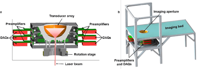

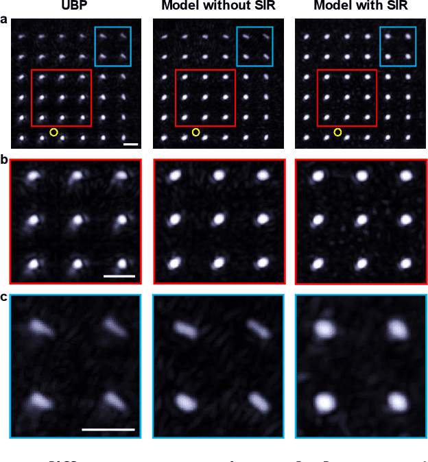
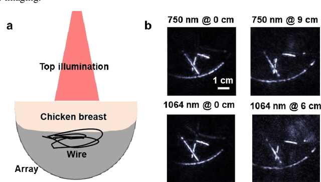
Abstract:Photoacoustic computed tomography (PACT) is emerging as a new technique for functional brain imaging, primarily due to its capabilities in label-free hemodynamic imaging. Despite its potential, the transcranial application of PACT has encountered hurdles, such as acoustic attenuations and distortions by the skull and limited light penetration through the skull. To overcome these challenges, we have engineered a PACT system that features a densely packed hemispherical ultrasonic transducer array with 3072 channels, operating at a central frequency of 1 MHz. This system allows for single-shot 3D imaging at a rate equal to the laser repetition rate, such as 20 Hz. We have achieved a single-shot light penetration depth of approximately 9 cm in chicken breast tissue utilizing a 750 nm laser (withstanding 3295-fold light attenuation and still retaining an SNR of 74) and successfully performed transcranial imaging through an ex vivo human skull using a 1064 nm laser. Moreover, we have proven the capacity of our system to perform single-shot 3D PACT imaging in both tissue phantoms and human subjects. These results suggest that our PACT system is poised to unlock potential for real-time, in vivo transcranial functional imaging in humans.
Quantification of cervical elasticity during pregnancy based on transvaginal ultrasound imaging and stress measurement
Mar 10, 2023Abstract:Objective: Strain elastography and shear wave elastography are two commonly used methods to quantify cervical elasticity; however, they have limitations. Strain elastography is effective in showing tissue elasticity distribution in a single image, but the absence of stress information causes difficulty in comparing the results acquired from different imaging sessions. Shear wave elastography is effective in measuring shear wave speed (an intrinsic tissue property correlated with elasticity) in relatively homogeneous tissue, such as in the liver. However, for inhomogeneous tissue in the cervix, the shear wave speed measurement is less robust. To overcome these limitations, we develop a quantitative cervical elastography system by adding a stress sensor to an ultrasound imaging system. Methods: In an imaging session for quantitative cervical elastography, we use the transvaginal ultrasound imaging system to record B-mode images of the cervix showing its deformation and use the stress sensor to record the probe-surface stress simultaneously. We develop a correlation-based automatic feature tracking algorithm to quantify the deformation, from which the strain is quantified. After each imaging session, we calibrate the stress sensor and transform its measurement to true stress. Applying a linear regression to the stress and strain, we obtain an approximation of the cervical Young's modulus. Results: We validate the accuracy and robustness of this elastography system using phantom experiments. Applying this system to pregnant participants, we observe significant softening of the cervix during pregnancy (p-value < 0.001) with the cervical Young's modulus decreasing 3.95% per week. We estimate that geometric mean values of cervical Young's moduli during the first (11 to 13 weeks), second, and third trimesters are 13.07 kPa, 7.59 kPa, and 4.40 kPa, respectively.
Photoacoustic vector tomography for deep hemodynamic imaging
Sep 19, 2022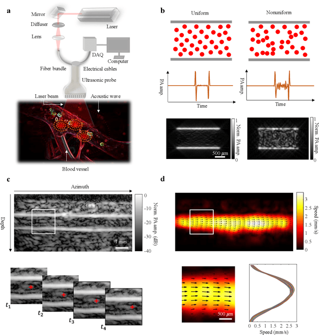
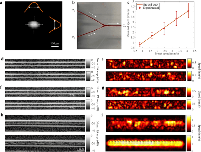
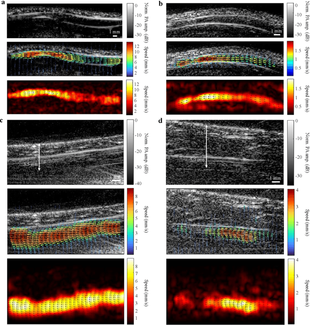
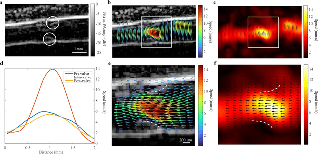
Abstract:Non-invasive imaging of deep blood vessels for mapping hemodynamics remains an open quest in biomedical optical imaging. Although pure optical imaging techniques offer rich optical contrast of blood and have been reported to measure blood flow, they are generally limited to surface imaging within the optical diffusion limit of about one millimeter. Herein, we present photoacoustic vector tomography (PAVT), breaking through the optical diffusion limit to image deep blood flow with speed and direction quantification. PAVT synergizes the spatial heterogeneity of blood and the photoacoustic contrast; it compiles successive single-shot, wide-field photoacoustic images to directly visualize the frame-to-frame propagation of the blood with pixel-wise flow velocity estimation. We demonstrated in vivo that PAVT allows hemodynamic quantification of deep blood vessels at five times the optical diffusion limit (more than five millimeters), leading to vector mapping of blood flow in humans. By offering the capability for deep hemodynamic imaging with optical contrast, PAVT may become a powerful tool for monitoring and diagnosing vascular diseases and mapping circulatory system function.
Transcranial photoacoustic computed tomography of human brain function
Jun 01, 2022

Abstract:Herein we report the first in-human transcranial imaging of brain function using photoacoustic computed tomography. Functional responses to benchmark motor tasks were imaged on both the skull-less and the skull-intact hemispheres of a hemicraniectomy patient. The observed brain responses in these preliminary results demonstrate the potential of photoacoustic computed tomography for achieving transcranial functional imaging.
 Add to Chrome
 Add to Firefox
 Add to Edge