Joshua Olick-Gibson
Functional photoacoustic noninvasive Doppler angiography in humans
Jun 22, 2024Abstract:Optical imaging of blood flow yields critical functional insights into the circulatory system, but its clinical implementation has typically been limited to shallow depths (~1 millimeter) due to light scattering in biological tissue. Here, we present photoacoustic noninvasive Doppler angiography (PANDA) for deep blood flow imaging. PANDA synergizes the photoacoustic and Doppler effects to generate color Doppler velocity and power Doppler blood flow maps of the vascular lumen. Our results demonstrate PANDA's ability to measure blood flow in vivo up to one centimeter in depth, marking approximately an order of magnitude improvement over existing high-resolution pure optical modalities. PANDA enhances photoacoustic flow imaging by increasing depth and enabling cross-sectional blood vessel imaging. We also showcase PANDA's clinical feasibility through three-dimensional imaging of blood flow in healthy subjects and a patient with varicose veins. By integrating the imaging system onto a mobile platform, we have designed PANDA to be a portable modality that is primed for expedient clinical translation. PANDA offers noninvasive, single modality imaging of hemoglobin and blood flow with three-dimensional capability, facilitating comprehensive assessment of deep vascular dynamics in humans.
Photoacoustic vector tomography for deep hemodynamic imaging
Sep 19, 2022


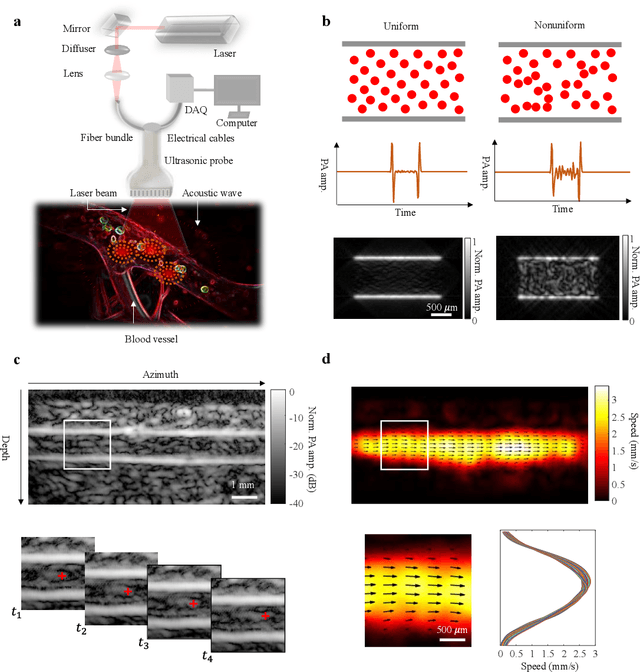
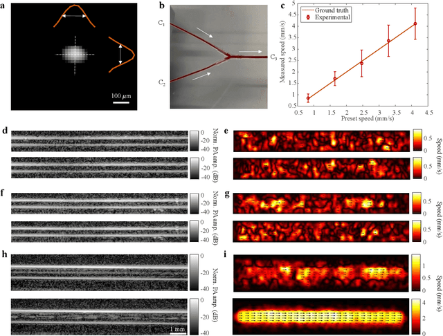
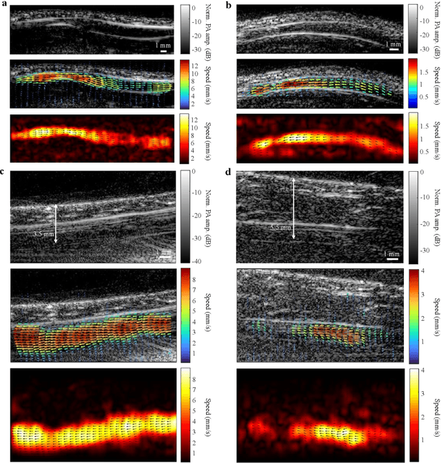
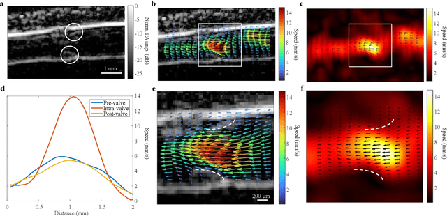
Abstract:Non-invasive imaging of deep blood vessels for mapping hemodynamics remains an open quest in biomedical optical imaging. Although pure optical imaging techniques offer rich optical contrast of blood and have been reported to measure blood flow, they are generally limited to surface imaging within the optical diffusion limit of about one millimeter. Herein, we present photoacoustic vector tomography (PAVT), breaking through the optical diffusion limit to image deep blood flow with speed and direction quantification. PAVT synergizes the spatial heterogeneity of blood and the photoacoustic contrast; it compiles successive single-shot, wide-field photoacoustic images to directly visualize the frame-to-frame propagation of the blood with pixel-wise flow velocity estimation. We demonstrated in vivo that PAVT allows hemodynamic quantification of deep blood vessels at five times the optical diffusion limit (more than five millimeters), leading to vector mapping of blood flow in humans. By offering the capability for deep hemodynamic imaging with optical contrast, PAVT may become a powerful tool for monitoring and diagnosing vascular diseases and mapping circulatory system function.
 Add to Chrome
 Add to Firefox
 Add to Edge