 Add to Chrome
 Add to Firefox
 Add to Edge
Rotational ultrasound and photoacoustic tomography of the human body
Apr 22, 2025Imaging the human body's morphological and angiographic information is essential for diagnosing, monitoring, and treating medical conditions. Ultrasonography performs the morphological assessment of the soft tissue based on acoustic impedance variations, whereas photoacoustic tomography (PAT) can visualize blood vessels based on intrinsic hemoglobin absorption. Three-dimensional (3D) panoramic imaging of the vasculature is generally not practical in conventional ultrasonography with limited field-of-view (FOV) probes, and PAT does not provide sufficient scattering-based soft tissue morphological contrast. Complementing each other, fast panoramic rotational ultrasound tomography (RUST) and PAT are integrated for hybrid rotational ultrasound and photoacoustic tomography (RUS-PAT), which obtains 3D ultrasound structural and PAT angiographic images of the human body quasi-simultaneously. The RUST functionality is achieved in a cost-effective manner using a single-element ultrasonic transducer for ultrasound transmission and rotating arc-shaped arrays for 3D panoramic detection. RUST is superior to conventional ultrasonography, which either has a limited FOV with a linear array or is high-cost with a hemispherical array that requires both transmission and receiving. By switching the acoustic source to a light source, the system is conveniently converted to PAT mode to acquire angiographic images in the same region. Using RUS-PAT, we have successfully imaged the human head, breast, hand, and foot with a 10 cm diameter FOV, submillimeter isotropic resolution, and 10 s imaging time for each modality. The 3D RUS-PAT is a powerful tool for high-speed, 3D, dual-contrast imaging of the human body with potential for rapid clinical translation.
Functional photoacoustic noninvasive Doppler angiography in humans
Jun 22, 2024Optical imaging of blood flow yields critical functional insights into the circulatory system, but its clinical implementation has typically been limited to shallow depths (~1 millimeter) due to light scattering in biological tissue. Here, we present photoacoustic noninvasive Doppler angiography (PANDA) for deep blood flow imaging. PANDA synergizes the photoacoustic and Doppler effects to generate color Doppler velocity and power Doppler blood flow maps of the vascular lumen. Our results demonstrate PANDA's ability to measure blood flow in vivo up to one centimeter in depth, marking approximately an order of magnitude improvement over existing high-resolution pure optical modalities. PANDA enhances photoacoustic flow imaging by increasing depth and enabling cross-sectional blood vessel imaging. We also showcase PANDA's clinical feasibility through three-dimensional imaging of blood flow in healthy subjects and a patient with varicose veins. By integrating the imaging system onto a mobile platform, we have designed PANDA to be a portable modality that is primed for expedient clinical translation. PANDA offers noninvasive, single modality imaging of hemoglobin and blood flow with three-dimensional capability, facilitating comprehensive assessment of deep vascular dynamics in humans.
Transcranial photoacoustic computed tomography of human brain function
Jun 01, 2022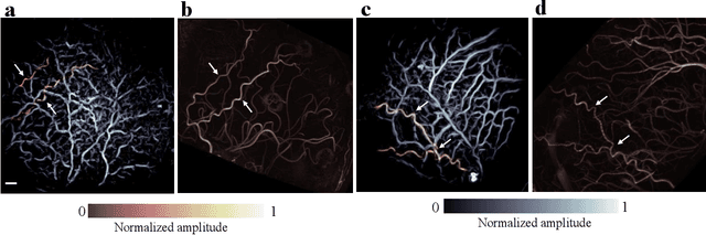
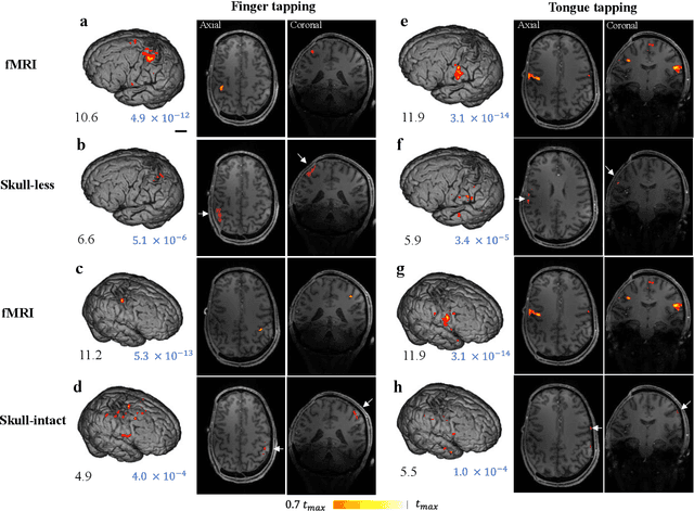
Herein we report the first in-human transcranial imaging of brain function using photoacoustic computed tomography. Functional responses to benchmark motor tasks were imaged on both the skull-less and the skull-intact hemispheres of a hemicraniectomy patient. The observed brain responses in these preliminary results demonstrate the potential of photoacoustic computed tomography for achieving transcranial functional imaging.