Ashutosh Jadhav
Towards Automatic Prediction of Outcome in Treatment of Cerebral Aneurysms
Nov 18, 2022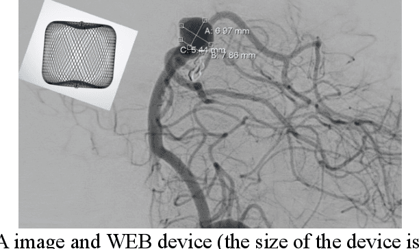
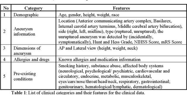

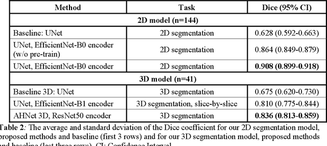
Abstract:Intrasaccular flow disruptors treat cerebral aneurysms by diverting the blood flow from the aneurysm sac. Residual flow into the sac after the intervention is a failure that could be due to the use of an undersized device, or to vascular anatomy and clinical condition of the patient. We report a machine learning model based on over 100 clinical and imaging features that predict the outcome of wide-neck bifurcation aneurysm treatment with an intravascular embolization device. We combine clinical features with a diverse set of common and novel imaging measurements within a random forest model. We also develop neural network segmentation algorithms in 2D and 3D to contour the sac in angiographic images and automatically calculate the imaging features. These deliver 90% overlap with manual contouring in 2D and 83% in 3D. Our predictive model classifies complete vs. partial occlusion outcomes with an accuracy of 75.31%, and weighted F1-score of 0.74.
* 10 pages
Extracting and Learning Fine-Grained Labels from Chest Radiographs
Nov 18, 2020


Abstract:Chest radiographs are the most common diagnostic exam in emergency rooms and intensive care units today. Recently, a number of researchers have begun working on large chest X-ray datasets to develop deep learning models for recognition of a handful of coarse finding classes such as opacities, masses and nodules. In this paper, we focus on extracting and learning fine-grained labels for chest X-ray images. Specifically we develop a new method of extracting fine-grained labels from radiology reports by combining vocabulary-driven concept extraction with phrasal grouping in dependency parse trees for association of modifiers with findings. A total of 457 fine-grained labels depicting the largest spectrum of findings to date were selected and sufficiently large datasets acquired to train a new deep learning model designed for fine-grained classification. We show results that indicate a highly accurate label extraction process and a reliable learning of fine-grained labels. The resulting network, to our knowledge, is the first to recognize fine-grained descriptions of findings in images covering over nine modifiers including laterality, location, severity, size and appearance.
Receptivity of an AI Cognitive Assistant by the Radiology Community: A Report on Data Collected at RSNA
Sep 13, 2020


Abstract:Due to advances in machine learning and artificial intelligence (AI), a new role is emerging for machines as intelligent assistants to radiologists in their clinical workflows. But what systematic clinical thought processes are these machines using? Are they similar enough to those of radiologists to be trusted as assistants? A live demonstration of such a technology was conducted at the 2016 Scientific Assembly and Annual Meeting of the Radiological Society of North America (RSNA). The demonstration was presented in the form of a question-answering system that took a radiology multiple choice question and a medical image as inputs. The AI system then demonstrated a cognitive workflow, involving text analysis, image analysis, and reasoning, to process the question and generate the most probable answer. A post demonstration survey was made available to the participants who experienced the demo and tested the question answering system. Of the reported 54,037 meeting registrants, 2,927 visited the demonstration booth, 1,991 experienced the demo, and 1,025 completed a post-demonstration survey. In this paper, the methodology of the survey is shown and a summary of its results are presented. The results of the survey show a very high level of receptiveness to cognitive computing technology and artificial intelligence among radiologists.
Chest X-ray Report Generation through Fine-Grained Label Learning
Jul 27, 2020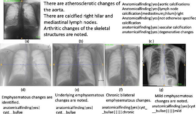

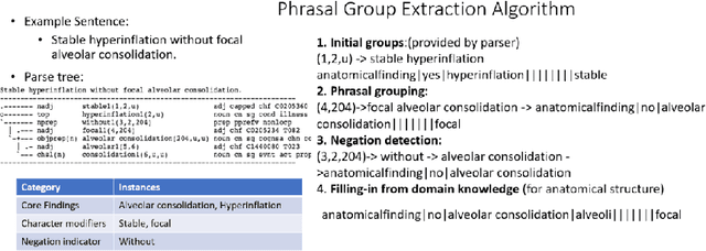

Abstract:Obtaining automated preliminary read reports for common exams such as chest X-rays will expedite clinical workflows and improve operational efficiencies in hospitals. However, the quality of reports generated by current automated approaches is not yet clinically acceptable as they cannot ensure the correct detection of a broad spectrum of radiographic findings nor describe them accurately in terms of laterality, anatomical location, severity, etc. In this work, we present a domain-aware automatic chest X-ray radiology report generation algorithm that learns fine-grained description of findings from images and uses their pattern of occurrences to retrieve and customize similar reports from a large report database. We also develop an automatic labeling algorithm for assigning such descriptors to images and build a novel deep learning network that recognizes both coarse and fine-grained descriptions of findings. The resulting report generation algorithm significantly outperforms the state of the art using established score metrics.
 Add to Chrome
 Add to Firefox
 Add to Edge