Ali Gooya
CardioMorphNet: Cardiac Motion Prediction Using a Shape-Guided Bayesian Recurrent Deep Network
Aug 28, 2025Abstract:Accurate cardiac motion estimation from cine cardiac magnetic resonance (CMR) images is vital for assessing cardiac function and detecting its abnormalities. Existing methods often struggle to capture heart motion accurately because they rely on intensity-based image registration similarity losses that may overlook cardiac anatomical regions. To address this, we propose CardioMorphNet, a recurrent Bayesian deep learning framework for 3D cardiac shape-guided deformable registration using short-axis (SAX) CMR images. It employs a recurrent variational autoencoder to model spatio-temporal dependencies over the cardiac cycle and two posterior models for bi-ventricular segmentation and motion estimation. The derived loss function from the Bayesian formulation guides the framework to focus on anatomical regions by recursively registering segmentation maps without using intensity-based image registration similarity loss, while leveraging sequential SAX volumes and spatio-temporal features. The Bayesian modelling also enables computation of uncertainty maps for the estimated motion fields. Validated on the UK Biobank dataset by comparing warped mask shapes with ground truth masks, CardioMorphNet demonstrates superior performance in cardiac motion estimation, outperforming state-of-the-art methods. Uncertainty assessment shows that it also yields lower uncertainty values for estimated motion fields in the cardiac region compared with other probabilistic-based cardiac registration methods, indicating higher confidence in its predictions.
FractMorph: A Fractional Fourier-Based Multi-Domain Transformer for Deformable Image Registration
Aug 17, 2025Abstract:Deformable image registration (DIR) is a crucial and challenging technique for aligning anatomical structures in medical images and is widely applied in diverse clinical applications. However, existing approaches often struggle to capture fine-grained local deformations and large-scale global deformations simultaneously within a unified framework. We present FractMorph, a novel 3D dual-parallel transformer-based architecture that enhances cross-image feature matching through multi-domain fractional Fourier transform (FrFT) branches. Each Fractional Cross-Attention (FCA) block applies parallel FrFTs at fractional angles of 0{\deg}, 45{\deg}, 90{\deg}, along with a log-magnitude branch, to effectively extract local, semi-global, and global features at the same time. These features are fused via cross-attention between the fixed and moving image streams. A lightweight U-Net style network then predicts a dense deformation field from the transformer-enriched features. On the ACDC cardiac MRI dataset, FractMorph achieves state-of-the-art performance with an overall Dice Similarity Coefficient (DSC) of 86.45%, an average per-structure DSC of 75.15%, and a 95th-percentile Hausdorff distance (HD95) of 1.54 mm on our data split. We also introduce FractMorph-Light, a lightweight variant of our model with only 29.6M parameters, which maintains the superior accuracy of the main model while using approximately half the memory. Our results demonstrate that multi-domain spectral-spatial attention in transformers can robustly and efficiently model complex non-rigid deformations in medical images using a single end-to-end network, without the need for scenario-specific tuning or hierarchical multi-scale networks. The source code of our implementation is available at https://github.com/shayankebriti/FractMorph.
Deep-Motion-Net: GNN-based volumetric organ shape reconstruction from single-view 2D projections
Jul 09, 2024


Abstract:We propose Deep-Motion-Net: an end-to-end graph neural network (GNN) architecture that enables 3D (volumetric) organ shape reconstruction from a single in-treatment kV planar X-ray image acquired at any arbitrary projection angle. Estimating and compensating for true anatomical motion during radiotherapy is essential for improving the delivery of planned radiation dose to target volumes while sparing organs-at-risk, and thereby improving the therapeutic ratio. Achieving this using only limited imaging available during irradiation and without the use of surrogate signals or invasive fiducial markers is attractive. The proposed model learns the mesh regression from a patient-specific template and deep features extracted from kV images at arbitrary projection angles. A 2D-CNN encoder extracts image features, and four feature pooling networks fuse these features to the 3D template organ mesh. A ResNet-based graph attention network then deforms the feature-encoded mesh. The model is trained using synthetically generated organ motion instances and corresponding kV images. The latter is generated by deforming a reference CT volume aligned with the template mesh, creating digitally reconstructed radiographs (DRRs) at required projection angles, and DRR-to-kV style transferring with a conditional CycleGAN model. The overall framework was tested quantitatively on synthetic respiratory motion scenarios and qualitatively on in-treatment images acquired over full scan series for liver cancer patients. Overall mean prediction errors for synthetic motion test datasets were 0.16$\pm$0.13 mm, 0.18$\pm$0.19 mm, 0.22$\pm$0.34 mm, and 0.12$\pm$0.11 mm. Mean peak prediction errors were 1.39 mm, 1.99 mm, 3.29 mm, and 1.16 mm.
An End-to-End Deep Learning Generative Framework for Refinable Shape Matching and Generation
Mar 10, 2024


Abstract:Generative modelling for shapes is a prerequisite for In-Silico Clinical Trials (ISCTs), which aim to cost-effectively validate medical device interventions using synthetic anatomical shapes, often represented as 3D surface meshes. However, constructing AI models to generate shapes closely resembling the real mesh samples is challenging due to variable vertex counts, connectivities, and the lack of dense vertex-wise correspondences across the training data. Employing graph representations for meshes, we develop a novel unsupervised geometric deep-learning model to establish refinable shape correspondences in a latent space, construct a population-derived atlas and generate realistic synthetic shapes. We additionally extend our proposed base model to a joint shape generative-clustering multi-atlas framework to incorporate further variability and preserve more details in the generated shapes. Experimental results using liver and left-ventricular models demonstrate the approach's applicability to computational medicine, highlighting its suitability for ISCTs through a comparative analysis.
Weakly-supervised learning for image-based classification of primary melanomas into genomic immune subgroups
Feb 23, 2022

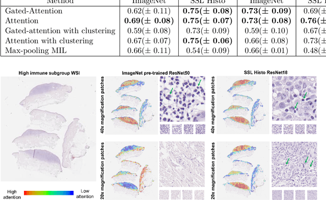

Abstract:Determining early-stage prognostic markers and stratifying patients for effective treatment are two key challenges for improving outcomes for melanoma patients. Previous studies have used tumour transcriptome data to stratify patients into immune subgroups, which were associated with differential melanoma specific survival and potential treatment strategies. However, acquiring transcriptome data is a time-consuming and costly process. Moreover, it is not routinely used in the current clinical workflow. Here we attempt to overcome this by developing deep learning models to classify gigapixel H&E stained pathology slides, which are well established in clinical workflows, into these immune subgroups. Previous subtyping approaches have employed supervised learning which requires fully annotated data, or have only examined single genetic mutations in melanoma patients. We leverage a multiple-instance learning approach, which only requires slide-level labels and uses an attention mechanism to highlight regions of high importance to the classification. Moreover, we show that pathology-specific self-supervised models generate better representations compared to pathology-agnostic models for improving our model performance, achieving a mean AUC of 0.76 for classifying histopathology images as high or low immune subgroups. We anticipate that this method may allow us to find new biomarkers of high importance and could act as a tool for clinicians to infer the immune landscape of tumours and stratify patients, without needing to carry out additional expensive genetic tests.
Nuclear Instance Segmentation using a Proposal-Free Spatially Aware Deep Learning Framework
Aug 27, 2019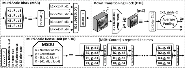
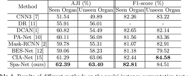

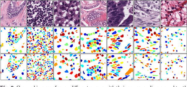
Abstract:Nuclear segmentation in histology images is a challenging task due to significant variations in the shape and appearance of nuclei. One of the main hurdles in nuclear instance segmentation is overlapping nuclei where a smart algorithm is needed to separate each nucleus. In this paper, we introduce a proposal-free deep learning based framework to address these challenges. To this end, we propose a spatially-aware network (SpaNet) to capture spatial information in a multi-scale manner. A dual-head variation of the SpaNet is first utilized to predict the pixel-wise segmentation and centroid detection maps of nuclei. Based on these outputs, a single-head SpaNet predicts the positional information related to each nucleus instance. Spectral clustering method is applied on the output of the last SpaNet, which utilizes the nuclear mask and the Gaussian-like detection map for determining the connected components and associated cluster identifiers, respectively. The output of the clustering method is the final nuclear instance segmentation mask. We applied our method on a publicly available multi-organ data set and achieved state-of-the-art performance for nuclear segmentation.
High Throughput Computation of Reference Ranges of Biventricular Cardiac Function on the UK Biobank Population Cohort
Jan 10, 2019


Abstract:The exploitation of large-scale population data has the potential to improve healthcare by discovering and understanding patterns and trends within this data. To enable high throughput analysis of cardiac imaging data automatically, a pipeline should comprise quality monitoring of the input images, segmentation of the cardiac structures, assessment of the segmentation quality, and parsing of cardiac functional indexes. We present a fully automatic, high throughput image parsing workflow for the analysis of cardiac MR images, and test its performance on the UK Biobank (UKB) cardiac dataset. The proposed pipeline is capable of performing end-to-end image processing including: data organisation, image quality assessment, shape model initialisation, segmentation, segmentation quality assessment, and functional parameter computation; all without any user interaction. To the best of our knowledge,this is the first paper tackling the fully automatic 3D analysis of the UKB population study, providing reference ranges for all key cardiovascular functional indexes, from both left and right ventricles of the heart. We tested our workflow on a reference cohort of 800 healthy subjects for which manual delineations, and reference functional indexes exist. Our results show statistically significant agreement between the manually obtained reference indexes, and those automatically computed using our framework.
Automatic Assessment of Full Left Ventricular Coverage in Cardiac Cine Magnetic Resonance Imaging with Fisher-Discriminative 3D CNN
Nov 08, 2018


Abstract:Cardiac magnetic resonance (CMR) images play a growing role in the diagnostic imaging of cardiovascular diseases. Full coverage of the left ventricle (LV), from base to apex, is a basic criterion for CMR image quality and necessary for accurate measurement of cardiac volume and functional assessment. Incomplete coverage of the LV is identified through visual inspection, which is time-consuming and usually done retrospectively in the assessment of large imaging cohorts. This paper proposes a novel automatic method for determining LV coverage from CMR images by using Fisher-discriminative three-dimensional (FD3D) convolutional neural networks (CNNs). In contrast to our previous method employing 2D CNNs, this approach utilizes spatial contextual information in CMR volumes, extracts more representative high-level features and enhances the discriminative capacity of the baseline 2D CNN learning framework, thus achieving superior detection accuracy. A two-stage framework is proposed to identify missing basal and apical slices in measurements of CMR volume. First, the FD3D CNN extracts high-level features from the CMR stacks. These image representations are then used to detect the missing basal and apical slices. Compared to the traditional 3D CNN strategy, the proposed FD3D CNN minimizes within-class scatter and maximizes between-class scatter. We performed extensive experiments to validate the proposed method on more than 5,000 independent volumetric CMR scans from the UK Biobank study, achieving low error rates for missing basal/apical slice detection (4.9\%/4.6\%). The proposed method can also be adopted for assessing LV coverage for other types of CMR image data.
Leveraging Transfer Learning for Segmenting Lesions and their Attributes in Dermoscopy Images
Sep 23, 2018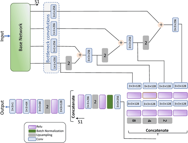

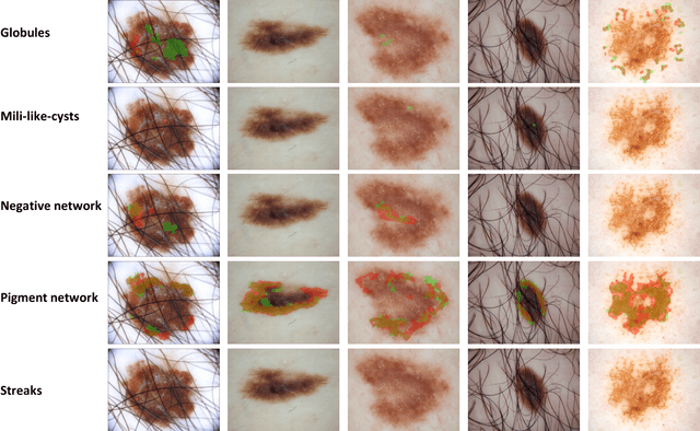

Abstract:Computer-aided diagnosis systems for classification of different type of skin lesions have been an active field of research in recent decades. It has been shown that introducing lesions and their attributes masks into lesion classification pipeline can greatly improve the performance. In this paper, we propose a framework by incorporating transfer learning for segmenting lesions and their attributes based on the convolutional neural networks. The proposed framework is inspired by the well-known UNet architecture. It utilizes a variety of pre-trained networks in the encoding path and generates the prediction map by combining multi-scale information in decoding path using a pyramid pooling manner. To circumvent the lack of training data and increase the proposed model generalization, an extensive set of novel augmentation routines have been applied during the training of the network. Moreover, for each task of lesion and attribute segmentation, a specific loss function has been designed to obviate the training phase difficulties. Finally, the prediction for each task is generated by ensembling the outputs from different models. The proposed approach achieves promising results on the cross-validation experiments on the ISIC2018- Task1 and Task2 data sets.
Nuclei Detection Using Mixture Density Networks
Aug 22, 2018


Abstract:Nuclei detection is an important task in the histology domain as it is a main step toward further analysis such as cell counting, cell segmentation, study of cell connections, etc. This is a challenging task due to the complex texture of histology image, variation in shape, and touching cells. To tackle these hurdles, many approaches have been proposed in the literature where deep learning methods stand on top in terms of performance. Hence, in this paper, we propose a novel framework for nuclei detection based on Mixture Density Networks (MDNs). These networks are suitable to map a single input to several possible outputs and we utilize this property to detect multiple seeds in a single image patch. A new modified form of a cost function is proposed for training and handling patches with missing nuclei. The probability maps of the nuclei in the individual patches are next combined to generate the final image-wide result. The experimental results show the state-of-the-art performance on complex colorectal adenocarcinoma dataset.
 Add to Chrome
 Add to Firefox
 Add to Edge