Rahman Attar
XAttn-BMD: Multimodal Deep Learning with Cross-Attention for Femoral Neck Bone Mineral Density Estimation
Nov 18, 2025Abstract:Poor bone health is a significant public health concern, and low bone mineral density (BMD) leads to an increased fracture risk, a key feature of osteoporosis. We present XAttn-BMD (Cross-Attention BMD), a multimodal deep learning framework that predicts femoral neck BMD from hip X-ray images and structured clinical metadata. It utilizes a novel bidirectional cross-attention mechanism to dynamically integrate image and metadata features for cross-modal mutual reinforcement. A Weighted Smooth L1 loss is tailored to address BMD imbalance and prioritize clinically significant cases. Extensive experiments on the data from the Hertfordshire Cohort Study show that our model outperforms the baseline models in regression generalization and robustness. Ablation studies confirm the effectiveness of both cross-attention fusion and the customized loss function. Experimental results show that the integration of multimodal data via cross-attention outperforms naive feature concatenation without cross-attention, reducing MSE by 16.7%, MAE by 6.03%, and increasing the R2 score by 16.4%, highlighting the effectiveness of the approach for femoral neck BMD estimation. Furthermore, screening performance was evaluated using binary classification at clinically relevant femoral neck BMD thresholds, demonstrating the model's potential in real-world scenarios.
3D Cardiac Shape Prediction with Deep Neural Networks: Simultaneous Use of Images and Patient Metadata
Jul 02, 2019


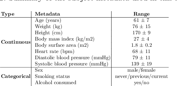
Abstract:Large prospective epidemiological studies acquire cardiovascular magnetic resonance (CMR) images for pre-symptomatic populations and follow these over time. To support this approach, fully automatic large-scale 3D analysis is essential. In this work, we propose a novel deep neural network using both CMR images and patient metadata to directly predict cardiac shape parameters. The proposed method uses the promising ability of statistical shape models to simplify shape complexity and variability together with the advantages of convolutional neural networks for the extraction of solid visual features. To the best of our knowledge, this is the first work that uses such an approach for 3D cardiac shape prediction. We validated our proposed CMR analytics method against a reference cohort containing 500 3D shapes of the cardiac ventricles. Our results show broadly significant agreement with the reference shapes in terms of the estimated volume of the cardiac ventricles, myocardial mass, 3D Dice, and mean and Hausdorff distance.
High Throughput Computation of Reference Ranges of Biventricular Cardiac Function on the UK Biobank Population Cohort
Jan 10, 2019


Abstract:The exploitation of large-scale population data has the potential to improve healthcare by discovering and understanding patterns and trends within this data. To enable high throughput analysis of cardiac imaging data automatically, a pipeline should comprise quality monitoring of the input images, segmentation of the cardiac structures, assessment of the segmentation quality, and parsing of cardiac functional indexes. We present a fully automatic, high throughput image parsing workflow for the analysis of cardiac MR images, and test its performance on the UK Biobank (UKB) cardiac dataset. The proposed pipeline is capable of performing end-to-end image processing including: data organisation, image quality assessment, shape model initialisation, segmentation, segmentation quality assessment, and functional parameter computation; all without any user interaction. To the best of our knowledge,this is the first paper tackling the fully automatic 3D analysis of the UKB population study, providing reference ranges for all key cardiovascular functional indexes, from both left and right ventricles of the heart. We tested our workflow on a reference cohort of 800 healthy subjects for which manual delineations, and reference functional indexes exist. Our results show statistically significant agreement between the manually obtained reference indexes, and those automatically computed using our framework.
2D Reconstruction of Small Intestine's Interior Wall
Mar 15, 2018


Abstract:Examining and interpreting of a large number of wireless endoscopic images from the gastrointestinal tract is a tiresome task for physicians. A practical solution is to automatically construct a two dimensional representation of the gastrointestinal tract for easy inspection. However, little has been done on wireless endoscopic image stitching, let alone systematic investigation. The proposed new wireless endoscopic image stitching method consists of two main steps to improve the accuracy and efficiency of image registration. First, the keypoints are extracted by Principle Component Analysis and Scale Invariant Feature Transform (PCA-SIFT) algorithm and refined with Maximum Likelihood Estimation SAmple Consensus (MLESAC) outlier removal to find the most reliable keypoints. Second, the optimal transformation parameters obtained from first step are fed to the Normalised Mutual Information (NMI) algorithm as an initial solution. With modified Marquardt-Levenberg search strategy in a multiscale framework, the NMI can find the optimal transformation parameters in the shortest time. The proposed methodology has been tested on two different datasets - one with real wireless endoscopic images and another with images obtained from Micro-Ball (a new wireless cubic endoscopy system with six image sensors). The results have demonstrated the accuracy and robustness of the proposed methodology both visually and quantitatively.
 Add to Chrome
 Add to Firefox
 Add to Edge