Marco Pereanez
Medical Image De-Identification Benchmark Challenge
Jul 31, 2025


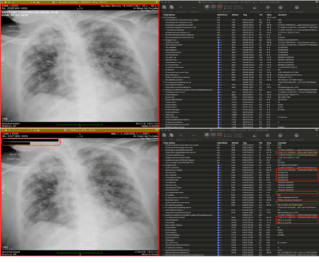
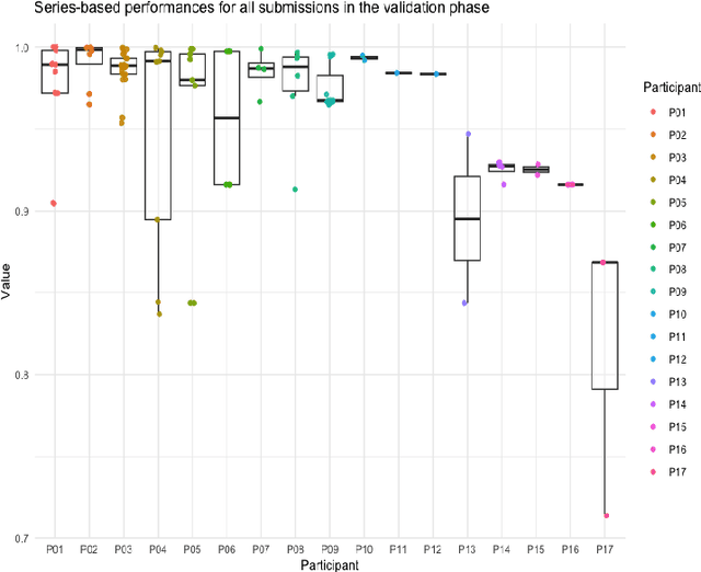
Abstract:The de-identification (deID) of protected health information (PHI) and personally identifiable information (PII) is a fundamental requirement for sharing medical images, particularly through public repositories, to ensure compliance with patient privacy laws. In addition, preservation of non-PHI metadata to inform and enable downstream development of imaging artificial intelligence (AI) is an important consideration in biomedical research. The goal of MIDI-B was to provide a standardized platform for benchmarking of DICOM image deID tools based on a set of rules conformant to the HIPAA Safe Harbor regulation, the DICOM Attribute Confidentiality Profiles, and best practices in preservation of research-critical metadata, as defined by The Cancer Imaging Archive (TCIA). The challenge employed a large, diverse, multi-center, and multi-modality set of real de-identified radiology images with synthetic PHI/PII inserted. The MIDI-B Challenge consisted of three phases: training, validation, and test. Eighty individuals registered for the challenge. In the training phase, we encouraged participants to tune their algorithms using their in-house or public data. The validation and test phases utilized the DICOM images containing synthetic identifiers (of 216 and 322 subjects, respectively). Ten teams successfully completed the test phase of the challenge. To measure success of a rule-based approach to image deID, scores were computed as the percentage of correct actions from the total number of required actions. The scores ranged from 97.91% to 99.93%. Participants employed a variety of open-source and proprietary tools with customized configurations, large language models, and optical character recognition (OCR). In this paper we provide a comprehensive report on the MIDI-B Challenge's design, implementation, results, and lessons learned.
3D Cardiac Shape Prediction with Deep Neural Networks: Simultaneous Use of Images and Patient Metadata
Jul 02, 2019


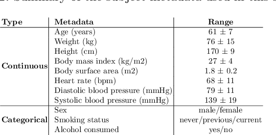
Abstract:Large prospective epidemiological studies acquire cardiovascular magnetic resonance (CMR) images for pre-symptomatic populations and follow these over time. To support this approach, fully automatic large-scale 3D analysis is essential. In this work, we propose a novel deep neural network using both CMR images and patient metadata to directly predict cardiac shape parameters. The proposed method uses the promising ability of statistical shape models to simplify shape complexity and variability together with the advantages of convolutional neural networks for the extraction of solid visual features. To the best of our knowledge, this is the first work that uses such an approach for 3D cardiac shape prediction. We validated our proposed CMR analytics method against a reference cohort containing 500 3D shapes of the cardiac ventricles. Our results show broadly significant agreement with the reference shapes in terms of the estimated volume of the cardiac ventricles, myocardial mass, 3D Dice, and mean and Hausdorff distance.
High Throughput Computation of Reference Ranges of Biventricular Cardiac Function on the UK Biobank Population Cohort
Jan 10, 2019


Abstract:The exploitation of large-scale population data has the potential to improve healthcare by discovering and understanding patterns and trends within this data. To enable high throughput analysis of cardiac imaging data automatically, a pipeline should comprise quality monitoring of the input images, segmentation of the cardiac structures, assessment of the segmentation quality, and parsing of cardiac functional indexes. We present a fully automatic, high throughput image parsing workflow for the analysis of cardiac MR images, and test its performance on the UK Biobank (UKB) cardiac dataset. The proposed pipeline is capable of performing end-to-end image processing including: data organisation, image quality assessment, shape model initialisation, segmentation, segmentation quality assessment, and functional parameter computation; all without any user interaction. To the best of our knowledge,this is the first paper tackling the fully automatic 3D analysis of the UKB population study, providing reference ranges for all key cardiovascular functional indexes, from both left and right ventricles of the heart. We tested our workflow on a reference cohort of 800 healthy subjects for which manual delineations, and reference functional indexes exist. Our results show statistically significant agreement between the manually obtained reference indexes, and those automatically computed using our framework.
Automatic Assessment of Full Left Ventricular Coverage in Cardiac Cine Magnetic Resonance Imaging with Fisher-Discriminative 3D CNN
Nov 08, 2018


Abstract:Cardiac magnetic resonance (CMR) images play a growing role in the diagnostic imaging of cardiovascular diseases. Full coverage of the left ventricle (LV), from base to apex, is a basic criterion for CMR image quality and necessary for accurate measurement of cardiac volume and functional assessment. Incomplete coverage of the LV is identified through visual inspection, which is time-consuming and usually done retrospectively in the assessment of large imaging cohorts. This paper proposes a novel automatic method for determining LV coverage from CMR images by using Fisher-discriminative three-dimensional (FD3D) convolutional neural networks (CNNs). In contrast to our previous method employing 2D CNNs, this approach utilizes spatial contextual information in CMR volumes, extracts more representative high-level features and enhances the discriminative capacity of the baseline 2D CNN learning framework, thus achieving superior detection accuracy. A two-stage framework is proposed to identify missing basal and apical slices in measurements of CMR volume. First, the FD3D CNN extracts high-level features from the CMR stacks. These image representations are then used to detect the missing basal and apical slices. Compared to the traditional 3D CNN strategy, the proposed FD3D CNN minimizes within-class scatter and maximizes between-class scatter. We performed extensive experiments to validate the proposed method on more than 5,000 independent volumetric CMR scans from the UK Biobank study, achieving low error rates for missing basal/apical slice detection (4.9\%/4.6\%). The proposed method can also be adopted for assessing LV coverage for other types of CMR image data.
 Add to Chrome
 Add to Firefox
 Add to Edge