Yunbi Liu
TAlignDiff: Automatic Tooth Alignment assisted by Diffusion-based Transformation Learning
Aug 06, 2025Abstract:Orthodontic treatment hinges on tooth alignment, which significantly affects occlusal function, facial aesthetics, and patients' quality of life. Current deep learning approaches predominantly concentrate on predicting transformation matrices through imposing point-to-point geometric constraints for tooth alignment. Nevertheless, these matrices are likely associated with the anatomical structure of the human oral cavity and possess particular distribution characteristics that the deterministic point-to-point geometric constraints in prior work fail to capture. To address this, we introduce a new automatic tooth alignment method named TAlignDiff, which is supported by diffusion-based transformation learning. TAlignDiff comprises two main components: a primary point cloud-based regression network (PRN) and a diffusion-based transformation matrix denoising module (DTMD). Geometry-constrained losses supervise PRN learning for point cloud-level alignment. DTMD, as an auxiliary module, learns the latent distribution of transformation matrices from clinical data. We integrate point cloud-based transformation regression and diffusion-based transformation modeling into a unified framework, allowing bidirectional feedback between geometric constraints and diffusion refinement. Extensive ablation and comparative experiments demonstrate the effectiveness and superiority of our method, highlighting its potential in orthodontic treatment.
Privacy-Preserving Federated Foundation Model for Generalist Ultrasound Artificial Intelligence
Nov 25, 2024


Abstract:Ultrasound imaging is widely used in clinical diagnosis due to its non-invasive nature and real-time capabilities. However, conventional ultrasound diagnostics face several limitations, including high dependence on physician expertise and suboptimal image quality, which complicates interpretation and increases the likelihood of diagnostic errors. Artificial intelligence (AI) has emerged as a promising solution to enhance clinical diagnosis, particularly in detecting abnormalities across various biomedical imaging modalities. Nonetheless, current AI models for ultrasound imaging face critical challenges. First, these models often require large volumes of labeled medical data, raising concerns over patient privacy breaches. Second, most existing models are task-specific, which restricts their broader clinical utility. To overcome these challenges, we present UltraFedFM, an innovative privacy-preserving ultrasound foundation model. UltraFedFM is collaboratively pre-trained using federated learning across 16 distributed medical institutions in 9 countries, leveraging a dataset of over 1 million ultrasound images covering 19 organs and 10 ultrasound modalities. This extensive and diverse data, combined with a secure training framework, enables UltraFedFM to exhibit strong generalization and diagnostic capabilities. It achieves an average area under the receiver operating characteristic curve of 0.927 for disease diagnosis and a dice similarity coefficient of 0.878 for lesion segmentation. Notably, UltraFedFM surpasses the diagnostic accuracy of mid-level ultrasonographers and matches the performance of expert-level sonographers in the joint diagnosis of 8 common systemic diseases. These findings indicate that UltraFedFM can significantly enhance clinical diagnostics while safeguarding patient privacy, marking an advancement in AI-driven ultrasound imaging for future clinical applications.
DArch: Dental Arch Prior-assisted 3D Tooth Instance Segmentation
Apr 25, 2022


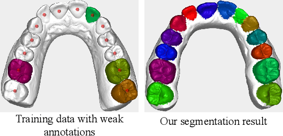
Abstract:Automatic tooth instance segmentation on 3D dental models is a fundamental task for computer-aided orthodontic treatments. Existing learning-based methods rely heavily on expensive point-wise annotations. To alleviate this problem, we are the first to explore a low-cost annotation way for 3D tooth instance segmentation, i.e., labeling all tooth centroids and only a few teeth for each dental model. Regarding the challenge when only weak annotation is provided, we present a dental arch prior-assisted 3D tooth segmentation method, namely DArch. Our DArch consists of two stages, including tooth centroid detection and tooth instance segmentation. Accurately detecting the tooth centroids can help locate the individual tooth, thus benefiting the segmentation. Thus, our DArch proposes to leverage the dental arch prior to assist the detection. Specifically, we firstly propose a coarse-to-fine method to estimate the dental arch, in which the dental arch is initially generated by Bezier curve regression, and then a graph-based convolutional network (GCN) is trained to refine it. With the estimated dental arch, we then propose a novel Arch-aware Point Sampling (APS) method to assist the tooth centroid proposal generation. Meantime, a segmentor is independently trained using a patch-based training strategy, aiming to segment a tooth instance from a 3D patch centered at the tooth centroid. Experimental results on $4,773$ dental models have shown our DArch can accurately segment each tooth of a dental model, and its performance is superior to the state-of-the-art methods.
 Add to Chrome
 Add to Firefox
 Add to Edge