 Add to Chrome
 Add to Firefox
 Add to Edge
Improved tissue sodium concentration quantification in breast cancer by reducing partial volume effects: a preliminary study
Mar 25, 2025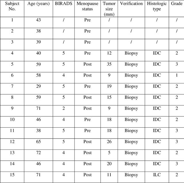
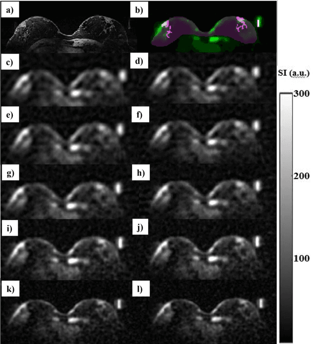


Introduction: In sodium (23Na) MRI, partial volume effects (PVE) are one of the most common causes of errors in the quantification of tissue sodium concentration (TSC) in vivo. Advanced image reconstruction algorithms, such as compressed sensing (CS), have been shown to potentially reduce PVE. Therefore, we investigated the feasibility of CS-based methods for image quality and TSC quantification accuracy improvement in patients with breast cancer (BC). Subjects and Methods: Three healthy participants and 12 female participants with BC were examined on a 7T MRI scanner in this study. We reconstructed 23Na-MRI images using the weighted total variation (wTV) and directional total variation (dTV), anatomically guided total variation (AG-TV), and adaptive combine (ADC) reconstruction and performed image quality assessment. We evaluated agreement in tumor volumes delineated on sodium data using the Dice score and performed TSC quantification for different image reconstruction approaches. Results: All methods provided sodium images of the breast with good quality. The mean Dice scores for wTV, dTV, and AG-TV were 65%, 72%, and 75%, respectively. In the breast tumors, average TSC values were 83.0, 72.0, 80.0, and 84.0 mmol/L, respectively. There was a significant difference between dTV and wTV (p<0.001), as well as between dTV and AG-TV (p<0.001) and dTV and ADC algorithm (p<0.001). Conclusion: The results of this study showed that there are differences in tumor appearance and TSC estimations that might be depending on the type of image reconstruction and parameters used, most likely due to differences in their robustness in reducing PVE.
Variational U-Net with Local Alignment for Joint Tumor Extraction and Registration (VALOR-Net) of Breast MRI Data Acquired at Two Different Field Strengths
Jan 23, 2025


Background: Multiparametric breast MRI data might improve tumor diagnostics, characterization, and treatment planning. Accurate alignment and delineation of images acquired at different field strengths such as 3T and 7T, remain challenging research tasks. Purpose: To address alignment challenges and enable consistent tumor segmentation across different MRI field strengths. Study type: Retrospective. Subjects: Nine female subjects with breast tumors were involved: six histologically proven invasive ductal carcinomas (IDC) and three fibroadenomas. Field strength/sequence: Imaging was performed at 3T and 7T scanners using post-contrast T1-weighted three-dimensional time-resolved angiography with stochastic trajectories (TWIST) sequence. Assessments: The method's performance for joint image registration and tumor segmentation was evaluated using several quantitative metrics, including signal-to-noise ratio (PSNR), structural similarity index (SSIM), normalized cross-correlation (NCC), Dice coefficient, F1 score, and relative sum of squared differences (rel SSD). Statistical tests: The Pearson correlation coefficient was used to test the relationship between the registration and segmentation metrics. Results: When calculated for each subject individually, the PSNR was in a range from 27.5 to 34.5 dB, and the SSIM was from 82.6 to 92.8%. The model achieved an NCC from 96.4 to 99.3% and a Dice coefficient of 62.9 to 95.3%. The F1 score was between 55.4 and 93.2% and the rel SSD was in the range of 2.0 and 7.5%. The segmentation metrics Dice and F1 Score are highly correlated (0.995), while a moderate correlation between NCC and SSIM (0.681) was found for registration. Data conclusion: Initial results demonstrate that the proposed method may be feasible in providing joint tumor segmentation and registration of MRI data acquired at different field strengths.
Predicting dynamic, motion-related changes in B0 field in the brain at a 7 T MRI using a subject-specific fine-tuned U-net
Apr 17, 2023Subject movement during the magnetic resonance examination is inevitable and causes not only image artefacts but also deteriorates the homogeneity of the main magnetic field (B0), which is a prerequisite for high quality data. Thus, characterization of changes to B0, e.g. induced by patient movement, is important for MR applications that are prone to B0 inhomogeneities. We propose a deep learning based method to predict such changes within the brain from the change of the head position to facilitate retrospective or even real-time correction. A 3D U-net was trained on in vivo brain 7T MRI data. The input consisted of B0 maps and anatomical images at an initial position, and anatomical images at a different head position (obtained by applying a rigid-body transformation on the initial anatomical image). The output consisted of B0 maps at the new head positions. We further fine-tuned the network weights to each subject by measuring a limited number of head positions of the given subject, and trained the U-net with these data. Our approach was compared to established dynamic B0 field mapping via interleaved navigators, which suffer from limited spatial resolution and the need for undesirable sequence modifications. Qualitative and quantitative comparison showed similar performance between an interleaved navigator-equivalent method and proposed method. We therefore conclude that it is feasible to predict B0 maps from rigid subject movement and, when combined with external tracking hardware, this information could be used to improve the quality of magnetic resonance acquisitions without the use of navigators.
Multi-scale Super-resolution Magnetic Resonance Spectroscopic Imaging with Adjustable Sharpness
Jun 17, 2022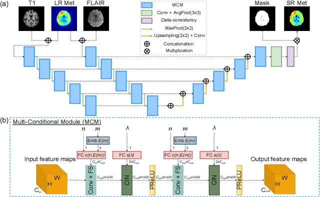
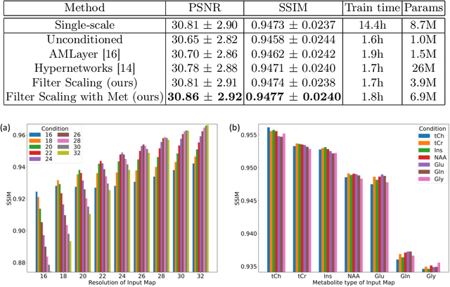
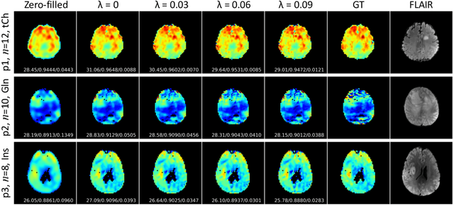
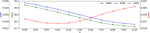
Magnetic Resonance Spectroscopic Imaging (MRSI) is a valuable tool for studying metabolic activities in the human body, but the current applications are limited to low spatial resolutions. The existing deep learning-based MRSI super-resolution methods require training a separate network for each upscaling factor, which is time-consuming and memory inefficient. We tackle this multi-scale super-resolution problem using a Filter Scaling strategy that modulates the convolution filters based on the upscaling factor, such that a single network can be used for various upscaling factors. Observing that each metabolite has distinct spatial characteristics, we also modulate the network based on the specific metabolite. Furthermore, our network is conditioned on the weight of adversarial loss so that the perceptual sharpness of the super-resolved metabolic maps can be adjusted within a single network. We incorporate these network conditionings using a novel Multi-Conditional Module. The experiments were carried out on a 1H-MRSI dataset from 15 high-grade glioma patients. Results indicate that the proposed network achieves the best performance among several multi-scale super-resolution methods and can provide super-resolved metabolic maps with adjustable sharpness.