 Add to Chrome
 Add to Firefox
 Add to Edge
Improved tissue sodium concentration quantification in breast cancer by reducing partial volume effects: a preliminary study
Mar 25, 2025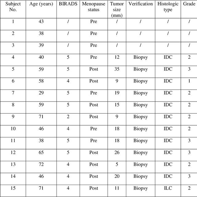
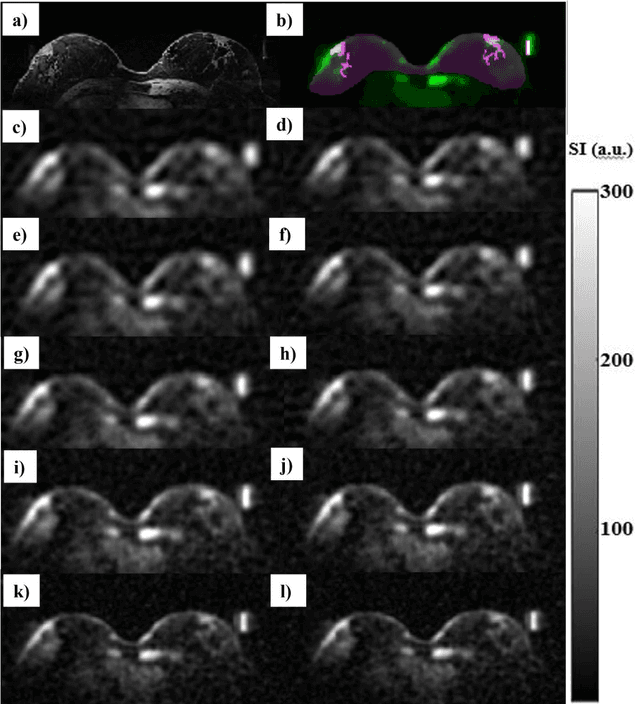


Introduction: In sodium (23Na) MRI, partial volume effects (PVE) are one of the most common causes of errors in the quantification of tissue sodium concentration (TSC) in vivo. Advanced image reconstruction algorithms, such as compressed sensing (CS), have been shown to potentially reduce PVE. Therefore, we investigated the feasibility of CS-based methods for image quality and TSC quantification accuracy improvement in patients with breast cancer (BC). Subjects and Methods: Three healthy participants and 12 female participants with BC were examined on a 7T MRI scanner in this study. We reconstructed 23Na-MRI images using the weighted total variation (wTV) and directional total variation (dTV), anatomically guided total variation (AG-TV), and adaptive combine (ADC) reconstruction and performed image quality assessment. We evaluated agreement in tumor volumes delineated on sodium data using the Dice score and performed TSC quantification for different image reconstruction approaches. Results: All methods provided sodium images of the breast with good quality. The mean Dice scores for wTV, dTV, and AG-TV were 65%, 72%, and 75%, respectively. In the breast tumors, average TSC values were 83.0, 72.0, 80.0, and 84.0 mmol/L, respectively. There was a significant difference between dTV and wTV (p<0.001), as well as between dTV and AG-TV (p<0.001) and dTV and ADC algorithm (p<0.001). Conclusion: The results of this study showed that there are differences in tumor appearance and TSC estimations that might be depending on the type of image reconstruction and parameters used, most likely due to differences in their robustness in reducing PVE.
Three-dimensional reconstruction and characterization of bladder deformations
Jan 18, 2023


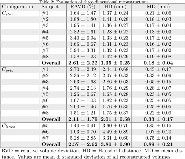
Background and Objective: Pelvic floor disorders are prevalent diseases and patient care remains difficult as the dynamics of the pelvic floor remains poorly known. So far, only 2D dynamic observations of straining exercises at excretion are available in the clinics and the understanding of three-dimensional pelvic organs mechanical defects is not yet achievable. In this context, we proposed a complete methodology for the 3D representation of the non-reversible bladder deformations during exercises, directly combined with synthesized 3D representation of the location of the highest strain areas on the organ surface. Methods: Novel image segmentation and registration approaches have been combined with three geometrical configurations of up-to-date rapid dynamic multi-slices MRI acquisition for the reconstruction of real-time dynamic bladder volumes. Results: For the first time, we proposed real-time 3D deformation fields of the bladder under strain from in-bore forced breathing exercises. The potential of our method was assessed on eight control subjects undergoing forced breathing exercises. We obtained average volume deviation of the reconstructed dynamic volume of bladders around 2.5\% and high registration accuracy with mean distance values of 0.4 $\pm$ 0.3 mm and Hausdorff distance values of 2.2 $\pm$ 1.1 mm. Conclusions: Immediately transferable to the clinics with rapid acquisitions, the proposed framework represents a real advance in the field of pelvic floor disorders as it provides, for the first time, a proper 3D+t spatial tracking of bladder non-reversible deformations. This work is intended to be extended to patients with cavities filling and excretion to better characterize the degree of severity of pelvic floor pathologies for diagnostic assistance or in preoperative surgical planning.