Pingli Ma
TOD-CNN: An Effective Convolutional Neural Network for Tiny Object Detection in Sperm Videos
Apr 18, 2022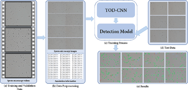


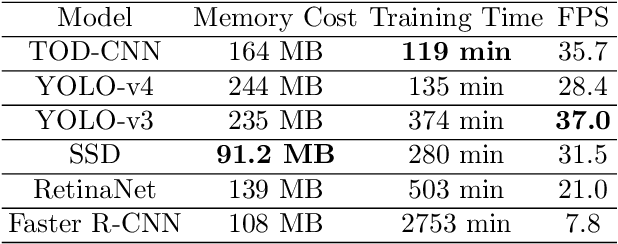
Abstract:The detection of tiny objects in microscopic videos is a problematic point, especially in large-scale experiments. For tiny objects (such as sperms) in microscopic videos, current detection methods face challenges in fuzzy, irregular, and precise positioning of objects. In contrast, we present a convolutional neural network for tiny object detection (TOD-CNN) with an underlying data set of high-quality sperm microscopic videos (111 videos, $>$ 278,000 annotated objects), and a graphical user interface (GUI) is designed to employ and test the proposed model effectively. TOD-CNN is highly accurate, achieving $85.60\%$ AP$_{50}$ in the task of real-time sperm detection in microscopic videos. To demonstrate the importance of sperm detection technology in sperm quality analysis, we carry out relevant sperm quality evaluation metrics and compare them with the diagnosis results from medical doctors.
A Comprehensive Survey with Quantitative Comparison of Image Analysis Methods for Microorganism Biovolume Measurements
Feb 18, 2022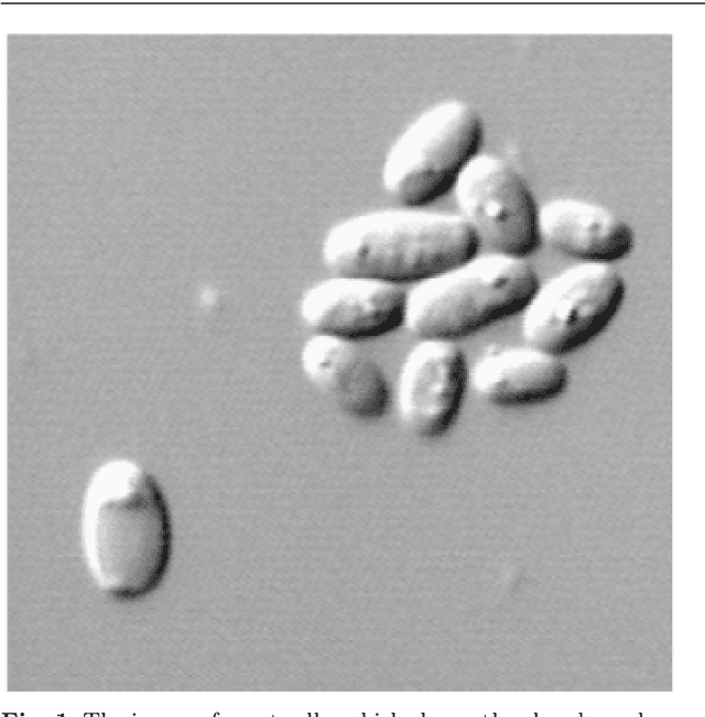

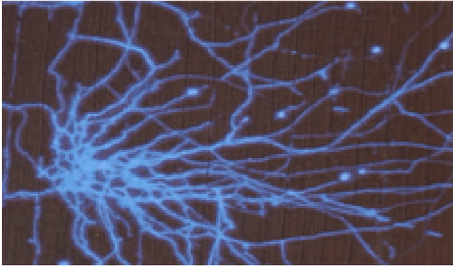
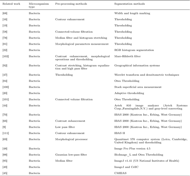
Abstract:With the acceleration of urbanization and living standards, microorganisms play increasingly important roles in industrial production, bio-technique, and food safety testing. Microorganism biovolume measurements are one of the essential parts of microbial analysis. However, traditional manual measurement methods are time-consuming and challenging to measure the characteristics precisely. With the development of digital image processing techniques, the characteristics of the microbial population can be detected and quantified. The changing trend can be adjusted in time and provided a basis for the improvement. The applications of the microorganism biovolume measurement method have developed since the 1980s. More than 60 articles are reviewed in this study, and the articles are grouped by digital image segmentation methods with periods. This study has high research significance and application value, which can be referred to microbial researchers to have a comprehensive understanding of microorganism biovolume measurements using digital image analysis methods and potential applications.
A Survey of Semen Quality Evaluation in Microscopic Videos Using Computer Assisted Sperm Analysis
Feb 17, 2022
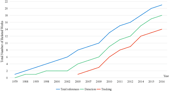
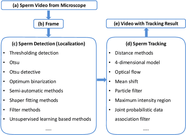
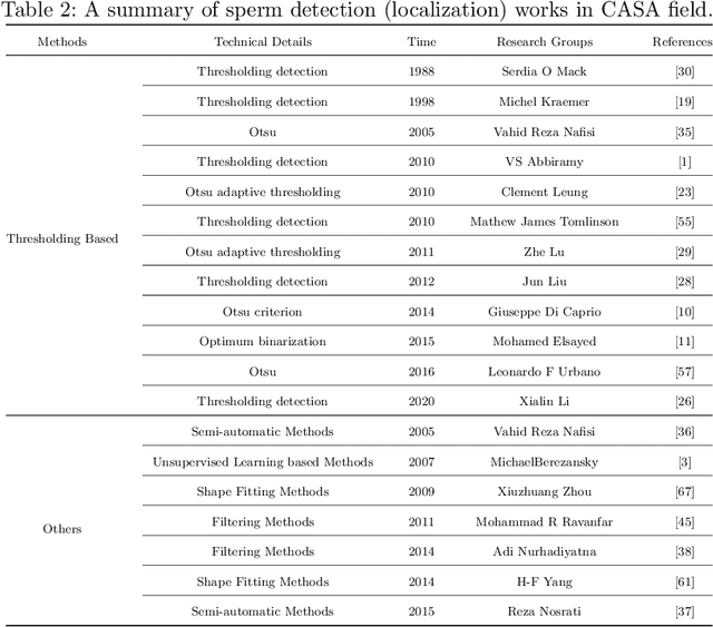
Abstract:The Computer Assisted Sperm Analysis (CASA) plays a crucial role in male reproductive health diagnosis and Infertility treatment. With the development of the computer industry in recent years, a great of accurate algorithms are proposed. With the assistance of those novel algorithms, it is possible for CASA to achieve a faster and higher quality result. Since image processing is the technical basis of CASA, including pre-processing,feature extraction, target detection and tracking, these methods are important technical steps in dealing with CASA. The various works related to Computer Assisted Sperm Analysis methods in the last 30 years (since 1988) are comprehensively introduced and analysed in this survey. To facilitate understanding, the methods involved are analysed in the sequence of general steps in sperm analysis. In other words, the methods related to sperm detection (localization) are first analysed, and then the methods of sperm tracking are analysed. Beside this, we analyse and prospect the present situation and future of CASA. According to our work, the feasible for applying in sperm microscopic video of methods mentioned in this review is explained. Moreover, existing challenges of object detection and tracking in microscope video are potential to be solved inspired by this survey.
EMDS-6: Environmental Microorganism Image Dataset Sixth Version for Image Denoising, Segmentation, Feature Extraction, Classification and Detection Methods Evaluation
Dec 14, 2021
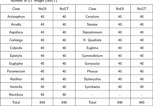

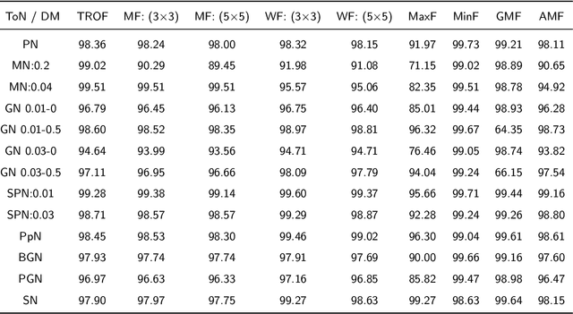
Abstract:Environmental microorganisms (EMs) are ubiquitous around us and have an important impact on the survival and development of human society. However, the high standards and strict requirements for the preparation of environmental microorganism (EM) data have led to the insufficient of existing related databases, not to mention the databases with GT images. This problem seriously affects the progress of related experiments. Therefore, This study develops the Environmental Microorganism Dataset Sixth Version (EMDS-6), which contains 21 types of EMs. Each type of EM contains 40 original and 40 GT images, in total 1680 EM images. In this study, in order to test the effectiveness of EMDS-6. We choose the classic algorithms of image processing methods such as image denoising, image segmentation and target detection. The experimental result shows that EMDS-6 can be used to evaluate the performance of image denoising, image segmentation, image feature extraction, image classification, and object detection methods.
EMDS-7: Environmental Microorganism Image Dataset Seventh Version for Multiple Object Detection Evaluation
Oct 28, 2021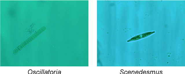
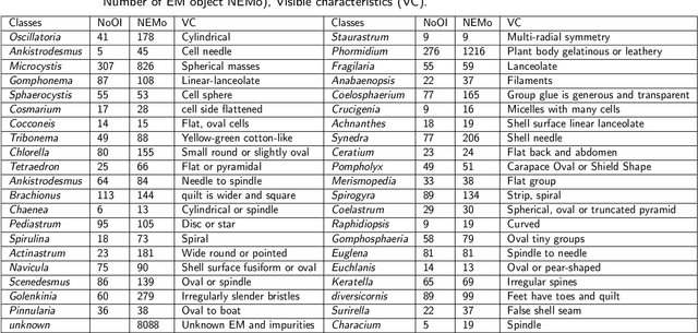
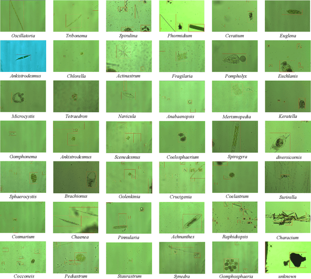
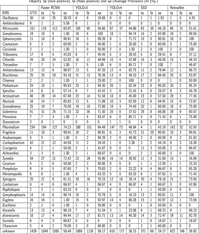
Abstract:The Environmental Microorganism Image Dataset Seventh Version (EMDS-7) is a microscopic image data set, including the original Environmental Microorganism images (EMs) and the corresponding object labeling files in ".XML" format file. The EMDS-7 data set consists of 41 types of EMs, which has a total of 2365 images and 13216 labeled objects. The EMDS-7 database mainly focuses on the object detection. In order to prove the effectiveness of EMDS-7, we select the most commonly used deep learning methods (Faster-RCNN, YOLOv3, YOLOv4, SSD and RetinaNet) and evaluation indices for testing and evaluation. EMDS-7 is freely published for non-commercial purpose at: https://figshare.com/articles/dataset/EMDS-7_DataSet/16869571
A State-of-the-art Survey of Object Detection Techniques in Microorganism Image Analysis: from Traditional Image Processing and Classical Machine Learning to Current Deep Convolutional Neural Networks and Potential Visual Transformers
May 07, 2021


Abstract:Microorganisms play a vital role in human life. Therefore, microorganism detection is of great significance to human beings. However, the traditional manual microscopic detection methods have the disadvantages of long detection cycle, low detection accuracy in large orders, and great difficulty in detecting uncommon microorganisms. Therefore, it is meaningful to apply computer image analysis technology to the field of microorganism detection. Computer image analysis can realize high-precision and high-efficiency detection of microorganisms. In this review, first,we analyse the existing microorganism detection methods in chronological order, from traditional image processing and traditional machine learning to deep learning methods. Then, we analyze and summarize these existing methods and introduce some potential methods, including visual transformers. In the end, the future development direction and challenges of microorganism detection are discussed. In general, we have summarized 137 related technical papers from 1985 to the present. This review will help researchers have a more comprehensive understanding of the development process, research status, and future trends in the field of microorganism detection and provide a reference for researchers in other fields.
A Comprehensive Review of Image Analysis Methods for Microorganism Counting: From Classical Image Processing to Deep Learning Approaches
Mar 25, 2021
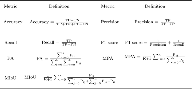


Abstract:Microorganisms such as bacteria and fungi play essential roles in many application fields, like biotechnique, medical technique and industrial domain. Microorganism counting techniques are crucial in microorganism analysis, helping biologists and related researchers quantitatively analyze the microorganisms and calculate their characteristics, such as biomass concentration and biological activity. However, traditional microorganism manual counting methods are time consuming and subjective, which cannot be applied in large-scale applications. In order to improve this situation, image analysis-based microorganism counting systems are developed since 1980s, which consists of digital image processing, image segmentation, image classification and so on. Moreover, image analysis-based microorganism counting methods are efficient comparing with traditional plate counting methods. In this article, we have studied the development of microorganism counting methods using digital image analysis. Firstly, the microorganisms are grouped as bacteria and other microorganisms. Then, the related articles are summarized based on image segmentation methods. Each part of articles are reviewed by time periods. Moreover, commonly used image processing methods for microorganism counting are summarized and analyzed to find technological common points. More than 142 papers are summarized in this article. In conclusion, this article can be referred to researchers to determine the development trend in microorganism counting field and further analyze the potential applications
 Add to Chrome
 Add to Firefox
 Add to Edge