 Add to Chrome
 Add to Firefox
 Add to Edge
Learning Robust and Task-Invariant Functional Representation from fMRI through Siamese Self-Supervised Learning
May 27, 2026Functional magnetic resonance imaging (fMRI) is a powerful tool for investigating human brain function. However, the high cost of data acquisition and the inherent subjectivity of psychiatric rating scales often lead to datasets with small sample sizes and variable label quality, especially when targeting a specific neurological condition. Combined with the inherently high dimensionality of fMRI data, these limitations substantially increase the risk of model overfitting. Recent years have seen growing interest in developing fMRI foundation models by combining multiple datasets; however, the computational resources needed for pretraining and fine-tuning are often prohibitive. We show that a lightweight self-supervised framework yields representations that generalize across diverse downstream tasks, outperforming fully supervised baselines and approaching the performance of large-scale models. We introduce BrainSimSiam, a data-efficient self-supervised representation learning framework that leverages positive-only data pairs to learn robust and generalizable features. We demonstrate that the learned representations achieve strong performance across multiple downstream classification and regression tasks, highlighting the potential of BrainSimSiam for data-limited neuroimaging applications.
FM-fMRI: Event Conditioned Flow Matching for Rest-to-Task fMRI Time-Series Synthesis
May 26, 2026Task-based fMRI provides a direct readout of task-evoked neural dynamics, but it is expensive and difficult to acquire at scale, motivating rest-to-task synthesis from widely available resting-state fMRI (rsfMRI). We propose FM-fMRI, an event-conditioned flow-matching model that learns a continuous-time conditional vector field to generate task ROI time series from a subject's rsfMRI and the task event information. The formulation enables fast ODE-based sampling and flexible conditioning over heterogeneous event schedules. Rather than optimizing for pointwise reconstruction, we evaluated generated signals using complementary criteria that probe temporal and spectral structure, subject and group-level connectome consistency, and distributional alignment. On the public Human Connectome Project and internal BioPoint autism cohort, FM-fMRI achieves the strongest spectral and connectivity agreement and improved distribution-level matching over conditional diffusion, generative adversarial networks (GANs), and variational autoencoders (VAEs) baselines. Furthermore, we augment the BioPoint cohort by synthesizing task-fMRI ROI time series with our method, improving downstream autism classification and demonstrating practical utility in data-limited clinical settings. The code will be available on GitHub.
BioFact-MoE: Biologically Factorized Mixture of Experts for Vision-Language Prognostic Modeling in Hepatocellular Carcinoma
May 25, 2026Hepatocellular carcinoma (HCC) is biologically heterogeneous, shaped by the interplay between hepatic functional reserve and tumor-related oncologic factors; thus, similar survival outcomes may reflect fundamentally different underlying biological processes. Prognostic modeling in HCC is informed by rich multimodal information from multiparametric MRI and radiology reports from routine clinical practice. Existing prognostic vision-language models (VLMs) learn a single entangled latent representation that blends hepatic and tumor-related factors, limiting both accuracy and biological interpretability. We present BioFact-MoE, a biologically factorized Mixture of Experts (MoE) framework that explicitly decomposes liver and tumor factors via biologically supervised experts within a residual MoE survival architecture. On a HCC cohort of N=588 patients (pretrained on 4,582 3D MRI image-report pairs), BioFact-MoE consistently improves survival prediction over all baselines across time horizons, achieving 12-, 18-, and 24-month AUCs of 75.33%, 75.85%, and 73.96%. Beyond scalar risk prediction, gated expert weights enable phenotype-aware risk stratification. Pathway-informed gating uncovers clinically meaningful treatment-associated survival heterogeneity. In held-out validation, hepatic and tumor embeddings show selective associations with liver function and tumor burden markers, respectively (p<0.05), without supervision. The code is available at https://github.com/jy-639/BioFact-MoE.
Causal Modeling of fMRI Time-series for Interpretable Autism Spectrum Disorder Classification
Feb 21, 2025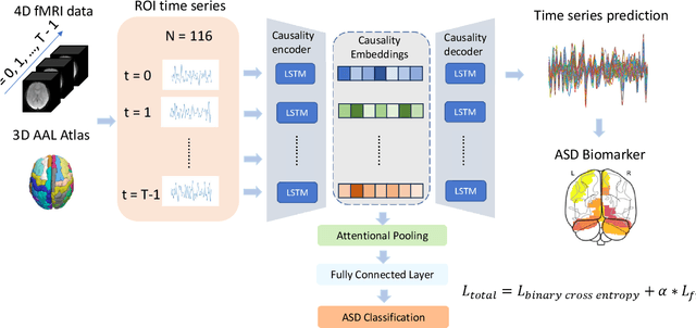
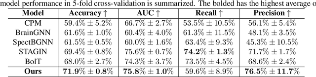
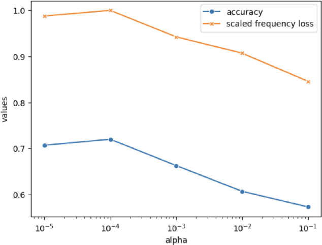
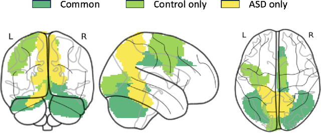
Autism spectrum disorder (ASD) is a neurological and developmental disorder that affects social and communicative behaviors. It emerges in early life and is generally associated with lifelong disabilities. Thus, accurate and early diagnosis could facilitate treatment outcomes for those with ASD. Functional magnetic resonance imaging (fMRI) is a useful tool that measures changes in brain signaling to facilitate our understanding of ASD. Much effort is being made to identify ASD biomarkers using various connectome-based machine learning and deep learning classifiers. However, correlation-based models cannot capture the non-linear interactions between brain regions. To solve this problem, we introduce a causality-inspired deep learning model that uses time-series information from fMRI and captures causality among ROIs useful for ASD classification. The model is compared with other baseline and state-of-the-art models with 5-fold cross-validation on the ABIDE dataset. We filtered the dataset by choosing all the images with mean FD less than 15mm to ensure data quality. Our proposed model achieved the highest average classification accuracy of 71.9% and an average AUC of 75.8%. Moreover, the inter-ROI causality interpretation of the model suggests that the left precuneus, right precuneus, and cerebellum are placed in the top 10 ROIs in inter-ROI causality among the ASD population. In contrast, these ROIs are not ranked in the top 10 in the control population. We have validated our findings with the literature and found that abnormalities in these ROIs are often associated with ASD.
STNAGNN: Spatiotemporal Node Attention Graph Neural Network for Task-based fMRI Analysis
Jun 17, 2024Task-based fMRI uses actions or stimuli to trigger task-specific brain responses and measures them using BOLD contrast. Despite the significant task-induced spatiotemporal brain activation fluctuations, most studies on task-based fMRI ignore the task context information aligned with fMRI and consider task-based fMRI a coherent sequence. In this paper, we show that using the task structures as data-driven guidance is effective for spatiotemporal analysis. We propose STNAGNN, a GNN-based spatiotemporal architecture, and validate its performance in an autism classification task. The trained model is also interpreted for identifying autism-related spatiotemporal brain biomarkers.
Efficient Annotation for Medical Image Analysis: A One-Pass Selective Annotation Approach
Sep 15, 2023


Annotating biomedical images for supervised learning is a complex and labor-intensive task due to data diversity and its intricate nature. In this paper, we propose an innovative method, the efficient one-pass selective annotation (EPOSA), that significantly reduces the annotation burden while maintaining robust model performance. Our approach employs a variational autoencoder (VAE) to extract salient features from unannotated images, which are subsequently clustered using the DBSCAN algorithm. This process groups similar images together, forming distinct clusters. We then use a two-stage sample selection algorithm, called representative selection (RepSel), to form a selected dataset. The first stage is a Markov Chain Monte Carlo (MCMC) sampling technique to select representative samples from each cluster for annotations. This selection process is the second stage, which is guided by the principle of maximizing intra-cluster mutual information and minimizing inter-cluster mutual information. This ensures a diverse set of features for model training and minimizes outlier inclusion. The selected samples are used to train a VGG-16 network for image classification. Experimental results on the Med-MNIST dataset demonstrate that our proposed EPOSA outperforms random selection and other state-of-the-art methods under the same annotation budget, presenting a promising direction for efficient and effective annotation in medical image analysis.
Rapid Brain Meninges Surface Reconstruction with Layer Topology Guarantee
Apr 13, 2023


The meninges, located between the skull and brain, are composed of three membrane layers: the pia, the arachnoid, and the dura. Reconstruction of these layers can aid in studying volume differences between patients with neurodegenerative diseases and normal aging subjects. In this work, we use convolutional neural networks (CNNs) to reconstruct surfaces representing meningeal layer boundaries from magnetic resonance (MR) images. We first use the CNNs to predict the signed distance functions (SDFs) representing these surfaces while preserving their anatomical ordering. The marching cubes algorithm is then used to generate continuous surface representations; both the subarachnoid space (SAS) and the intracranial volume (ICV) are computed from these surfaces. The proposed method is compared to a state-of-the-art deformable model-based reconstruction method, and we show that our method can reconstruct smoother and more accurate surfaces using less computation time. Finally, we conduct experiments with volumetric analysis on both subjects with multiple sclerosis and healthy controls. For healthy and MS subjects, ICVs and SAS volumes are found to be significantly correlated to sex (p<0.01) and age (p<0.03) changes, respectively.