Nchongmaje Ndipenoch
SegRap2023: A Benchmark of Organs-at-Risk and Gross Tumor Volume Segmentation for Radiotherapy Planning of Nasopharyngeal Carcinoma
Dec 15, 2023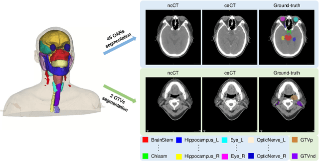

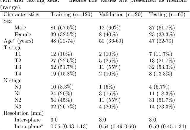
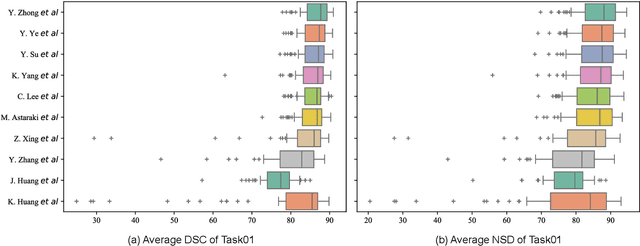
Abstract:Radiation therapy is a primary and effective NasoPharyngeal Carcinoma (NPC) treatment strategy. The precise delineation of Gross Tumor Volumes (GTVs) and Organs-At-Risk (OARs) is crucial in radiation treatment, directly impacting patient prognosis. Previously, the delineation of GTVs and OARs was performed by experienced radiation oncologists. Recently, deep learning has achieved promising results in many medical image segmentation tasks. However, for NPC OARs and GTVs segmentation, few public datasets are available for model development and evaluation. To alleviate this problem, the SegRap2023 challenge was organized in conjunction with MICCAI2023 and presented a large-scale benchmark for OAR and GTV segmentation with 400 Computed Tomography (CT) scans from 200 NPC patients, each with a pair of pre-aligned non-contrast and contrast-enhanced CT scans. The challenge's goal was to segment 45 OARs and 2 GTVs from the paired CT scans. In this paper, we detail the challenge and analyze the solutions of all participants. The average Dice similarity coefficient scores for all submissions ranged from 76.68\% to 86.70\%, and 70.42\% to 73.44\% for OARs and GTVs, respectively. We conclude that the segmentation of large-size OARs is well-addressed, and more efforts are needed for GTVs and small-size or thin-structure OARs. The benchmark will remain publicly available here: https://segrap2023.grand-challenge.org
Retinal Image Segmentation with Small Datasets
Mar 09, 2023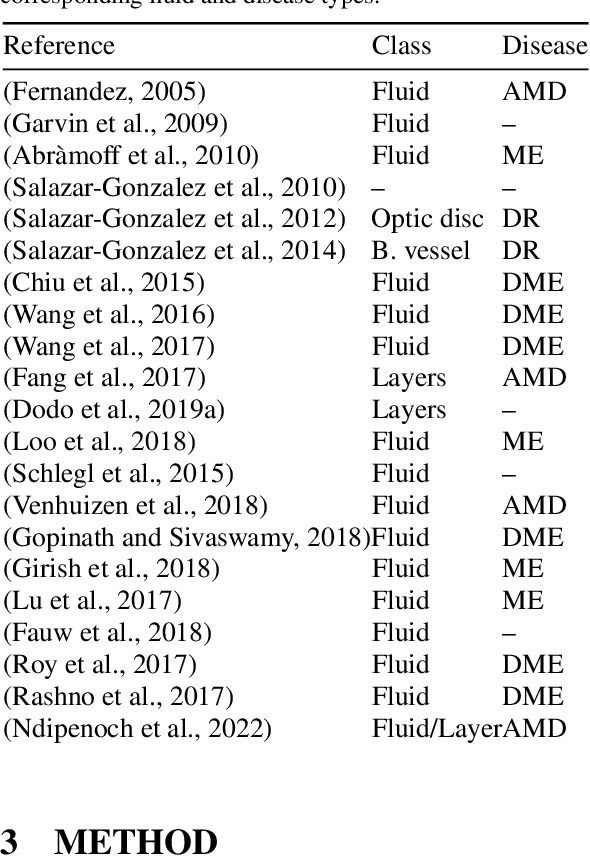
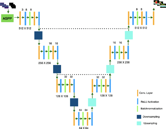

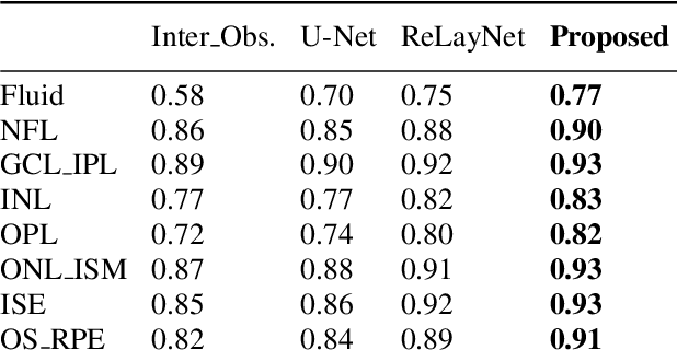
Abstract:Many eye diseases like Diabetic Macular Edema (DME), Age-related Macular Degeneration (AMD), and Glaucoma manifest in the retina, can cause irreversible blindness or severely impair the central version. The Optical Coherence Tomography (OCT), a 3D scan of the retina with high qualitative information about the retinal morphology, can be used to diagnose and monitor changes in the retinal anatomy. Many Deep Learning (DL) methods have shared the success of developing an automated tool to monitor pathological changes in the retina. However, the success of these methods depend mainly on large datasets. To address the challenge from very small and limited datasets, we proposed a DL architecture termed CoNet (Coherent Network) for joint segmentation of layers and fluids in retinal OCT images on very small datasets (less than a hundred training samples). The proposed model was evaluated on the publicly available Duke DME dataset consisting of 110 B-Scans from 10 patients suffering from DME. Experimental results show that the proposed model outperformed both the human experts' annotation and the current state-of-the-art architectures by a clear margin with a mean Dice Score of 88% when trained on 55 images without any data augmentation.
nnUNet RASPP for Retinal OCT Fluid Detection, Segmentation and Generalisation over Variations of Data Sources
Feb 25, 2023Abstract:Retinal Optical Coherence Tomography (OCT), a noninvasive cross-sectional scan of the eye with qualitative 3D visualization of the retinal anatomy is use to study the retinal structure and the presence of pathogens. The advent of the retinal OCT has transformed ophthalmology and it is currently paramount for the diagnosis, monitoring and treatment of many eye pathogens including Macular Edema which impairs vision severely or Glaucoma that can cause irreversible blindness. However the quality of retinal OCT images varies among device manufacturers. Deep Learning methods have had their success in the medical image segmentation community but it is still not clear if the level of success can be generalised across OCT images collected from different device vendors. In this work we propose two variants of the nnUNet [8]. The standard nnUNet and an enhanced vision call nnUnet_RASPP (nnU-Net with residual and Atrous Spatial Pyramid Pooling) both of which are robust and generalise with consistent high performance across images from multiple device vendors. The algorithm was validated on the MICCAI 2017 RETOUCH challenge dataset [1] acquired from 3 device vendors across 3 medical centers from patients suffering from 2 retinal disease types. Experimental results show that our algorithms outperform the current state-of-the-arts algorithms by a clear margin for segmentation obtaining a mean Dice Score (DS) of 82.3% for the 3 retinal fluids scoring 84.0%, 80.0%, 83.0% for Intraretinal Fluid (IRF), Subretinal Fluid (SRF), and Pigment Epithelium Detachments (PED) respectively on the testing dataset. Also we obtained a perfect Area Under the Curve (AUC) score of 100% for the detection of the presence of fluid for all 3 fluid classes on the testing dataset.
 Add to Chrome
 Add to Firefox
 Add to Edge