Mathias Maier
Dynamic Structural Recovery Parameters Enhance Prediction of Visual Outcomes After Macular Hole Surgery
Sep 11, 2025Abstract:Purpose: To introduce novel dynamic structural parameters and evaluate their integration within a multimodal deep learning (DL) framework for predicting postoperative visual recovery in idiopathic full-thickness macular hole (iFTMH) patients. Methods: We utilized a publicly available longitudinal OCT dataset at five stages (preoperative, 2 weeks, 3 months, 6 months, and 12 months). A stage specific segmentation model delineated related structures, and an automated pipeline extracted quantitative, composite, qualitative, and dynamic features. Binary logistic regression models, constructed with and without dynamic parameters, assessed their incremental predictive value for best-corrected visual acuity (BCVA). A multimodal DL model combining clinical variables, OCT-derived features, and raw OCT images was developed and benchmarked against regression models. Results: The segmentation model achieved high accuracy across all timepoints (mean Dice > 0.89). Univariate and multivariate analyses identified base diameter, ellipsoid zone integrity, and macular hole area as significant BCVA predictors (P < 0.05). Incorporating dynamic recovery rates consistently improved logistic regression AUC, especially at the 3-month follow-up. The multimodal DL model outperformed logistic regression, yielding higher AUCs and overall accuracy at each stage. The difference is as high as 0.12, demonstrating the complementary value of raw image volume and dynamic parameters. Conclusions: Integrating dynamic parameters into the multimodal DL model significantly enhances the accuracy of predictions. This fully automated process therefore represents a promising clinical decision support tool for personalized postoperative management in macular hole surgery.
EyeLS: Shadow-Guided Instrument Landing System for Intraocular Target Approaching in Robotic Eye Surgery
Nov 15, 2023Abstract:Robotic ophthalmic surgery is an emerging technology to facilitate high-precision interventions such as retina penetration in subretinal injection and removal of floating tissues in retinal detachment depending on the input imaging modalities such as microscopy and intraoperative OCT (iOCT). Although iOCT is explored to locate the needle tip within its range-limited ROI, it is still difficult to coordinate iOCT's motion with the needle, especially at the initial target-approaching stage. Meanwhile, due to 2D perspective projection and thus the loss of depth information, current image-based methods cannot effectively estimate the needle tip's trajectory towards both retinal and floating targets. To address this limitation, we propose to use the shadow positions of the target and the instrument tip to estimate their relative depth position and accordingly optimize the instrument tip's insertion trajectory until the tip approaches targets within iOCT's scanning area. Our method succeeds target approaching on a retina model, and achieves an average depth error of 0.0127 mm and 0.3473 mm for floating and retinal targets respectively in the surgical simulator without damaging the retina.
Towards Robotic Eye Surgery: Marker-free, Online Hand-eye Calibration using Optical Coherence Tomography Images
Aug 17, 2018


Abstract:Ophthalmic microsurgery is known to be a challenging operation, which requires very precise and dexterous manipulation. Image guided robot-assisted surgery (RAS) is a promising solution that brings significant improvements in outcomes and reduces the physical limitations of human surgeons. However, this technology must be further developed before it can be routinely used in clinics. One of the problems is the lack of proper calibration between the robotic manipulator and appropriate imaging device. In this work, we developed a flexible framework for hand-eye calibration of an ophthalmic robot with a microscope-integrated Optical Coherence Tomography (MIOCT) without any markers. The proposed method consists of three main steps: a) we estimate the OCT calibration parameters; b) with micro-scale displacements controlled by the robot, we detect and segment the needle tip in 3D-OCT volume; c) we find the transformation between the coordinate system of the OCT camera and the coordinate system of the robot. We verified the capability of our framework in ex-vivo pig eye experiments and compared the results with a reference method (marker-based). In all experiments, our method showed a small difference from the marker based method, with a mean calibration error of 9.2 $\mu$m and 7.0 $\mu$m, respectively. Additionally, the noise test shows the robustness of the proposed method.
Fast 5DOF Needle Tracking in iOCT
Feb 18, 2018


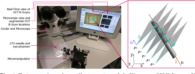
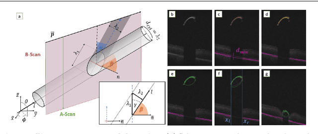
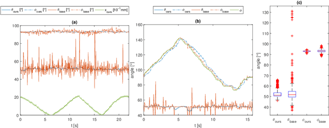
Abstract:Purpose. Intraoperative Optical Coherence Tomography (iOCT) is an increasingly available imaging technique for ophthalmic microsurgery that provides high-resolution cross-sectional information of the surgical scene. We propose to build on its desirable qualities and present a method for tracking the orientation and location of a surgical needle. Thereby, we enable direct analysis of instrument-tissue interaction directly in OCT space without complex multimodal calibration that would be required with traditional instrument tracking methods. Method. The intersection of the needle with the iOCT scan is detected by a peculiar multi-step ellipse fitting that takes advantage of the directionality of the modality. The geometric modelling allows us to use the ellipse parameters and provide them into a latency aware estimator to infer the 5DOF pose during needle movement. Results. Experiments on phantom data and ex-vivo porcine eyes indicate that the algorithm retains angular precision especially during lateral needle movement and provides a more robust and consistent estimation than baseline methods. Conclusion. Using solely crosssectional iOCT information, we are able to successfully and robustly estimate a 5DOF pose of the instrument in less than 5.5 ms on a CPU.
 Add to Chrome
 Add to Firefox
 Add to Edge