Jia Ding
The Llama 4 Herd: Architecture, Training, Evaluation, and Deployment Notes
Jan 15, 2026Abstract:This document consolidates publicly reported technical details about Metas Llama 4 model family. It summarizes (i) released variants (Scout and Maverick) and the broader herd context including the previewed Behemoth teacher model, (ii) architectural characteristics beyond a high-level MoE description covering routed/shared-expert structure, early-fusion multimodality, and long-context design elements reported for Scout (iRoPE and length generalization strategies), (iii) training disclosures spanning pre-training, mid-training for long-context extension, and post-training methodology (lightweight SFT, online RL, and lightweight DPO) as described in release materials, (iv) developer-reported benchmark results for both base and instruction-tuned checkpoints, and (v) practical deployment constraints observed across major serving environments, including provider-specific context limits and quantization packaging. The manuscript also summarizes licensing obligations relevant to redistribution and derivative naming, and reviews publicly described safeguards and evaluation practices. The goal is to provide a compact technical reference for researchers and practitioners who need precise, source-backed facts about Llama 4.
LlamaRL: A Distributed Asynchronous Reinforcement Learning Framework for Efficient Large-scale LLM Trainin
May 29, 2025Abstract:Reinforcement Learning (RL) has become the most effective post-training approach for improving the capabilities of Large Language Models (LLMs). In practice, because of the high demands on latency and memory, it is particularly challenging to develop an efficient RL framework that reliably manages policy models with hundreds to thousands of billions of parameters. In this paper, we present LlamaRL, a fully distributed, asynchronous RL framework optimized for efficient training of large-scale LLMs with various model sizes (8B, 70B, and 405B parameters) on GPU clusters ranging from a handful to thousands of devices. LlamaRL introduces a streamlined, single-controller architecture built entirely on native PyTorch, enabling modularity, ease of use, and seamless scalability to thousands of GPUs. We also provide a theoretical analysis of LlamaRL's efficiency, including a formal proof that its asynchronous design leads to strict RL speed-up. Empirically, by leveraging best practices such as colocated model offloading, asynchronous off-policy training, and distributed direct memory access for weight synchronization, LlamaRL achieves significant efficiency gains -- up to 10.7x speed-up compared to DeepSpeed-Chat-like systems on a 405B-parameter policy model. Furthermore, the efficiency advantage continues to grow with increasing model scale, demonstrating the framework's suitability for future large-scale RL training.
DARWIN: A Highly Flexible Platform for Imaging Research in Radiology
Sep 02, 2020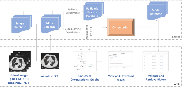
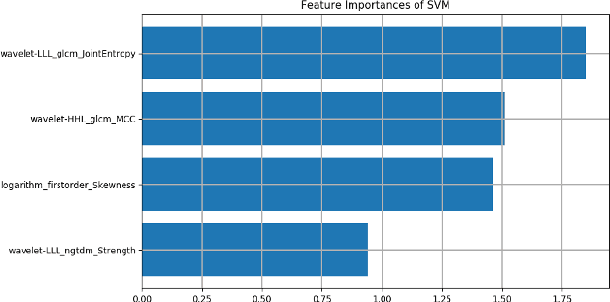
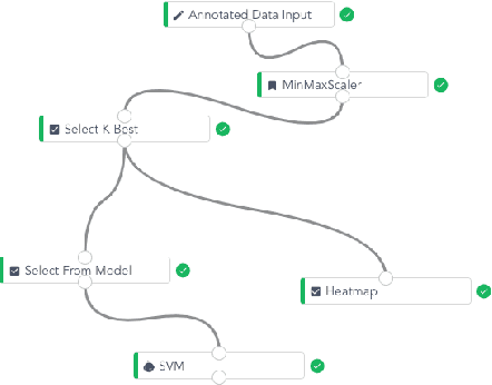
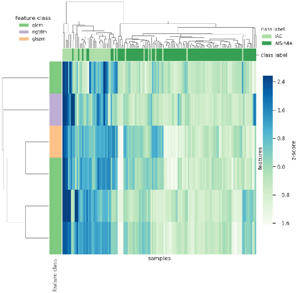
Abstract:To conduct a radiomics or deep learning research experiment, the radiologists or physicians need to grasp the needed programming skills, which, however, could be frustrating and costly when they have limited coding experience. In this paper, we present DARWIN, a flexible research platform with a graphical user interface for medical imaging research. Our platform is consists of a radiomics module and a deep learning module. The radiomics module can extract more than 1000 dimension features(first-, second-, and higher-order) and provided many draggable supervised and unsupervised machine learning models. Our deep learning module integrates state of the art architectures of classification, detection, and segmentation tasks. It allows users to manually select hyperparameters, or choose an algorithm to automatically search for the best ones. DARWIN also offers the possibility for users to define a custom pipeline for their experiment. These flexibilities enable radiologists to carry out various experiments easily.
Improve bone age assessment by learning from anatomical local regions
May 27, 2020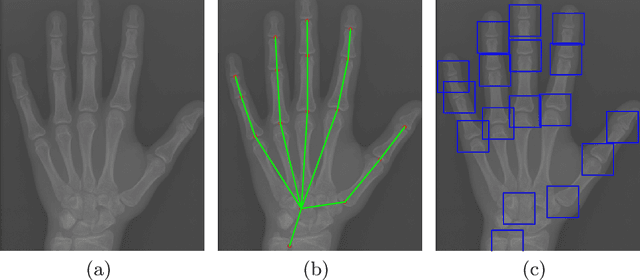
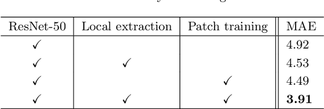
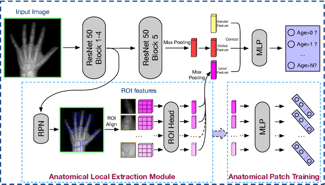

Abstract:Skeletal bone age assessment (BAA), as an essential imaging examination, aims at evaluating the biological and structural maturation of human bones. In the clinical practice, Tanner and Whitehouse (TW2) method is a widely-used method for radiologists to perform BAA. The TW2 method splits the hands into Region Of Interests (ROI) and analyzes each of the anatomical ROI separately to estimate the bone age. Because of considering the analysis of local information, the TW2 method shows accurate results in practice. Following the spirit of TW2, we propose a novel model called Anatomical Local-Aware Network (ALA-Net) for automatic bone age assessment. In ALA-Net, anatomical local extraction module is introduced to learn the hand structure and extract local information. Moreover, we design an anatomical patch training strategy to provide extra regularization during the training process. Our model can detect the anatomical ROIs and estimate bone age jointly in an end-to-end manner. The experimental results show that our ALA-Net achieves a new state-of-the-art single model performance of 3.91 mean absolute error (MAE) on the public available RSNA dataset. Since the design of our model is well consistent with the well recognized TW2 method, it is interpretable and reliable for clinical usage.
Accurate Pulmonary Nodule Detection in Computed Tomography Images Using Deep Convolutional Neural Networks
Aug 29, 2017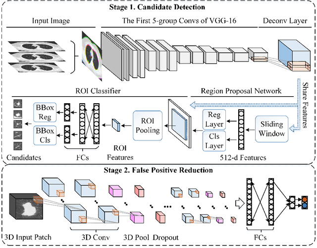
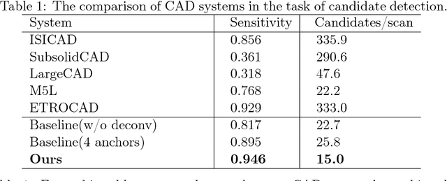
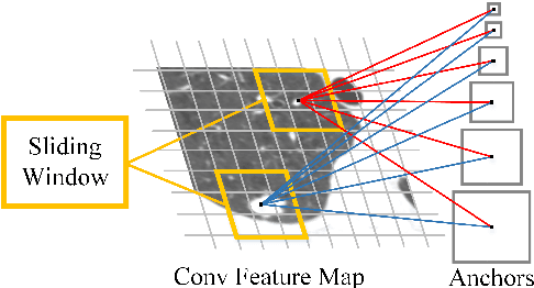

Abstract:Early detection of pulmonary cancer is the most promising way to enhance a patient's chance for survival. Accurate pulmonary nodule detection in computed tomography (CT) images is a crucial step in diagnosing pulmonary cancer. In this paper, inspired by the successful use of deep convolutional neural networks (DCNNs) in natural image recognition, we propose a novel pulmonary nodule detection approach based on DCNNs. We first introduce a deconvolutional structure to Faster Region-based Convolutional Neural Network (Faster R-CNN) for candidate detection on axial slices. Then, a three-dimensional DCNN is presented for the subsequent false positive reduction. Experimental results of the LUng Nodule Analysis 2016 (LUNA16) Challenge demonstrate the superior detection performance of the proposed approach on nodule detection(average FROC-score of 0.891, ranking the 1st place over all submitted results).
 Add to Chrome
 Add to Firefox
 Add to Edge